Cerebral Palsy

Published on 06/06/2015 by admin

Filed under Pediatrics

Last modified 22/04/2025

Print this page

rate 1 star rate 2 star rate 3 star rate 4 star rate 5 star
Your rating: none, Average: 0 (0 votes)

This article have been viewed 8163 times

77 Cerebral Palsy

Cerebral palsy (CP) is an “umbrella diagnosis” describing a group of chronic syndromes of nonprogressive motor and postural dysfunction caused by brain lesions occurring early in development. Although the disorder does not involve degeneration over time, the features may change over the course of a lifetime. This may relate to expected changes across neurodevelopment, evolving musculoskeletal contractures, or changes in symptoms related to comorbidities. CP has an estimated prevalence of about two to three per 1000, with an overall 30-year life expectancy of 90%.

Etiology and Pathogenesis

CP can arise from many different causes and is often multifactorial. The majority of children with CP have brain lesions arising prenatally or in the perinatal period. Approximately 15% of causes occur in the neonatal period or early in childhood. There is no consensus for an upper age limit when brain injury would no longer cause symptoms classified as CP. Although research groups sometimes set a maximum age of brain injury ranging from 2 to 5 years, such cutoffs are less important in clinical practice.

The most important prenatal and perinatal risk factors associated with CP are prematurity and low birth weight. About half of new cases of CP occur in children born weighing less than 1000 g. Intracranial hemorrhage, intrauterine growth retardation, and placental pathology are associated with higher rates of CP. Increasingly, infection (including maternal chorioamnionitis) and stroke have been recognized as contributors. Multiple pregnancy increases the risk for CP, with higher risk conferred with each multiple. Less common factors include congenital abnormalities, such as brain malformation and genetic disorders. In the past, many cases of CP were attributed to birth asphyxia. Although perinatal hypoxia or ischemia may be present in up to 15% of children with later CP, it is now thought that only a small number of cases are attributable to this alone. In fact, other pathologies are identified in more than 95% of cases of CP.

Causes of CP occurring after birth include infection, neonatal stroke, hypoxic brain injury, traumatic brain injury, and kernicterus. Infection may cause CP either through sepsis leading to secondary brain hypoperfusion or through direct central nervous system (CNS) infection. Kernicterus is the sequelae of severe hyperbilirubinemia; this typically causes a choreoathetoid movement disorder in conjunction with gaze abnormalities and hearing impairment.

Clinical Presentation

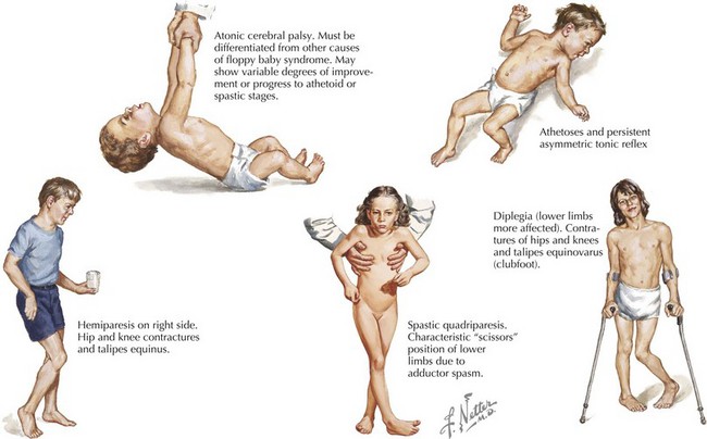
Some children with CP have recognized risk factors (e.g., prematurity and low birth weight) leading to early neurologic and developmental screening. In these children, follow-up visits or early intervention evaluations should include serial assessments of tone and motor development. In the first few months, early signs may include low tone or decreased movements in affected limbs (Figure 77-1). However, not all such clinical presentations progress to CP. Although severe CP may be evident in the first months of life, it is generally difficult to make a definitive diagnosis before 1 year of age. Some clinicians advise not giving the diagnosis to a child younger than 2 years of age. This is because many children identified as having tone or motor abnormalities before 12 months of age will be free of symptoms by school age. Caution is necessary to avoid overdiagnosis in infancy.

In children without recognized risk factors, CP most often presents as delayed motor development. In the first year, this may manifest as difficulty with use of an affected limb. Some children are identified when “toe walking” becomes obvious as toddlers. Delay in reaching gross and fine motor milestones or early asymmetry in limb posture or use should prompt consideration of CP in the differential diagnosis.

In all children with CP, a careful history must confirm a nonprogressive course. The pregnancy and birth history are particularly important in eliciting possible etiologies. If the child was born prematurely or received intensive care as a neonate, specific details should be obtained. Reports of any prior neuroimaging should be obtained. The clinician should assess for other factors causing functional impairment that may mimic motor dysfunction, such as musculoskeletal or sensory difficulties. Current medications must be inventoried, particularly any sedating medications that might affect tone or motor activity at the time of examination. A developmental history is useful in recognizing the trajectory of development, as well as whether nonmotor delays coexist. A family history of neurologic disease should raise suspicion for the possibility of a genetic or metabolic process.

It can be helpful to classify CP by the type and distribution of motor impairment. There is no universally recognized set of categories, and some providers may subtype CP by more specific features, severity, and etiology (when known). Overlap may occur, with at least 10% of cases considered “mixed” CP. Nonetheless, the history and physical examination allow the clinician to classify CP into the broad categories described here.

Spastic Cerebral Palsy

Spastic CP is the most common type, comprising about 70% of cases. It may be further subdivided according to the pattern of limb involvement. Spastic diplegia primarily involves the lower limbs and may be symmetric or asymmetric (see Figure 77-1). Parents often give a history of “tiptoeing” even when walking holding on to furniture. Spastic hemiparesis involves the upper and lower limb on one side, usually with the upper extremity more affected than the lower (see Figure 77-1). Parents may report early handedness (before 2 years) or that the child did not support his or her weight as well on the affected side when crawling. Spastic quadriparesis evenly involves all four limbs (see Figure 77-1).

In the clinical history, patients with spastic CP are more likely to have been at risk for a cortical brain injury. Certain types of spastic CP are more highly associated with certain risks for brain injury. Children with spastic diplegia have likely had injury to both cerebral hemispheres; this most often is periventricular leukomalacia related to premature birth. Children with spastic hemiparesis more likely had unilateral injury, such as prenatal or perinatal stroke. Risk factors for stroke, such as a family history of hypercoagulability, should be assessed. Children with spastic quadriparesis may be the most severely affected. Global brain injury has often occurred, and there is a higher rate of associated cognitive impairment, epilepsy, and other disabilities.

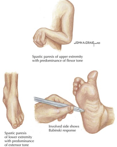
Spastic CP is characterized on physical examination by signs of an upper motor neuron syndrome, indicating a CNS process. On observation, the clinician may see a characteristic pattern in spastic limbs (Figure 77-2). Affected upper extremities have a tendency toward flexion at the elbow and wrist, with pronation of the forearm and fingers kept closed or fisted. Affected hips are adducted and are “scissored” in extreme cases. There is partial flexion at the knees and plantarflexion at the ankles. Torticollis and truncal hyperextension or twisting may also be present. After inspection, the clinician may perform passive range of motion, revealing increased tone in the affected limb. Typical of spasticity is “velocity dependence, meaning resistance increases with more rapid passive movement. In some cases, a spastic “catch” may be elicited in a limb with passive joint extension in which the increase in tone is momentarily reduced. Tone may also be increased in states of stress or excitement and decreased in sleep. In long-standing or severe cases, range of motion may be limited by secondary joint contractures, which typically evolve later in infancy or childhood. Strength may be intact, although this varies, and an element of weakness and motor fatigue is common. Reflex testing reveals hyperreflexia. This is the result of a hyperexcitable stretch reflex and in extreme forms presents as clonus. Extensor plantar responses (i.e., Babinski’s sign) may be present on the affected side. Children with spastic CP may experience difficulties with fine motor movements (e.g., sequential finger movements) or in isolated effortful movements. In mild cases, this may appear as “clumsiness” with easy fatigability. Observation of gait is helpful both in recognizing the pattern of involvement and in assessing functional impairment.

Dyskinetic Cerebral Palsy

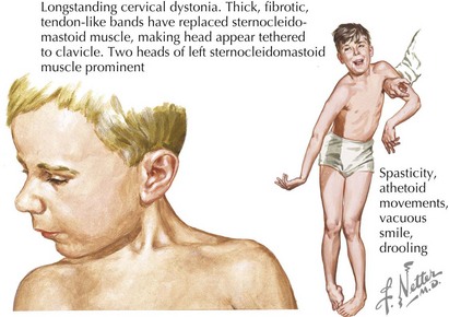
Dyskinetic CP comprises about 15% of CP. Involuntary movements interfering with voluntary motor function are characteristic. Dyskinetic CP may be further divided by primary movement type into choreoathetoid CP and dystonic CP (Figure 77-3). Choreoathetoid movements have a smooth, fluctuating dancelike quality (chorea) and a writhing or wriggling quality (athetosis) in the distal extremities. Dystonia is abnormal sustained movement or posturing in a repetitive, patterned fashion. Although dystonias are also quite common in spastic CP, dystonic CP refers to the case in which dystonia is the primary feature. Even in retrospect, dyskinetic CP may not be noticed until after age 2 or 3 years because infants without CP have some degree of choreoathetoid movement. The movement disorder in dyskinetic CP reflects injury to the basal ganglia. Clinical history should explore risk factors for such injury, including hyperbilirubinemia or hypoxic ischemic injury at term or postnatally. Children with dyskinetic CP are often described as having “fluctuating tone.” This reflects involuntary, often sustained muscle contraction that may be mistaken for intermittent spasticity. The physical examination should include observation of involuntary movements, recognizing that multiple movement types may be present. Sustained posture (e.g., extension of the arms) or distraction may elicit involuntary movements. Serial assessments of tone may be necessary to characterize whether spasticity is also present.

Evaluation and Management

CP is a clinical diagnosis; investigations may clarify the etiology. Neuroimaging is indicated for all children with CP who have not had prior studies (i.e., in the neonatal period). Magnetic resonance imaging (MRI) is the preferred method, revealing abnormalities in 68% to 100% of children with CP. In conjunction with clinical history, MRI can help determine the cause of the brain lesion causing CP. The severity and pattern of lesions on MRI may also be helpful in guiding the prognosis.

Metabolic and genetic testing is not indicated for all children with CP but should be considered if the history or examination is suggestive. Examples include dysmorphic features, a family history suggesting a genetic etiology, or characteristic abnormalities on neuroimaging to suggest a metabolic cause. In cases of hemiplegia or when neuroimaging is consistent with stroke, a prothrombotic screening may be considered. Electroencephalography is only recommended if there are features of the history suggestive of seizure.

The American Academy of Neurology and the Child Neurology Society suggest screening for common comorbidities as part of the initial assessment of children with CP. Indeed, 50% to 75% of children with CP have another disability. As part of the initial assessment, screening for mental retardation, vision and hearing impairment, speech and language disorders, and disorders of oromotor function is advised. Importantly, although such screening for comorbidities is necessary, care should be taken not to assume that there is related cognitive impairment, especially in children with spastic diplegia.

Initial and follow-up visits should include screens for other conditions commonly associated with CP (Table 77-1). The pediatrician should identify these conditions and refer the patient for further evaluation and management when appropriate. Although CP itself is not curable, many commonly associated conditions are highly amenable to treatment and may impact quality of life even more than the motor impairment itself. Furthermore, mortality in CP is almost always caused by secondary difficulties, such as aspiration.

Table 77-1 Conditions Commonly Associated with Cerebral Palsy

System Condition (Prevalence Among Children with Cerebral Palsy)
Neurodevelopmental Speech or language disorder (38%-59%)
  Seizures (25-45%)
  Cognitive impairment (25%-75%)
Sensory Strabismus or refractive error (50%)
  Vision impairment (10%-39%)
  Hearing impairment (4%-15%)
Nutrition Malnutrition
  Excess adipose tissue
Gastrointestinal Constipation (74%)
  Swallowing dysfunction (60%)
  Reflux (32%)
Respiratory Aspiration (41%)
Orthopedic Osteopenia
  Hip subluxation
  Scoliosis
Genitourinary Voiding dysfunction (30%)

Because CP affects multiple domains of function and has many comorbidities, special attention should be paid to the integration of care. Ideally, a multidisciplinary team coordinates the many aspects of a child’s care. An awareness of the psychosocial needs of the family is particularly important. This includes caregiver education, identification of educational needs, facilitation of accommodations, procurement and maintenance of adaptive devices, referral to appropriate community groups or resources (e.g., United Cerebral Palsy, WE MOVE), and discussions of respite care, when appropriate.

In managing the motor dysfunction of CP, many options exist. Unfortunately, there is a paucity of evidence to support specific treatments in children. The brain lesion causing CP is not correctable. Treatment instead targets specific symptoms affecting function or quality of life.

Spasticity

A cornerstone of symptomatic treatment for CP is spasticity management. Improving spasticity can facilitate motor function. This would be the case, for instance, when improved ankle flexion facilitates ambulation for a child with spastic diplegia. In other cases, spasticity management is needed to facilitate caregiving. An example is a child with spastic quadriparesis whose positioning, dressing, and changing become much easier as limb flexion and adduction decrease. Finally, improvements in spasticity may postpone or preclude later orthopedic procedures and joint contractures.

The first step in spasticity management is detailed assessment of the involved muscle groups and the degree of spasticity in each. The modified Ashworth Scale is a grading from 0 (no increased tone) to 4 (rigid) that quantifies the subjective assessment. A quantitative record of spasticity may help track subtle responses to treatment that are gradual over time. The functional impairment the patient and family identify should guide the targets of spasticity management. If spasticity is present but causes no functional limitation, treatment need not be as aggressive.

When spasticity is focal, limited to a few muscle groups, injectable botulinum toxin can be particularly helpful. Botulinum toxin blocks neuromuscular transmission and thus relieves spasticity by partially weakening the targeted muscle. Evidence has supported its efficacy in quantitative measures of spasticity, particularly in the lower extremities, although there is conflicting evidence regarding longer term functional impact. Injections must be repeated every 3 to 8 months and should be done in conjunction with physical therapy (PT), which has a synergistic benefit. A more permanent effect is seen with phenol or ethanol injection, which causes chemodenervation of the targeted muscle.

For global spasticity, oral medications may be used. These include benzodiazepines, baclofen, tizanidine, and dantrolene. Benzodiazepines are systemic sedatives acting on GABA (γ-aminobutyric acid) in the CNS. Diazepam is among the most commonly used. Baclofen likewise works through GABA pathways but through a different mechanism. Tizanidine is an α-adrenergic medication that has been used widely but has no published evidence supporting its use in children. In each of these medications, sedation is a common adverse effect. Children with truncal low tone or drooling may also have a worsening of these symptoms. Frequently, adverse effects limit oral medications’ usefulness for treatment of spasticity and argues for slow titration. Dantrolene acts directly upon muscle and thus does not have associated sedation. However, a potentially fatal hepatotoxicity has been associated with dantrolene, thus limiting its use. When baclofen is effective but associated sedation is limiting, intrathecal baclofen is an option. Intrathecal baclofen may be infused into the cerebrospinal fluid through a surgically placed pump and catheter terminating in the T10 region of the spinal cord. A single test dose infused through a standard lumbar puncture is performed initially to demonstrate benefit. Although this avoids cognitive sedation, pump-related difficulties may occur in 20% to 50% of children.

In some cases, surgical options target spasticity. This includes selective dorsal rhizotomy (SDR). SDR is partial transection of the dorsal lumbosacral spinal roots, interrupting sensory input to selected levels of the spinal cord, which in turn diminishes the motor output at that level. This is most useful for children with spasticity in the lower extremities who are old enough to have achieved a stable gait. Tendon release is another common surgical intervention, which decreases the mechanical tension across a joint that contributes to spasticity.

Finally, PT and occupation therapy (OT) are established components of spasticity management programs despite uncertainty as to the best regimen. PT focuses primarily upon lower limb motor impairment and ambulation issues, and OT addresses mainly upper limb and fine motor concerns. In many cases, a trained professional initiates this therapy and teaches parents exercises to continue at home. These customized regimens seek to build strength, maintain range of motion, reduce excessive muscle tone, and improve motor function. Caregivers should understand that medication and surgical interventions are to be used in conjunction with physical therapy, not as substitutes. Speech therapy (ST) is used to address oromotor and speech-related issues, as previously described.

Persuant to a U.S. federal law, all states are required to have an “early intervention” program for at-risk infants and children from birth to 3 years old, with screening and delivery of PT, OT, ST services usually in the home setting. At age 3 years, a transition to a local school-based system occurs. Physicians need to be aware of these services and promote them for patients with CP and other developmental disorders.