Cerebral hemisphere

Published on 17/03/2015 by admin

Filed under Basic Science

Last modified 22/04/2025

Print this page

rate 1 star rate 2 star rate 3 star rate 4 star rate 5 star
Your rating: none, Average: 0 (0 votes)

This article have been viewed 11065 times

CHAPTER 23 Cerebral hemisphere

The two cerebral hemispheres are the largest major divisions of the brain. Each hemisphere consists of an external highly-convoluted cortex, beneath which lies an extensive internal mass of white matter that partly encloses the basal ganglia. Each hemisphere also contains a lateral ventricle that is continuous with the third ventricle through the interventricular foramen. The two hemispheres are linked by the commissural fibres of the corpus callosum.

The cerebral hemisphere contains primary motor and sensory cortical areas. These represent the highest level at which motor activities are controlled and the highest level to which general and special sensory systems project, providing the neural substrate for conscious experience of sensory stimuli. Association areas are modality-specific and also multimodal, and they enable complex analysis of the internal and external environment and the relationship of the individual to the external world. Parts of the hemisphere, termed the limbic system, have an ancient lineage. They are concerned with memory and the emotional aspects of behaviour, and provide an affective overtone to conscious experience as well as interfacing with subcortical areas, such as the hypothalamus, through which widespread physiological activities are integrated. Other cortical areas, primarily within the frontal region, are concerned with the highest aspects of cognitive function, and contribute to personality, foresight and planning.

The cerebral hemispheres are separated by a deep median cleft, the great longitudinal fissure, which contains a crescent-shaped fold of the dura mater, the falx cerebri (see Ch. 27).

Each cerebral hemisphere may be considered to have superolateral, medial and inferior (basal) surfaces or aspects. The superolateral surface follows the concavity of the cranial vault. The medial surface is flat and vertical, separated from the opposite hemisphere by the great longitudinal fissure and falx cerebri. The inferior surface is irregular and divided into orbital and tentorial regions. The orbital part is concave and lies above the orbital and nasal roofs. The tentorial part lies in the middle cranial fossa; posteriorly it lies above the tentorium cerebelli, which separates it from the superior surface of the cerebellum.

GYRI, SULCI AND LOBES

The surface of the cerebral hemisphere shows a complex pattern of convolutions, or gyri, which are separated by furrows of varying depth known as fissures, or sulci (Fig. 23.1, Fig. 23.2). Some of these are consistently located, others less so. They provide part of the basis for the division of the hemisphere into lobes. The frontal, parietal, temporal and occipital lobes approximately correspond in surface extent to the overlying cranial bones from which they take their names. The anterior and posterior extremities of the hemispheres form the frontal and occipital poles respectively, and the temporal pole is the anterior extremity of the temporal lobe.

image

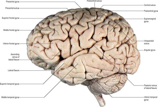
Fig. 23.1 Lateral aspect of the left cerebral hemisphere indicating the major gyri and sulci.

(Dissection by EL Rees; photograph by Kevin Fitzpatrick on behalf of GKT School of Medicine, London; figure enhanced by B. Crossman.)

image

Fig. 23.2 Sagittal section of the brain, with the brain stem removed, showing the major gyri and sulci on the medial aspect of the left cerebral hemisphere.

(Photograph by Kevin Fitzpatrick on behalf of GKT School of Medicine, London; figure enhanced by B. Crossman.)

The insula is a cortical region hidden within the depths of the lateral fissure by overhanging parts (opercula) of the frontal, parietal and temporal lobes. A complex of gyri on the medial aspect of the hemisphere makes up the limbic lobe.

The area of the adult cerebral cortex is approximately 2200 cm2: its convoluted form increases the cortical volume to three times greater than it would be if the surface were smooth.

Two prominent furrows, the lateral (Sylvian) fissure and the central sulcus, are the main features on the superolateral cerebral surface, and they determine its surface divisions.

The lateral fissure is a deep cleft on the lateral and inferior surfaces. It separates the frontal and parietal lobes above from the temporal lobe below and accommodates the middle cerebral vessels. It commences inferiorly at the anterior perforated substance, extending laterally between the orbital surface of the frontal lobe and the anterior pole of the temporal lobe and accommodating the sphenoparietal venous sinus. Reaching the lateral surface of the hemisphere it gives rise to an ascending ramus which runs into the inferior frontal gyrus. The larger posterior ramus runs posteriorly and slightly upwards, across the lateral surface of the hemisphere to end in the parietal lobe. The insula lies in the depths of the lateral fissure.

The central sulcus is the boundary between the frontal and parietal lobes and demarcates the primary motor and somatosensory areas of the cortex, located in the precentral and postcentral gyri, respectively. It starts in or near the superomedial border of the hemisphere, a little behind the midpoint between the frontal and occipital poles. It runs sinuously downwards and forwards to end a little above the posterior ramus of the lateral sulcus.

The superior frontal gyrus, above the superior frontal sulcus, is continuous over the superomedial margin with the medial frontal gyrus and may be incompletely divided. The middle frontal gyrus lies between the superior and inferior frontal sulci. The inferior frontal gyrus lies below the inferior frontal sulcus which is invaded by the ascending ramus of the lateral fissure. In the left hemisphere, the cortical areas around this ramus make up the motor speech area (Broca’s area; areas 44 and 45).

The medial cerebral surface lies within the great longitudinal fissure. The commissural fibres of the corpus callosum lie in the depths of the fissure: the curved anterior part of the corpus callosum, the genu, is continuous below with the rostrum and narrows rapidly as it passes back to the upper end of the lamina terminalis. The cortex immediately below the rostrum is the subcallosal area (parolfactory gyrus). The genu of the corpus callosum continues above into the trunk or body, which arches up and back to a thick, rounded posterior extremity, the splenium. The bilateral vertical laminae of the septum pellucidum are attached to the concave surfaces of the trunk, genu and rostrum, occupying the interval between them and the fornix.

The anterior region of the medial surface of the hemisphere is divided into outer and inner zones by the curved cingulate sulcus, which starts below the rostrum and passes first forwards, then up and finally backwards, conforming to the callosal curvature. Its posterior end turns up to the superomedial margin of the hemisphere approximately 4 cm behind its midpoint, and is posterior to the upper end of the central sulcus. The outer zone, except for its posterior extremity, is part of the frontal lobe, and is subdivided into anterior and posterior areas by a short sulcus, which ascends from the cingulate sulcus above the midpoint of the corpus callosum. The larger anterior area is the medial frontal gyrus; the posterior is the paracentral lobule. The superior end of the central sulcus usually invades the paracentral lobule posteriorly and the precentral gyrus is continuous with the lobule. This area is concerned with movements of the contralateral lower limb and perineal region – clinical evidence suggests that it exercises voluntary control over defaecation and micturition.

The zone under the cingulate sulcus is the cingulate gyrus. Starting below the rostrum, this gyrus follows the callosal curve, separated by the callosal sulcus. It continues round the splenium to the inferior surface, and then into the parahippocampal gyrus, through the narrow isthmus.

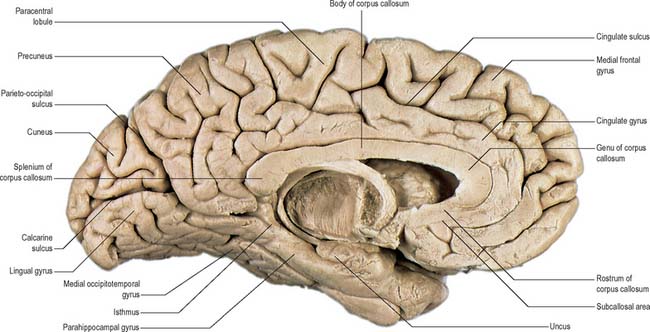
The posterior region of the medial surface is traversed by the parieto-occipital and the calcarine sulci. These two deep sulci converge anteriorly to meet a little posterior to the splenium. The parieto-occipital sulcus marks the boundary between parietal and occipital lobes. It starts on the superomedial margin of the hemisphere approximately 5 cm anterior to the occipital pole, sloping down and slightly forwards to the calcarine sulcus. The calcarine sulcus starts near the occipital pole. Though usually restricted to the medial surface, its posterior end may reach the lateral surface. Directed anteriorly, it joins the parieto-occipital sulcus at an acute angle behind the splenium. Continuing forwards, it crosses the inferomedial margin of the hemisphere, and forms the inferior boundary of the isthmus, which connects the cingulate with the parahippocampal gyrus. The visual cortex lies above and below the posterior part of the calcarine sulcus, behind the junction with the parieto-occipital. The calcarine sulcus is deep and produces an elevation, the calcar avis, in the wall of the posterior horn of the lateral ventricle.

The area posterior to the upturned end of the cingulate sulcus, and anterior to the parieto-occipital sulcus, is the precuneus. It forms the medial surface of the parietal lobe with the part of the paracentral lobule behind the central sulcus. The medial surface of the occipital lobe is formed by the cuneus, a wedge of cortex bounded in front by the parieto-occipital sulcus, below by the calcarine sulcus, and above by the superomedial margin.

The inferior cerebral surface is divided by the stem of the lateral fissure into a small anterior and larger posterior part (Fig. 23.3, Fig. 23.4). The anterior part constitutes the orbital region of the frontal lobe. It is concave and lies above the cribriform plate of the ethmoid, the orbital plate of the frontal, and the lesser wing of the sphenoid bone. A rostrocaudal olfactory sulcus traverses it near its medial margin, overlapped by the olfactory bulb and tract. The medial strip thus demarked is the gyrus rectus. The rest of this surface bears irregular orbital sulci, generally H-shaped, which divide it into the anterior, medial, posterior and lateral orbital gyri.

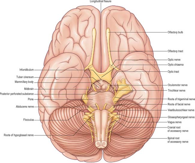
The larger, posterior region of the inferior cerebral surface is partly superior to the tentorium but also to the middle cranial fossa. The collateral sulcus starts near the occipital pole, and extends anteriorly and parallel to the calcarine sulcus, separated from it by the lingual gyrus. The lingual gyrus, between the calcarine and collateral sulci, passes into the parahippocampal gyrus, which begins at the isthmus where it is continuous with the cingulate gyrus. Anteriorly, the parahippocampal gyrus continues into the hook-shaped uncus, which lies lateral to the midbrain and posterolateral to the anterior perforated substance. The uncus is part of the piriform cortex of the olfactory system, phylogenetically one of the oldest parts of the cortex, and is separated from the temporal pole by the rhinal sulcus (fissure) which marks the lateral limit of the entorhinal cortex (area) (Fig. 23.4).

The occipitotemporal sulcus is parallel and lateral to the collateral sulcus, does not usually reach the occipital pole, and is frequently divided. The medial occipitotemporal gyrus extends from the occipital to the temporal pole and is limited medially by the collateral and rhinal sulci and laterally by the occipitotemporal sulcus. The lateral occipitotemporal gyrus is continuous, round the inferolateral margin of the hemisphere, with the inferior temporal gyrus.

CEREBRAL CORTEX

The cerebral cortex is sometimes described as consisting of a phylogenetically old allocortex, which includes archicortex and palaeocortex, and a newer neocortex. These distinctions are reflected in the organizational arrangement of neuronal elements in different regions.

MICROSTRUCTURE

The microscopic structure of the cerebral cortex is an intricate blend of nerve cells and fibres, neuroglia and blood vessels. The neocortex essentially consists of three neuronal cell types. Pyramidal cells are the most abundant. Non-pyramidal cells, also called stellate or granule cells are divided into spiny and non-spiny neurones. All types have been subdivided on the basis of size and shape (Fig. 23.5).

Pyramidal cells have a flask-shaped or triangular cell body ranging from 10 to 80 μm in diameter. The soma gives rise to a single thick apical dendrite and multiple basal dendrites. The apical dendrite ascends towards the cortical surface, tapering and branching, to end in a spray of terminal twigs in the most superficial lamina, the molecular layer. From the basal surface of the cell body, dendrites spread more horizontally, for distances up to 1 mm for the largest pyramidal cells. Like the apical dendrite, the basal dendrites branch profusely along their length. All pyramidal cell dendrites are studded with a myriad of dendritic spines. These become more numerous as distance from the parent cell soma increases. A single slender axon arises from the axon hillock, which is usually situated centrally on the basal surface of the pyramidal neurone. Ultimately, in the vast majority of, if not in all, cases, the axon leaves the cortical grey matter to enter the white matter. Pyramidal cells are thus, perhaps universally, projection neurones. They use an excitatory amino acid, either glutamate or aspartate, as their neurotransmitter.

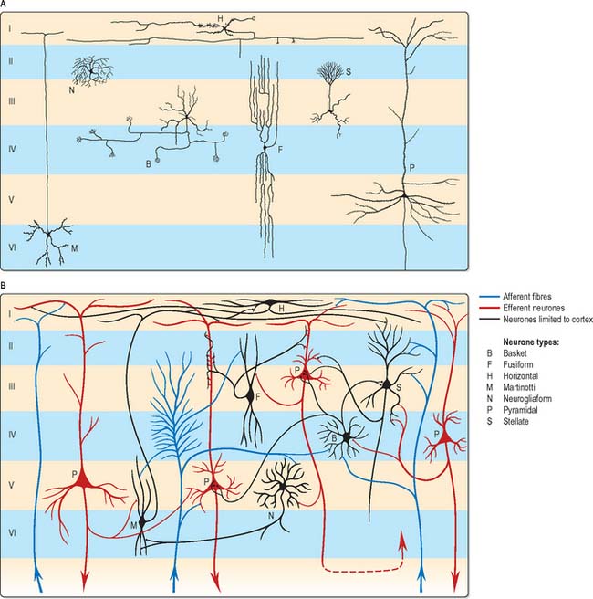
Spiny stellate cells are the second most numerous cell type in the neocortex and for the most part occupy lamina IV. They have relatively small multipolar cell bodies, commonly 6 to 10 μm in diameter. Several primary dendrites, profusely covered in spines, radiate for variable distances from the cell body. Their axons ramify within the grey matter predominantly in the vertical plane. Spiny stellate cells probably use glutamate as their neurotransmitter.

The smallest group comprises the heterogeneous non-spiny or sparsely spinous stellate cells. All are interneurones, and their axons are confined to grey matter. In morphological terms, this is not a single class of cell, but a multitude of different forms, including basket, chandelier, double bouquet, neurogliaform, bipolar/fusiform and horizontal cells. Various types may have horizontally, vertically or radially ramifying axons.

Neurones with mainly horizontally dispersed axons include basket and horizontal cells. Basket cells have a short, vertical axon, which rapidly divides into horizontal collaterals, and these end in large terminal sprays synapsing with the somata and proximal dendrites of pyramidal cells. The cell bodies of horizontal cells lie mainly at the superficial border of lamina II, occasionally deep in lamina I (the molecular or plexiform layer). They are small and fusiform, and their dendrites spread short distances in two opposite directions in lamina I. Their axons often stem from a dendrite, then divide into two branches, which travel away from each other for great distances in the same layer.

Neurones with an axonal arborization predominantly perpendicular to the pial surface include chandelier, double bouquet and bipolar/fusiform cells. Chandelier cells have a variable morphology, although most are ovoid or fusiform and their dendrites arise from the upper and lower poles of the cell body. The axonal arborization, which emerges from the cell body or a proximal dendrite, is characteristic and identifies these neurones. A few cells in the more superficial laminae (II and IIIa) have descending axons, deeper cells (laminae IIIc and IV) have ascending axons, and intermediate neurones (IIIb) often have both. The axons ramify close to the parent cell body and terminate in numerous vertically oriented strings, which run alongside the axon hillocks of pyramidal cells with which they synapse. Double bouquet (or bitufted) cells are found in laminae II and III and their axons traverse laminae II and V. Generally, these neurones have two or three main dendrites, which give rise to a superficial and deep dendritic tuft. A single axon arises usually from the oval or spindle-shaped cell soma and rapidly divides into an ascending and descending branch. These branches collateralize extensively, but the axonal arbor is confined to a perpendicularly extended, but horizontally confined, cylinder, approximately 50 to 80 μm across. Bipolar cells are ovoid with a single ascending and a single descending dendrite, which arise from the upper and lower poles, respectively. These primary dendrites branch sparsely and their branches run vertically to produce a narrow dendritic tree, rarely more than 10 μm across, which may extend through most of the cortical thickness. Commonly, the axon originates from one of the primary dendrites, and rapidly branches to give a vertically elongated, horizontally confined, axonal arbor, which closely parallels the dendritic tree in extent.

The principal recognizable neuronal type is the neurogliaform or spiderweb cell. These small spherical cells, 10 to 12 μm in diameter, are found mainly in laminae II to IV, depending on cortical area. Seven to ten thin dendrites typically radiate out from the cell soma, some branching once or twice to form a spherical dendritic field of approximately 100 to 150 μm diameter. The slender axon arises from the cell body or a proximal dendrite. Almost immediately, it branches profusely within the vicinity of the dendritic field (and usually somewhat beyond), to give a spherical axonal arbor up to 350 μm in diameter.

The majority of non-spiny or sparsely spinous non-pyramidal cells probably use GABA as their principal neurotransmitter. This is almost certainly the case for basket, chandelier, double bouquet, neurogliaform and bipolar cells. Some are also characterized by the coexistence of one or more neuropeptides, including neuropeptide Y, vasoactive intestinal polypeptide (VIP), cholecystokinin, somatostatin and substance P. Acetylcholine is present in a subpopulation of bipolar cells, which may additionally be GABAergic and contain VIP.

Laminar organization

The most apparent microscopical feature of the neocortex when it has been stained for cell bodies or for fibres is its horizontal lamination. The significance of this for understanding cortical functional organization is debatable, but the use of cytoarchitectonic description to identify regions of cortex is common.

Typical neocortex is described as having six layers or laminae lying parallel to the surface (Fig. 23.6).

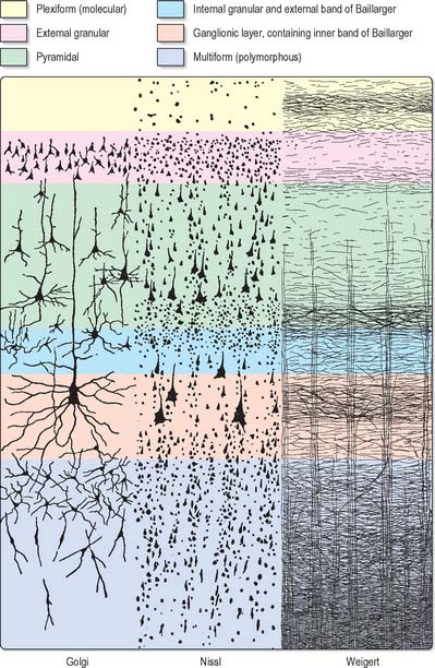
I The molecular or plexiform layer is cell sparse, containing only scattered horizontal cells and their processes enmeshed in a compacted mass of tangential, principally horizontal axons and dendrites. These are afferent fibres, which arise from outside the cortical area, together with intrinsic fibres from cortical interneurones, and the apical dendritic arbors of virtually all pyramidal neurones of the cerebral cortex. In histological sections stained to show myelin, layer I appears as a narrow horizontal band of fibres.

II The external granular lamina contains a varying density of small neuronal cell bodies including both small pyramidal and non-pyramidal cells; the latter may predominate. Myelin fibre stains show mainly vertically arranged processes traversing the layer.

III The external pyramidal lamina contains pyramidal cells of varying sizes, together with scattered non-pyramidal neurones. The size of the pyramidal cells is smallest in the most superficial part of the layer and greatest in the deepest part. This lamina is frequently further subdivided into IIIa, IIIb and IIIc, with IIIa most superficial and IIIc deepest. As in layer II, myelin stains reveal a mostly vertical organization of fibres.

IV The internal granular lamina is usually the narrowest of the cellular laminae. It contains densely packed, small, round cell bodies of non-pyramidal cells, notably spiny-stellate cells and some small pyramidal cells. Within the lamina, in myelin stained sections, a prominent band of horizontal fibres (outer band of Baillarger) is seen.

V The internal pyramidal (ganglionic) lamina typically contains the largest pyramidal cells in any cortical area, though actual sizes vary considerably from area to area. Scattered non-pyramidal cells are also present. In myelin stains, the lamina is traversed by ascending and descending vertical fibres, and also contains a prominent central band of horizontal fibres (inner band of Baillarger).

VI The multiform (or fusiform/pleiomorphic) layer consists of neurones with a variety of shapes, including recognizable pyramidal, spindle, ovoid and many other shaped somata. Typically, most cells are small to medium in size. This lamina blends gradually with the underlying white matter, and a clear demarcation of its deeper boundary is not always possible.

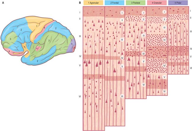
Five regional variations are described in neocortical structure (Fig. 23.7). While all are said to develop from the same six-layered pattern, two types, granular and agranular, are regarded as virtually lacking certain laminae, and are referred to as heterotypical. Homotypical variants, in which all six laminae are found, are called frontal, parietal and polar, names that link them with specific cortical regions in a somewhat misleading manner (e.g. the frontal type also occurs in parietal and temporal lobes).

The agranular type is considered to have diminished, or absent, granular laminae (II and IV), but always contains scattered stellate somata. Large pyramidal neurones are found in the greatest densities in agranular cortex, which is typified by the numerous efferent projections of pyramidal cell axons. Although it is often equated with motor cortical areas such as the precentral gyrus (area 4), agranular cortex also occurs elsewhere, e.g. areas 6, 8 and 44 and parts of the limbic system.

In the granular type of cortex the granular layers are maximally developed, and contain densely packed stellate cells, among which small pyramidal neurones are dispersed. Laminae III and IV are poorly developed or unidentifiable. This type of cortex is particularly associated with afferent projections. However, it does receive efferent fibres, derived from the scattered pyramidal cells, although they are less numerous than elsewhere. Granular cortex occurs in the postcentral gyrus (somatosensory area), striate area (visual area) and superior temporal gyrus (acoustic area), and in small areas of the parahippocampal gyrus. Despite its very high density of stellate cells, especially in the striate area, it is almost the thinnest of the five main types. In the striate cortex the external band of Baillarger (lamina IV) is well defined as the stria (white line) of Gennari.

The other three types of cortex are intermediate forms. In the frontal type, large numbers of small- and medium-sized pyramidal neurones appear in laminae III and V, and granular layers (II and IV) are less prominent. The relative prominence of these major forms of neurone vary reciprocally wherever this form of cortex exists.

The parietal type of cortex contains pyramidal cells, which are mostly smaller in size than in the frontal type. In marked contrast, the granular laminae are wider and contain more of the stellate cells: this kind of cortex occupies large areas in the parietal and temporal lobes. The polar type is classically identified with small areas near the frontal and occipital poles and is the thinnest form of cortex. All six laminae are represented, but the pyramidal layer (III) is reduced in thickness and not so extensively invaded by stellate cells as it is in the granular type of cortex. In both polar and granular types, the multiform layer (VI) is more highly organized than in other types.

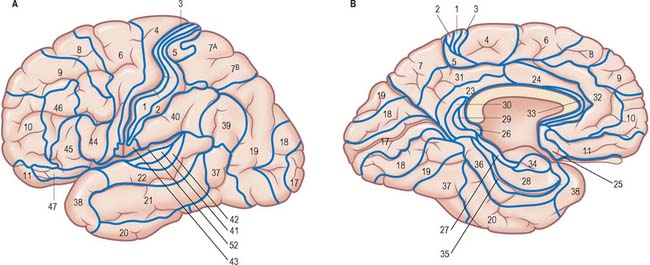
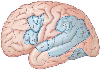
It is customary to refer to some discrete cortical territories not only by their anatomical location in relation to gyri and sulci, but also in relation to their cytoarchitectonic characteristics (Brodmann’s areas) (Fig. 23.8). Some of the areas so defined, e.g. the primary sensory and motor cortices, have clear relevance in terms of anatomical connections and functional significance, others less so.

OVERVIEW OF CORTICAL CONNECTIVITY

All neocortical areas have axonal connections with other cortical areas on the same side (association fibres), the opposite side (commissural fibres), and with subcortical structures (projection fibres).

The primary somatosensory, visual and auditory areas give rise to ipsilateral corticocortical connections to the association areas of the parietal, occipital and temporal lobes, respectively, which then progressively project towards the medial temporal limbic areas, notably the parahippocampal gyrus, entorhinal cortex and hippocampus. Thus, the first (primary) somatic sensory area (SI) projects to the superior parietal cortex (Brodmann’s area 5), which in turn projects to the inferior parietal cortex (area 7). From here connections pass to cortex in the walls of the superior temporal sulcus, and so on to the posterior parahippocampal gyrus, and on into limbic cortex. Similarly, for the visual system, the primary visual cortex (area 17) projects to the parastriate cortex (area 18), which in turn projects to the peristriate region (area 19). Information then flows to inferotemporal cortex (area 20), to cortex in the walls of the superior temporal sulcus, then to medial temporal cortex in the posterior parahippocampal gyrus, and so to limbic areas. The auditory system shows a similar progression from primary auditory cortex to temporal association cortex and so to the medial temporal lobe.

In addition to this stepwise outward progression from sensory areas through posterior association cortex, connections also occur at each stage with parts of the frontal cortex. Thus, taking the somatic sensory system as an example, primary somatic sensory cortex (SI) in the postcentral gyrus is reciprocally connected with the primary motor cortex (area 4) in the precentral gyrus. The next step in the outward progression, the superior parietal lobule (area 5), is interconnected with the premotor cortex (area 6), and this in turn is connected with area 7 in the inferior parietal lobule. This has reciprocal connections with prefrontal association cortex on the lateral surface of the hemisphere (areas 9 and 46), and temporal association areas, which connect with more anterior prefrontal association areas, and, ultimately in the sequence, with orbitofrontal cortex. Similar stepwise links exist between areas on the visual and auditory association pathways in the occipitotemporal lobe and areas of the frontal association cortex. The connections between sensory and association areas are reciprocal.

All neocortical areas are connected with subcortical regions although their density varies between areas. First among these are connections with the thalamus. All areas of the neocortex receive afferents from more than one thalamic nucleus, and all such connections are reciprocal. The vast majority of, if not all, cortical areas project to the striatum, tectum, pons and brain stem reticular formation. Additionally, all cortical areas are reciprocally connected with the claustrum; the frontal cortex connects with the anterior part and the occipital lobe with the posterior part.

All cortical areas receive topographically organized cholinergic projections from the basal forebrain, noradrenergic fibres from the locus coeruleus, serotoninergic fibres from the midbrain raphe nuclei, dopaminergic fibres from the ventral midbrain and histaminergic fibres from the posterior hypothalamus.

Different cortical areas have widely different afferent and efferent connections. Some have connections that are unique, e.g. the corticospinal tract arises from pyramidal cells in a restricted area around the central sulcus.

Widely separated, but functionally interconnected, areas of cortex share common patterns of connections with subcortical nuclei, and within the neocortex. For example, contiguous zones of the striatum, thalamus, claustrum, cholinergic basal forebrain, superior colliculus and pontine nuclei connect with anatomically widely separated areas in the prefrontal and parietal cortex, which are themselves interconnected. In contrast, other functionally distinct cortical regions, e.g. areas in the temporal and parietal cortex, do not share such contiguity in their subcortical connections.

Cortical lamination and cortical connections

The cortical laminae represent, to some extent, horizontal aggregations of neurones with common connections. This is most clearly seen in the lamination of cortical efferent (pyramidal) cells. The internal pyramidal lamina, layer V, gives rise to cortical projection fibres, most notably corticostriate, corticobulbar (including corticopontine) and corticospinal axons. In addition, a significant proportion of feedback corticocortical axons arise from cells in this layer, as do some corticothalamic fibres. Layer VI, the multiform lamina, is the major source of corticothalamic fibres. Supragranular pyramidal cells, predominantly layer III, but also lamina II, give rise primarily to both association and commissural corticocortical pathways. Generally, short corticocortical fibres arise more superficially, and long corticocortical (both association and commissural) axons come from cells in the deeper parts of layer III. Major afferents to a cortical area tend to terminate in layers I, IV and VI. Quantitatively lesser projections end either in the intervening laminae II/III and V, or sparsely throughout the depth of the cortex. Numerically, the largest input to a cortical area tends to terminate mainly in layer IV. This pattern of termination is seen in the major thalamic input to visual and somatic sensory cortex. In general, non-thalamic subcortical afferents to the neocortex, which are shared by widespread areas, tend to terminate throughout all cortical layers, but the laminar pattern of their endings still varies considerably from area to area.

FRONTAL LOBE

The frontal lobe is the rostral region of the hemisphere, anterior to the central sulcus and above the lateral fissure. The precentral gyrus runs parallel to the central sulcus on the superolateral surface and extends onto the medial surface, and is limited anteriorly by the precentral sulcus. The area of the frontal lobe anterior to the precentral sulcus is divided into the superior, middle and inferior frontal gyri (Fig. 23.1). The frontal pole lies in front of these gyri. The ventral surface of the frontal lobe, the orbitofrontal cortex, overlies the bony orbit. The medial surface extends from the frontal pole to the paracentral lobule and consists of the medial frontal cortex and the anterior cingulate cortex.

Primary motor cortex

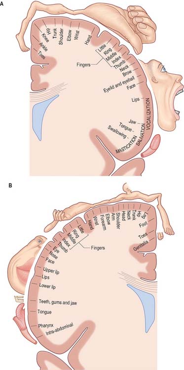
The primary motor cortex (MI) corresponds to the precentral gyrus (area 4), and is the area of cortex with the lowest threshold for eliciting contralateral muscle contraction by electrical stimulation. It contains a detailed topographically organized map (motor homunculus) of the opposite body half, with the head represented most laterally, and the leg and foot represented on the medial surface of the hemisphere in the paracentral lobule (Fig. 23.9). A striking feature is the disproportionate representation of body parts in relation to their physical size: large areas represent the muscles of the face and hand, which are capable of finely controlled or fractionated movements.

The cortex of area 4 is agranular, and layers II and IV are difficult to identify. The most characteristic feature is the presence in lamina V of some extremely large pyramidal cell bodies, Betz cells, which may approach 80 μm in diameter. These neurones project their axons into the corticospinal and corticobulbar tracts.

The major thalamic connections of area 4 are with the ventral posterolateral nucleus (VPL), which in turn receives afferents from the deep cerebellar nuclei. The VPL nucleus also contains a topographic representation of the contralateral body, which is preserved in its point-to-point projection to area 4, where it terminates largely in lamina IV. Other thalamic connections of area 4 are with the centromedian and parafascicular nuclei. These appear to provide the only route through which output from the basal ganglia, routed via the thalamus, reaches the primary motor cortex, since the projection of the internal segment of the globus pallidus to the ventrolateral nucleus of the thalamus is confined to the anterior division, and there is no overlap with cerebellothalamic territory. The anterior part of the ventrolateral nucleus projects to the premotor and supplementary motor areas of cortex with no projection to area 4.

The ipsilateral somatosensory cortex (SI) projects in a topographically organized way to area 4, and the connection is reciprocal. The projection to the motor cortex arises in areas 1 and 2, with little or no contribution from area 3b. Fibres from SI terminate in layers II and III of area 4, where they contact mainly pyramidal neurones. Evidence suggests that neurones activated monosynaptically by fibres from SI, as well as those activated polysynaptically, make contact with layer V pyramidal cells, including Betz cells, which give rise to corticospinal fibres. Movement-related neurones in the motor cortex which can be activated from SI tend to have a late onset of activity, mainly during the execution of movement. It has been suggested that this pathway plays a role primarily in making motor adjustments during a movement. Additional ipsilateral corticocortical fibres to area 4 from behind the central sulcus come from the second somatic sensory area (SII).

Neurones in area 4 are responsive to peripheral stimulation, and have receptive fields similar to those in the primary sensory cortex. Cells located posteriorly in the motor cortex have cutaneous receptive fields, whereas more anteriorly situated neurones respond to stimulation of deep tissues.

The motor cortex receives major frontal lobe association fibres from the premotor cortex and the supplementary motor area and also fibres from the insula. It is probable that these pathways modulate motor cortical activity in relation to the preparation, guidance and temporal organization of movements. Area 4 sends fibres to, and receives fibres from, its contralateral counterpart, and also projects to the contralateral supplementary motor cortex.

Apart from its contribution to the corticospinal tract, the motor cortex has diverse subcortical projections. The connections to the striatum and pontine nuclei are heavy. It also projects to the subthalamic nucleus. The motor cortex sends projections to all nuclei in the brain stem, which are themselves the origin of descending pathways to the spinal cord, namely the reticular formation, the red nucleus, the superior colliculus, the vestibular nuclei and the inferior olivary nucleus.

Corticospinal tract

The corticospinal or pyramidal tract provides direct control by the cerebral cortex over motor centres of the spinal cord. An homologous pathway to the brain stem, the corticobulbar projection, fulfils a similar function in relation to motor nuclei of the brain stem. The corticospinal tract does not originate solely from the motor cortex, but is conveniently considered in conjunction with it.

The percentage of corticospinal fibres that arise from the primary motor cortex may actually be quite small, probably in the region of 20 to 30%. They arise from pyramidal cells in layer V and give rise to the largest diameter corticospinal axons. There is also a widespread origin from other parts of the frontal lobe, including the premotor cortex and the supplementary motor area. Many axons from the frontal cortex, notably the motor cortex, terminate in the ventral horn of the spinal cord. In cord segments mediating dexterous hand and finger movements they terminate in the lateral part of the ventral horn, in close relationship to motor neuronal groups. A small percentage establish direct monosynaptic connections with α motor neurones.

Between 40 and 60% of pyramidal tract axons arise from parietal areas, including area 3a, area 5 of the superior parietal lobe, and SII in the parietal operculum. The majority of parietal fibres to the spinal cord terminate in the deeper layers of the dorsal horn.

Motor cortical neurones are active in relation to the force of contraction of agonist muscles; their relation to amplitude of movement is less clear. Their activity precedes the onset of electromyographic activity by 50 to 100 milliseconds, suggesting a role for cortical activation in generating rather than monitoring movement.

Premotor cortex

Immediately in front of the primary motor cortex lies Brodmann’s area 6. It extends onto the medial surface, where it becomes contiguous with area 24 in the cingulate gyrus, anterior and inferior to the paracentral lobule. A number of functional motor areas are contained within this cortical region. Lateral area 6, the area over most of the lateral surface of the hemisphere, corresponds to the premotor cortex.

The premotor cortex is divided into a dorsal and a ventral area (PMd and PMv respectively) on functional grounds, and on the basis of ipsilateral corticocortical association connections.

The major thalamic connections of the premotor cortex are with the anterior division of the ventrolateral nucleus and with the centromedian, parafascicular and centrolateral components of the intralaminar nuclei. Subcortical projections to the striatum and pontine nuclei are prominent, and this area also projects to the superior colliculus and the reticular formation. Both dorsal and ventral areas contribute to the corticospinal tract. Commissural connections are with the contralateral premotor, motor and superior parietal (area 5) cortex. Ipsilateral corticocortical connections with area 5 in the superior parietal cortex, and inferior parietal area 7b are common to both dorsal and ventral subdivisions of the premotor cortex, and both send a major projection to the primary motor cortex. The dorsal premotor area also receives fibres from the posterior superior temporal cortex and projects to the supplementary motor cortex. The frontal eye field (area 8) projects to the dorsal subdivision. Perhaps the greatest functionally significant difference in connectivity between the two premotor area subdivisions is that the dorsal premotor area receives fibres from the dorsolateral prefrontal cortex, whereas the ventral subdivision receives fibres from the ventrolateral prefrontal cortex. All of these association connections are likely to be, or are known to be, reciprocal.

Neuronal activity in the premotor cortex in relation to both preparation for movement and movement itself has been extensively studied experimentally. Direction selectivity for movement is a common feature of many premotor neurones. In behavioural tasks, neurones in the dorsal premotor cortex show anticipatory activity and task-related discharge as well as direction selectivity, but little or no stimulus-related changes. The dorsal premotor cortex is probably important in establishing a motor set or intention, contributing to motor preparation in relation to internally guided movement. In contrast, ventral premotor cortex is more related to the execution of externally (especially visually) guided movements in relation to a specific external stimulus.

Frontal eye field

The frontal eye field corresponds to parts of Brodmann’s areas 6, 8 and 9 (Fig. 23.10). It receives its major thalamic projection from the parvocellular mediodorsal nucleus, with additional afferents from the medial pulvinar, the ventral anterior nucleus and the suprageniculate–limitans complex, and connects with the paracentral nucleus of the intralaminar group. The thalamocortical pathways to the frontal eye field form part of a pathway from the superior colliculus, the substantia nigra and the dentate nucleus of the cerebellum. The frontal eye field has extensive ipsilateral corticocortical connections, receiving fibres from several visual areas in the occipital, parietal and temporal lobes, including the medial temporal area (V5) and area 7a. There is also a projection from the superior temporal gyrus, which is auditory rather than visual in function. From within the frontal lobe, the frontal eye field receives fibres from the ventrolateral and dorsolateral prefrontal cortices. It projects to the dorsal and ventral premotor cortices and to the medial motor area, probably to the supplementary eye field adjacent to the supplementary motor area proper. It projects prominently to the superior colliculus, to the pontine gaze centre within the pontine reticular formation, and to other oculomotor related nuclei in the brain stem. As its name implies, it is important in the control of eye movements.

Supplementary motor cortex

The supplementary motor area (SMA; MII) lies medial to area 6, and extends from the most superolateral part to the medial surface of the hemisphere. Area 24 in the cingulate gyrus adjacent to area 6 contains several motor areas, which are termed cingulate motor areas. An additional functional subdivision, the preSMA, lies anterior to the supplementary motor area on the medial surface of the cortex. In the present discussion, these additional medial motor areas are included with the supplementary motor cortex.

The supplementary motor area receives its major thalamic input from the anterior part of the ventral lateral nucleus, which in turn is the major recipient of fibres from the internal segment of the globus pallidus. Additional thalamic afferents are from the ventral anterior nucleus, the intralaminar nuclei, notably the centrolateral and centromedial nuclei, and also from the mediodorsal nucleus. The connections with the thalamus are reciprocal. The supplementary motor cortex receives connections from widespread regions of the ipsilateral frontal lobe, including from the primary motor cortex, the dorsal premotor area, the dorsolateral and ventrolateral prefrontal, medial prefrontal and orbitofrontal cortex and the frontal eye field. These connections are reciprocal, but the major ipsilateral efferent pathway is to the motor cortex. Parietal lobe connections of the supplementary motor cortex are with the superior parietal area 5 and possibly inferior parietal area 7b. Contralateral connections are with the supplementary motor area, and motor and premotor cortices of the contralateral hemisphere. Subcortical connections, other than with the thalamus, pass to the striatum, subthalamic nucleus and pontine nuclei, the brain stem reticular formation and the inferior olivary nucleus. The supplementary motor area makes a substantial contribution to the corticospinal tract, contributing as much as 40% of the fibres from the frontal lobe.

The supplementary motor area contains a representation of the body in which the leg is posterior and the face anterior, with the upper limb between them. Its role in the control of movement is primarily in complex tasks which require temporal organization of sequential movements and in the retrieval of motor memory.

Stimulation of the supplementary motor area in conscious patients has been reported to elicit the sensation of an urge to move, or of anticipation that a movement is about to occur. A region anterior to the supplementary motor area for face representation (areas 44, 45) is important in vocalization and speech production (Fig. 23.10).

Prefrontal cortex

The prefrontal cortex on the lateral surface of the hemisphere comprises predominantly Brodmann’s areas 9, 46 and 45 (Fig. 23.8). In non-human primates, two subdivisions of the lateral prefrontal cortex are recognized, a dorsal area equivalent to area 9, and perhaps including the superior part of area 46, and a ventral area, consisting of the inferior part of area 46 and area 45. Areas 44 and 45 are particularly notable in man since, in the dominant hemisphere, they constitute the motor speech area (Broca’s area). Both the dorsolateral and ventrolateral prefrontal areas receive their major thalamic afferents from the mediodorsal nucleus, and there are additional contributions from the medial pulvinar, the ventral anterior nucleus and from the paracentral nucleus of the anterior intralaminar group. The dorsolateral area receives long association fibres from the posterior and middle superior temporal gyrus (including auditory association areas), from parietal area 7a, and from much of the middle temporal cortex. From within the frontal lobe it also receives projections from the frontal pole (area 10), and from the medial prefrontal cortex (area 32) on the medial surface of the hemisphere. It projects to the supplementary motor area, the dorsal premotor cortex and the frontal eye field. All these thalamic and corticocortical connections are reciprocal. Commissural connections are with the homologous area, and with the contralateral inferior parietal cortex. The ventrolateral prefrontal area receives long association fibres from both area 7a and area 7b of the parietal lobe, from auditory association areas of the temporal operculum, from the insula and from the anterior part of the lower bank of the superior temporal sulcus. From within the frontal lobe it receives fibres from the anterior orbitofrontal cortex and projects to the frontal eye field and the ventral premotor cortex. It connects with the contralateral homologous area via the corpus callosum. These connections are probably all reciprocal.

The cortex of the frontal pole (area 10) receives thalamic input from the mediodorsal nucleus, the medial pulvinar and the paracentral nucleus. It is reciprocally connected with the cortex of the temporal pole, the anterior orbitofrontal cortex, and the dorsolateral prefrontal cortex. The orbitofrontal cortex connects with the mediodorsal, anteromedial, ventral anterior, medial pulvinar, paracentral and midline nuclei of the thalamus. Cortical association pathways come from the inferotemporal cortex, the anterior superior temporal gyrus and the temporal pole. Within the frontal lobe it has connections with the medial prefrontal cortex, the ventrolateral prefrontal cortex and medial motor areas. Commissural and other connections follow the general pattern for all neocortical areas.

The medial prefrontal cortex is connected with the mediodorsal, ventral anterior, anterior medial pulvinar, paracentral, midline and suprageniculate–limitans nuclei of the thalamus. It receives fibres from the anterior cortex of the superior temporal gyrus. Within the frontal lobe, it has connections with the orbitofrontal cortex, and the medial motor areas of the dorsolateral prefrontal cortex.

PARIETAL LOBE

The parietal lobe lies posterior to the central sulcus. On the medial aspect of the hemisphere its boundary with the occipital lobe is clearly demarcated by the deep parieto-occipital sulcus. On the lateral aspect of the hemisphere its boundaries with the occipital and temporal lobes are less distinct and somewhat arbitrary. The inferior boundary is the posterior ramus of the lateral fissure and its imaginary posterior prolongation.

The lateral aspect of the parietal lobe is divided into three areas by postcentral and intraparietal sulci (Fig. 23.1). The postcentral sulcus, often divided into upper and lower parts, is posterior and parallel to the central sulcus. Inferiorly, it ends above the posterior ramus of the lateral fissure. The postcentral gyrus or primary somatosensory cortex lies between the central and postcentral sulci. Posterior to the postcentral sulcus there is a large area, subdivided by the intraparietal sulcus. It usually starts in the postcentral sulcus near its midpoint and extends posteroinferiorly across the parietal lobe, dividing the latter into superior and inferior parietal lobules.

The superior parietal lobule, between the superomedial margin of the hemisphere and the intraparietal sulcus, is continuous anteriorly with the postcentral gyrus round the upper end of the postcentral sulcus.

The inferior parietal lobule, below the intraparietal sulcus and behind the lower part of the postcentral sulcus, is divided into three. The anterior part is the supramarginal gyrus, which arches over the upturned end of the lateral fissure. It is continuous anteriorly with the lower part of the postcentral gyrus and posteroinferiorly with the superior temporal gyrus. The middle part of the inferior parietal lobule, called the angular gyrus, arches over the end of the superior temporal sulcus and is continuous posteroinferiorly with the middle temporal gyrus. The posterior part of the inferior parietal lobule arches over the upturned end of the inferior temporal sulcus on to the occipital lobe.

Somatosensory cortex

The postcentral gyrus corresponds to the primary somtosensory cortex (SI; Brodmann’s areas 3a, 3b, 1 and 2). Area 3a lies most anteriorly, apposing area 4, the primary motor cortex of the frontal lobe; area 3b is buried in the posterior wall of the central sulcus; area 1 lies along the posterior lip of the central sulcus; and area 2 occupies the crown of the postcentral gyrus.

The primary somatosensory cortex contains within it a topographical map of the contralateral half of the body. The face, tongue and lips are represented inferiorly, the trunk and upper limb are represented on the superolateral aspect, and the lower limb on the medial aspect of the hemisphere, giving rise to the familiar ‘homunculus’ map (Fig. 23.9).

The somatosensory properties of SI depend on its thalamic input from the ventral posterior nucleus of the thalamus, which in turn receives the medial lemniscal, spinothalamic and trigeminothalamic pathways. The nucleus is divided into a ventral posterolateral part, which receives information from the trunk and limbs, and a ventral posteromedial part, in which the head is represented. Within the ventral posterior nucleus, neurones in the central core respond to cutaneous stimuli and those in the most dorsal anterior and posterior parts, which arch as a ‘shell’ over this central core, respond to deep stimuli. This is reflected in the differential projections to SI: the cutaneous central core projects to 3b, the deep tissue-responsive neurones send fibres to areas 3a and 2, and an intervening zone projects to area 1. Within the ventral posterior nucleus, anteroposterior rods of cells respond with similar modality and somatotopic properties. They appear to project to restricted focal patches in SI of approximately 0.5 mm width, which form narrow strips mediolaterally along SI. The laminar termination of thalamocortical axons from the ventral posterior nucleus is different in the separate cytoarchitectonic subdivisions of SI. In 3a and 3b, these axons terminate mainly in layer IV and the adjacent deep part of layer III, whereas in areas 1 and 2 they end in the deeper half of layer III, avoiding lamina IV. Additional thalamocortical fibres to SI arise from the intralaminar system, notably the centrolateral nucleus.

There is a complex internal connectivity within SI. An apparently stepwise hierarchical progression of information processing occurs from area 3b through area 1 to area 2. Outside the postcentral gyrus, SI has ipsilateral corticocortical association connections with a second somatosensory area (SII); area 5 in the superior parietal lobe; area 4, the motor cortex, in the precentral gyrus; and the supplementary motor cortex in the medial part of area 6 of the frontal lobe.

SI has reciprocal commissural connections with its contralateral homologue, with the exception that the cortices containing the representation of the distal extremities are relatively devoid of such connections. Callosal fibres in SI arise mainly from the deep part of layer III and terminate in layers I to IV. Pyramidal cells contributing callosal projections receive monosynaptic thalamic and commissural connections.

SI has reciprocal subcortical connections with the thalamus and claustrum, and receives afferents from the basal nucleus of Meynert, the locus coeruleus and the midbrain raphe. It has other prominent subcortical projections. Corticostriatal fibres, arising in layer V, pass mainly to the putamen of the same side. Corticopontine and corticotectal fibres from SI arise in layer V. SI projects to the main pontine nuclei, and to the pontine tegmental reticular nucleus. In addition, axons arising in SI pass to the dorsal column nuclei and the spinal cord. Corticospinal pyramidal cells are found in layer V of SI. The topographical representation in the cortex is preserved in terms of the spinal segments to which different parts of the postcentral gyrus project. Thus, the arm representation projects to the cervical enlargement, the leg representation to the lumbosacral enlargement, and so on. Within the grey matter of the spinal cord, fibres from SI terminate in the dorsal horn, in Rexed’s laminae 3 to 5: fibres from 3b and 1 end more dorsally, and those from area 2 more ventrally.

The second somatosensory area (SII) lies along the upper bank of the lateral fissure, posterior to the central sulcus. SII contains a somatotopic representation of the body, with the head and face most anteriorly, adjacent to SI, and the sacral regions most posteriorly. SII is reciprocally connected with the ventral posterior nucleus of the thalamus in a topographically organized fashion. Some thalamic neurones probably project to both SI and SII via axon collaterals. Other thalamic connections of SII are with the posterior group of nuclei and with the intralaminar central lateral nucleus. SII also projects to laminae IV to VII of the dorsal horn of the cervical and thoracic spinal cord, the dorsal column nuclei, the principal trigeminal nucleus, and the periaqueductal grey matter of the midbrain.

Within the cortex, SII is reciprocally connected with SI in a topographically organized manner, and projects to the primary motor cortex. SII also projects in a topographically organized way to the lateral part of area 7 (area 7b) in the superior parietal lobe, and makes connections with the posterior cingulate gyrus. Both right and left SII areas are interconnected across the corpus callosum, although distal limb representations are probably excluded. There are additional callosal projections to SI and area 7b.

Experimental studies show that neurones in SII respond particularly to transient cutaneous stimuli, e.g. brush strokes or tapping, which are characteristic of the responses of Pacinian corpuscles in the periphery. They show little response to maintained stimuli.

Superior and inferior parietal lobules

Posterior to the postcentral gyrus, the superior part of the parietal lobe is composed of areas 5, 7a and 7b (Fig. 23.8). Area 5 receives a dense feedforward projection from all cytoarchitectonic areas of SI in a topographically organized manner. The thalamic afferents to this area come from the lateral posterior nucleus and from the central lateral nucleus of the intralaminar group. Ipsilateral corticocortical fibres from area 5 go to area 7, the premotor and supplementary motor cortices, the posterior cingulate gyrus, and the insular granular cortex. Commissural connections between area 5 on both sides tend to avoid the areas of representation of the distal limbs. The response properties of cells in area 5 are more complex than in SI, with larger receptive fields and evidence of submodality convergence. Area 5 contributes to the corticospinal tract.

In non-human primates, the inferior parietal lobe is area 7. In man, this area is more superior, and areas 39 and 40 intervene inferiorly. The counterparts for the latter areas in monkeys are unclear and little experimental evidence is available on their connections and functions. Their role in human cerebral processing is discussed below. In the monkey, area 7b receives somatosensory inputs from area 5 and SII. Connections pass to the posterior cingulate gyrus (area 23), insula and temporal cortex. Area 7b is reciprocally connected with area 46 in the prefrontal cortex and the lateral part of the premotor cortex. Commissural connections of area 7b are with the contralateral homologous area, and with SII, the insular granular cortex and area 5. Thalamic connections are with the medial pulvinar nucleus and the intralaminar paracentral nucleus.

In monkeys, area 7a is not related to the cortical pathways for somatosensory processing, but instead forms part of a dorsal cortical pathway for spatial vision. The major ipsilateral corticocortical connections to area 7a are derived from visual areas in the occipital and temporal lobes. In the ipsilateral hemisphere, area 7a has connections with the posterior cingulate cortex (area 24) and with areas 8 and 46 of the frontal lobe. Commissural connections are with its contralateral homologue. Area 7a is connected with the medial pulvinar and intralaminar paracentral nuclei of the thalamus. In experimental studies, neurones within area 7a are visually responsive: they relate largely to peripheral vision, respond to stimulus movement, and are modulated by eye movement.

TEMPORAL LOBE

The temporal lobe is inferior to the lateral fissure. It is limited behind by an arbitrary line from the preoccipital incisure to the parieto-occipital sulcus which meets the superomedial margin of the hemisphere approximately 5 cm from the occipital pole. Its lateral surface is divided into three parallel gyri by the superior and inferior temporal sulci.

The superior temporal sulcus begins near the temporal pole and slopes slightly up and backwards parallel to the posterior ramus of the lateral sulcus. Its end curves up into the parietal lobe. The inferior temporal sulcus is subjacent and parallel to the superior and is often broken into two or three short sulci. Its posterior end also ascends into the parietal lobe, posterior and parallel to the upturned end of the superior sulcus.

The three parallel gyri on the lateral surface of the temporal lobe are the superior (area 22); middle (area 21); and inferior (area 20) temporal gyri. The temporal pole (area 38) lies in front of the termination of these gyri. Along its superior margin the superior temporal gyrus is continuous with gyri in the floor of the posterior ramus of the lateral sulcus. These vary in number, and extend obliquely anterolaterally from the circular sulcus around the insula as the transverse temporal gyri of Heschl (Fig. 23.11). The anterior transverse temporal gyrus and adjoining part of the superior temporal gyrus are auditory in function, and are considered to be Brodmann’s area 42. The anterior gyrus is approximately area 41.

The cortex of the medial temporal lobe includes major subdivisions of the limbic system, such as the hippocampus and entorhinal cortex. Areas of neocortex adjacent to these limbic regions are grouped together as medial temporal association cortex. The temporal and frontal lobes are expanded enormously in man, which poses a problem when relating physiological and anatomical studies of non-human primates to human brain topography. In general, the commonly studied old world monkeys lack a middle temporal gyrus.

Auditory cortex

The temporal operculum houses the primary auditory cortex, AI. This is coextensive with the granular area 41 in the transverse temporal gyri. Surrounding areas constitute auditory association cortex. The primary auditory cortex is reciprocally connected with all subdivisions of the medial geniculate nucleus, and may receive additional thalamocortical projections from the medial pulvinar. The geniculocortical fibres terminate densely in layer IV. AI contains a tonotopic representation of the cochlea in which high frequencies are represented posteriorly, and low frequencies anteriorly. Single-cell responses are to single tones of a narrow frequency band. Cells in single vertical electrode penetrations share an optimum frequency response.

The auditory cortex interconnects with prefrontal cortex, though the projections from AI are small. In general, posterior parts of the operculum project to areas 8 and 9. Central parts project to areas 8, 9 and 46. More anterior regions project to areas 9 and 46, to area 12 on the orbital surface of the hemisphere, and to the anterior cingulate gyrus on the medial surface. Contralateral corticocortical connections are with the same and adjacent regions in the other hemisphere. Onward connections of the auditory association pathway converge with those of the other sensory association pathways in cortical regions within the superior temporal sulcus.

Evidence suggests that area 21, the middle temporal cortex, is polysensory in man, and that it connects with auditory, somatosensory and visual cortical association pathways. The auditory association areas of the superior temporal gyrus project in a complex ordered fashion to the middle temporal gyrus, as does the parietal cortex. The middle temporal gyrus connects with the frontal lobe: the most posterior parts project to posterior prefrontal cortex, areas 8 and 9, while intermediate regions connect more anteriorly with areas 19 and 46. Further forwards, the middle temporal region has connections with anterior prefrontal areas 10 and 46, and with anterior orbitofrontal areas 11 and 14. The most anterior middle temporal cortex is connected with the posterior orbitofrontal cortex, area 12, and with the medial surface of the frontal pole. Further forwards, this middle temporal region projects to the temporal pole and the entorhinal cortex. Thalamic connections are with the pulvinar nuclei and the intralaminar group. Other subcortical connections follow the general pattern for all cortical areas. Some projections (e.g. to the pons), particularly from anteriorly in the temporal lobe, are minimal. Physiological responses of cells in this middle temporal region show convergence of different sensory modalities, and many neurones respond to faces.

The inferior temporal cortex, area 20, is a higher visual association area. The posterior inferior temporal cortex receives major ipsilateral corticocortical fibres from occipitotemporal visual areas, notably V4. It contains a coarse retinotopic representation of the contralateral visual field, and sends a major feedforward pathway to the anterior part of the inferior temporal cortex. The anterior inferior temporal cortex projects onto the temporal pole and to paralimbic areas on the medial surface of the temporal lobe. Additional ipsilateral association connections of the inferior temporal cortex are with the anterior middle temporal cortex, in the walls of the superior temporal gyrus, and with visual areas of the parietotemporal cortex. Frontal lobe connections are with area 46 in the dorsolateral prefrontal cortex (posterior inferior temporal), and with the orbitofrontal cortex (anterior inferior temporal). The posterior area also connects with the frontal eye field. Reciprocal thalamic connections are with the pulvinar nuclei; the posterior part is related mainly to the inferior and lateral nuclei, and the anterior part to the medial and adjacent lateral pulvinar. Intralaminar connections are with the paracentral and central medial nuclei. Other subcortical connections conform to the general pattern of all cortical regions. Callosal connections are between corresponding areas and the adjacent visual association areas of each hemisphere.

The cortex of the temporal pole receives feedforward projections from widespread areas of temporal association cortex which are immediately posterior to it. The dorsal part receives predominantly auditory input from the anterior part of the superior temporal gyrus. The inferior part receives visual input from the anterior area of the inferior temporal cortex. Other ipsilateral connections are with the anterior insular, the posterior and medial orbitofrontal, and the medial prefrontal cortices. The temporal pole projects onwards into limbic and paralimbic areas. Thalamic connections are mainly with the medial pulvinar nucleus, and with intralaminar and midline nuclei. Other subcortical connections are as for the cortex in general, although some projections, such as that to the pontine nuclei, are very small. Physiological responses of cells in this and more medial temporal cortex correspond particularly to behavioural performance and to the recognition of high-level aspects of social stimuli.

Nuclei of the amygdala project to, and receive fibres from, neocortical areas, predominantly of the temporal lobe, and possibly inferior parietal cortex. The density of these pathways increases towards the temporal pole.

INSULA

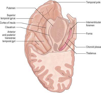
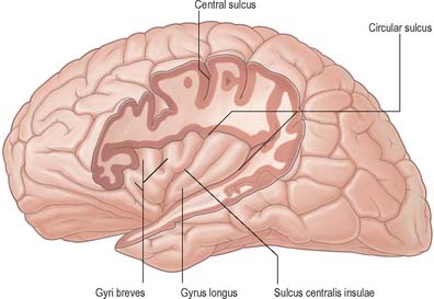
The insula (Fig. 23.12) lies deep in the floor of the lateral fissure, almost surrounded by a circular sulcus, and overlapped by adjacent cortical areas, the opercula. The frontal operculum is between the anterior and ascending rami of the lateral fissure, forming a triangular division of the inferior frontal gyrus. The frontoparietal operculum, between ascending and posterior rami of the lateral fissure, consists of the posterior part of the inferior frontal gyrus, the lower ends of the precentral and postcentral gyri, and the lower end of the anterior part of the inferior parietal lobule. The temporal operculum, below the posterior ramus of the lateral fissure, is formed by superior temporal and transverse temporal gyri. Anteriorly, the inferior region of the insula adjoins the orbital part of the inferior frontal gyrus.

When the opercula are removed, the insula appears as a pyramidal area, its apex beneath and near the anterior perforated substance, where the circular sulcus is deficient. The medial part of the apex is termed the limen insulae (gyrus ambiens). A central insular sulcus, which slants posterosuperiorly from the apex, divides the insular surface into a large anterior and a small posterior part. The anterior part is divided by shallow sulci into three or four short gyri, whereas the posterior part is one long gyrus, often divided at its upper end. The cortex of the insula is continuous with that of its opercula in the circular sulcus. The insula is approximately coextensive with the subjacent claustrum and putamen.

Cytoarchitectonically, three zones are recognized within the insula. Anteriorly, and extending caudally into the central insula, the cortex is agranular. It is surrounded by a belt of dysgranular cortex, in which laminae II and III can be recognized, and this in turn is surrounded by an outer zone of homotypical granular cortex which extends to the caudal limit of the insula.

Thalamic afferents to the insula come from subdivisions of the ventral posterior nucleus and of the medial geniculate body, from the oral and medial parts of the pulvinar, the suprageniculate/nucleus limitans complex, the mediodorsal nucleus and the nuclei of the intralaminar and midline groups. It appears that the anterior (agranular) cortex is connected predominantly with the mediodorsal and ventroposterior nuclei, while the posterior (granular) cortex is connected predominantly with the pulvinar and the ventral posterior nuclei. The other nuclear groups appear to connect with all areas.

Ipsilateral cortical connections of the insula are diverse. Somatosensory connections are with SI, SII and surrounding areas, area 5 of the superior parietal lobe and area 7b of the inferior parietal lobe. The insular cortex also has connections with the orbitofrontal cortex. Several auditory regions in the temporal lobe interconnect with the posterior granular insula and the dysgranular cortex more anteriorly. Connections with visual areas are virtually absent. The anterior agranular cortex of the insula appears to have connections primarily with olfactory, limbic and paralimbic structures, including, most prominently, the amygdala. Little is known about the functions of the human insula other than that the anterior insular cortex appears to have a role in olfaction and taste and the posterior part in somatosensory functions. The latter has also been implicated in language functions, which resonates with the possibility that higher order auditory association pathways may pass via areas in the insula.

Claustrum

The claustrum (see Fig. 23.27) is a thin sheet of grey matter lying deep to the insula. It is approximately coextensive with the insula, from which it is separated by the extreme capsule. Medially, the claustrum is separated from the putamen by the external capsule. It is thickest anteriorly and inferiorly, where it becomes continuous with the anterior perforated substance, amygdala and prepiriform cortex. In animals, it has reciprocal, topographically organized, connections with many regions of the neocortex. Little is known about the connections and functional significance of the claustrum in the human brain.

OCCIPITAL LOBE

The occipital lobe lies behind an arbitrary line joining the preoccipital incisure and the parieto-occipital sulcus (Fig. 23.1, Fig. 23.2) and is comprised almost entirely of Brodmann’s areas 17, 18 and 19. Area 17, the striate cortex, is the primary visual cortex (VI). A host of other distinct visual areas reside in the occipital and temporal cortex. Functional subdivisions V2, V3 (dorsal and ventral) and V3A lie within Brodmann’s area 18. Other functional areas at the junction of the occipital cortex with the parietal or temporal lobes lie wholly or partly in area 19.

The primary visual cortex is mostly located on the medial aspect of the occipital lobe, and is coextensive with the subcortical nerve fibre stria of Gennari in layer IV; hence its alternative name, the striate cortex. It occupies the upper and lower lips and depths of the posterior part of the calcarine sulcus and extends into the cuneus and lingual gyrus.

The primary visual cortex receives afferent fibres from the lateral geniculate nucleus via the optic radiation (see Fig. 23.27). The latter curves posteriorly and spreads through the white matter of the occipital lobe. Its fibres terminate in strict point-to-point fashion in the striate area. The cortex of each hemisphere receives impulses from two hemi-retinae, which represent the contralateral half of the binocular visual field. Superior and inferior retinal quadrants are connected with corresponding areas of the striate cortex. Thus, the superior retinal quadrants (representing the inferior half of the visual field) are connected with the visual cortex above the calcarine sulcus, and the inferior retinal quadrants (representing the upper half of the visual field) are connected with the visual cortex below the calcarine sulcus. The peripheral parts of the retinae activate the most anterior parts in the visual cortex. The macula impinges upon a disproportionately large posterior part around the occipital pole.

The striate cortex is granular. Layer IV, bearing the stria of Gennari, is commonly divided into three sublayers. Passing from superficial to deep, these are IVA, IVB (which contains the stria), and IVC. The densely cellular IVC is further subdivided into a superficial IVCα and a deep IVCβ. Layer IVB contains only sparse, mainly non-pyramidal, neurones. The input to area 17 from the lateral geniculate nucleus terminates predominantly in layers IVA and IVC. Other thalamic afferents, from the inferior pulvinar nucleus and the intralaminar group, pass to layers I and VI. Geniculocortical fibres terminate in alternating bands. Axons from geniculate laminae which receive information from the ipsilateral eye (laminae 2, 3 and 5), are segregated from those of laminae which receive input from the contralateral eye (laminae 1, 4 and 6). Neurones within layer IVC are monocular, i.e. they respond to stimulation of either the ipsilateral or contralateral eye, but not both. This horizontal segregation forms the anatomical basis of the ocular dominance column in that neurones encountered in a vertical strip from pia to white matter, although binocular outside layer IV, exhibit a preference for stimulation of one or other eye. The other major functional basis for visual cortical columnar organization is the orientation column. This describes the observation that an electrode passing through the depth of the cortex at right angles to the plane from pia to white matter, encounters neurones that all respond preferentially to either a stationary or a moving straight line of a given orientation within the visual field. Cells with simple, complex and hypercomplex receptive fields occur in area 17. Simple cells respond optimally to lines in a narrowly defined position. Complex cells respond to a line anywhere within a receptive field, but with a specific orientation. Hypercomplex cells are similar to complex cells except that the length of the line or bar stimulus is also critical for an optimal response. There is a relationship between the complexity of response and the position of cells in relation to the cortical laminae. Simple cells are mainly in layer IV and complex and hypercomplex cells predominate in either layers II and III or layers V and VI.

Ipsilateral corticocortical fibres pass from area 17 to a variety of functional areas in areas 18 and 19 and in the parietal and temporal cortices. Fibres from area 17 pass to area 18 (which contains visual areas V2, V3 and V3a); area 19 (which contains V4); the posterior intraparietal and the parieto-occipital areas; and to parts of the posterior temporal lobe, the middle temporal area and the medial superior temporal area. Subcortical efferents of the striate cortex pass to the superior colliculus, pretectum and parts of the brain stem reticular formation. Projections to the striatum (notably the tail of the caudate nucleus), and to the pontine nuclei, are sparse. Geniculo- and claustrocortical projections are reciprocated by prominent descending projections, which arise in layer VI.

The second visual area (V2) occupies much of area 18 but is not coextensive with it. It contains a complete retinotopic representation of the visual hemifield, which is a mirror image of that in area 17, with the vertical meridian represented most posteriorly along the border between areas 17 and 18. The major ipsilateral corticocortical feedforward projection to V2 comes from V1. Feedforward projections from V2 pass to several other visual areas (and are reciprocated by feedback connections) including the third visual area (V3) and its various subdivisions (V3/V3d; VP/V3v; V3a); the fourth visual area (V4); areas in the temporal and parietal association cortices; and the frontal eye field. Thalamic afferents to V2 come from the lateral geniculate nucleus, the inferior and lateral pulvinar nuclei and parts of the intralaminar group of nuclei. Additional subcortical afferents are as for cortical areas in general. Subcortical efferents arise predominantly in layers V and VI. They pass to the thalamus, claustrum, superior colliculus, pretectum, brain stem reticular formation, striatum and pons. As for area 17, the callosal connections of V2 are restricted predominantly to the cortex, which contains the representation of the vertical meridian.

The third visual area (V3) is a narrow strip adjoining the anterior margin of V2, probably still within area 18 of Brodmann. V3 has been subdivided into dorsal (V3/V3d) and ventral (VP/V3v) regions on the basis of its afferents from area V1, myeloarchitecture, callosal and association connections, and receptive field properties. The dorsal subdivision receives from V1, whereas the ventral does not. Functionally, the dorsal part shows less wavelength selectivity, greater direction selectivity and smaller receptive fields than does the ventral subdivision. Both areas receive a feedforward projection from V2, and are interconnected by association fibres. A further visual area, area V3a, lies anterior to the dorsal subdivision of V3. It receives afferent association connections from V1, V2, V3/V3d and VP/V3v, and has a complex and irregular topographic organization. All subdivisions project to diverse visual areas in the parietal, occipital and temporal cortices, including V4, and to the frontal eye fields.

The fourth visual area, V4, lies within area 19 anterior to the V3 complex. It receives a major ipsilateral feedforward projection from V2. Colour selectivity as well as orientation selectivity may be transmitted to V4 and bilateral damage causes achromatopsia. V4 is more complex than a simple colour discrimination area because it is also involved in the discrimination of orientation, form and movement. It sends a feedforward projection to the inferior temporal cortex and receives a feedback projection. It also connects with other visual areas that lie more dorsally in the temporal lobe, and in the parietal lobe. Thalamocortical connections are with the lateral and inferior pulvinar and the intralaminar nuclei. Other subcortical connections conform to the general pattern for all cortical areas. Callosal connections are with the contralateral V4 and other occipital visual areas.

A fifth visual area, V5 or the middle temporal area, is found in non-human primates towards the posterior end of the superior temporal sulcus. It receives ipsilateral association connections from areas VI, V2, V3 and V4, in a topographically organized way. Other lesser projections are received from widespread visual areas in the temporal and parieto-occipital lobes and from the frontal eye fields. V5 is primarily a movement detection or discrimination area, and contains a high proportion of movement-sensitive, direction-selective, cells. Feedforward projections go to surrounding temporal and parietal areas, and to the frontal eye field. Thalamic connections are with the lateral and inferior pulvinar and intralaminar group of nuclei. Other connections follow the general pattern of all neocortical areas.

Current concepts of visual processing in inferior temporal and temporoparietal cortices suggest that two parallel pathways (dorsal and ventral) emanate from the occipital lobe. The dorsal pathway, concerned primarily with visuospatial discrimination, projects from V1 and V2 to the superior temporal and surrounding parietotemporal areas and ultimately to area 7a of the parietal cortex. The fourth visual area, V4, is a key relay station for the ventral pathway, which is related to perception and object recognition. Its connections pass sequentially along the inferior temporal gyrus in a feedforward manner, from V4 to posterior, intermediate and then anterior, inferior temporal cortices. Ultimately they feed into the temporal polar and medial temporal areas and so interface with the limbic system.

LIMBIC LOBE

The limbic lobe (Fig. 23.13) includes large parts of the cortex on the medial wall of the hemisphere, principally the subcallosal, cingulate and parahippocampal gyri. It also includes the hippocampal formation, which consists of the hippocampus proper (Ammon’s horn or ‘cornu ammonis’), the dentate gyrus, the subicular complex (subiculum, presubiculum, parasubiculum) and the entorhinal cortex (area 28). There is a close relationship between these phylogenetically old cortical structures and the termination of the olfactory tract in the frontal and medial temporal lobes.

On the basis of emotional disturbances displayed by patients who presented with damage to the hippocampus and cingulate gyrus, Papez (1937) described a closed circuit (the Papez circuit), which linked the hippocampus with the cingulate cortex, via the mammillary bodies and anterior thalamus. He proposed that emotional expression is organized in the hippocampus, experienced in the cingulate gyrus and expressed via the mammillary bodies. The hypothalamus was considered to be the site where hippocampal processes gain access to the autonomic outflow that controls the peripheral expression of emotional states. The Papez circuit is now widely accepted to be involved with cognitive processes, including mnemonic functions and spatial short-term memory.

Later the term ‘limbic system’ became popular to describe the limbic lobe, together with closely associated subcortical nuclei including the amygdala, septum, hypothalamus, habenula, anterior thalamic nuclei and parts of the basal ganglia.

The cingulate gyrus may be divided rostrocaudally into several cytoarchitectonically discrete areas. These are the prelimbic (area 32) and infralimbic (area 25) cortices, the anterior cingulate cortex (areas 23 and 24) and part of the posterior cingulate or retrosplenial cortex (area 29).

The cingulate gyrus, which is related to the medial surfaces of the frontal lobe, contains specific motor areas, and has extensive connections with neocortical areas of the frontal lobe. The cingulate gyrus on the medial surface of the parietal lobe has equally extensive connections with somatosensory and visual-association areas of the parietal, occipital and temporal lobes. These afferents to the cingulate gyrus are predominantly from neocortical areas on the lateral surface of the hemisphere. Within the cingulate cortex, most projections pass caudally, ultimately into the posterior parahippocampal gyrus. Through this system, afferents from widespread areas of association cortex converge upon the medial temporal lobe and hippocampal formation. There are other parallel stepwise routes to these targets through cortical areas on the lateral surface.

The cingulate gyrus is the area that shows the most consistent pain-evoked changes in synaptic activity related to regional cerebral blood flow (rCBF) as measured by either positron emission tomography (PET) or functional magnetic resonance imaging (fMRI) (Derbyshire et al 1997). It must be remembered that pain evokes a multidimensional response in the brain including cognitive, emotional, autonomic and motor components, and so it is not always possible to assign specific functions to parts of the brain that generate PET or fMRI signals in response to pain (Peyron et al 2000). However, in many experimental paradigms, a combination of signals from the cingulate gyrus, somatosensory area SII and the insula appears to be involved in the conscious appreciation of nociception and neuropathic pain.

The parahippocampal gyrus includes areas 27, 28 (entorhinal cortex), 35, 36, 48, 49 and temporal cortical fields. It has complex interconnections with the cingulate cortex and with the hippocampal formation. In monkeys, the infralimbic cortex (area 25) has been shown to project to areas 24a and 24b. Area 25 also has reciprocal connections with the entorhinal cortex. Projections between the paralimbic area 32 and the limbic cortex (anterior, retrosplenial and entorhinal cortex) are somewhat less prominent. Areas 24 and 29 are connected with the paralimbic posterior cingulate area 23. A pattern of cortical connections running outwards from the hippocampus, via the entorhinal cortex to the perirhinal cortex, caudal parahippocampal gyrus and posterior cingulate gyrus, is considered to be of great functional importance so far as the hippocampus is concerned. The parahippocampal gyrus projects to virtually all association areas of the cortex in primates and also provides the major funnel through which polymodal sensory inputs converge on the hippocampus.

Olfactory pathways

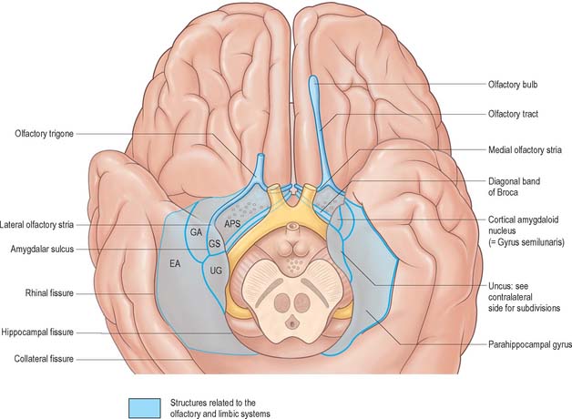
The organization of the olfactory system reflects its phylogenetically ancient lineage: afferent olfactory pathways proceed directly to the cerebral cortex, bypassing the thalamus, and its terminal fields are primitive cortical areas that are considered to be parts of the limbic system. Details of the relationship between the olfactory pathways and the limbic system are shown in Fig. 23.4.

The olfactory nerves arise from olfactory receptor neurones in the olfactory mucosa (see Ch. 32). The axons collect into numerous small bundles ensheathed by a population of unique glia and surrounded by layers of meninges, and enter the anterior cranial fossa by passing through the foramina in the cribriform plate of the ethmoid bone. They attach to the inferior surface of the olfactory bulb, which is situated at the anterior end of the olfactory sulcus on the orbital surface of the frontal lobe, and terminate in the bulb. Olfactory receptor neurones are continually replaced throughout life by differentiation of stem cells in the olfactory mucosa. The olfactory bulb is continuous posteriorly with the olfactory tract, through which the output of the bulb passes directly to the olfactory cortex.

There is a clear laminar structure in the olfactory bulb (Fig. 23.14). From the surface inwards the laminae are the olfactory nerve layer, glomerular layer, external plexiform layer, mitral cell layer, internal plexiform layer and granule cell layer. The olfactory nerve layer consists of the unmyelinated axons of the olfactory neurones. The continuous turnover of receptor cells means that axons in this layer are at different stages of growth, maturity or degeneration. The glomerular layer consists of a thin sheet of glomeruli where the incoming olfactory axons divide and synapse on terminal dendrites of secondary olfactory neurones, i.e. mitral, tufted and periglomerular cells. The external plexiform layer contains the principal and secondary dendrites of mitral and tufted cells. The mitral cell layer is a thin sheet composed of the cell bodies of mitral cells, each of which sends a single principal dendrite to a glomerulus, secondary dendrites to the external plexiform layer, and a single axon to the olfactory tract. It also contains a few granule cell bodies. The internal plexiform layer contains axons, recurrent and deep collaterals of mitral and tufted cells, and granule cell bodies. The granule cell layer contains the majority of the granule cells and their superficial and deep processes, together with numerous centripetal and centrifugal nerve fibres which pass through the layer.

The principal neurones in the olfactory bulb are the mitral and tufted cells: their axons form its output via the olfactory tract. These cells are morphologically similar and most use an excitatory amino acid, probably glutamate or aspartate, as their neurotransmitter. The mitral cell spans the layers of the bulb, and receives the sensory input superficially at its glomerular tuft. The axons of mitral and tufted cells appear to be parallel output pathways from the olfactory bulb.

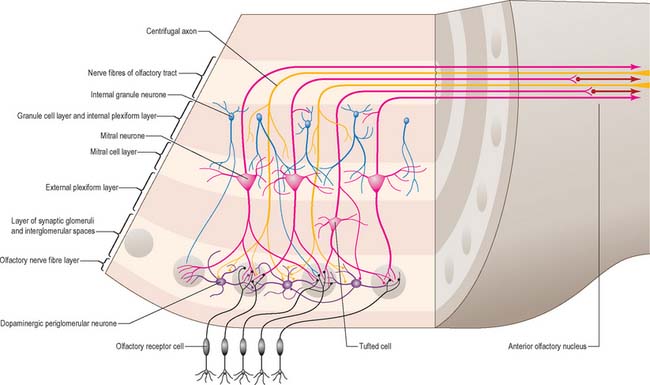
The main types of interneurones in the olfactory bulb are the periglomerular cells and granule cells. The majority of periglomerular cells are dopaminergic (cell group A15), but some are GABAergic: their axons are distributed laterally and terminate within extraglomerular regions. Granule cells are similar in size to periglomerular cells. Their most characteristic feature is the absence of an axon, and they therefore resemble amacrine cells in the retina. Granule cells have two principal spine-bearing dendrites which pass radially in the bulb: they appear to be GABAergic. The granule cell is likely to be a powerful inhibitory influence on the output neurones of the olfactory bulb.

Centrifugal inputs to the olfactory bulb arise from a variety of central sites. Neurones of the anterior olfactory nucleus and collaterals of pyramidal neurones in the olfactory cortex project to the granule cells of the olfactory bulb. Cholinergic neurones in the horizontal limb nucleus of the diagonal band of Broca, part of the basal forebrain cholinergic system, project to the granule cell layer and also to the glomerular layer. Other afferents to the granule cell layer and the glomeruli arise from the pontine locus coeruleus and the mesencephalic raphe nucleus.

The granule cell layer of the bulb is extended into the olfactory tract as scattered medium-sized multipolar neurones that constitute the anterior olfactory nucleus. Many centripetal axons from mitral and tufted cells relay in, or give collaterals to, the anterior olfactory nucleus; the axons from the nucleus continue with the remaining direct fibres from the bulb into the olfactory striae.

As the olfactory tract approaches the anterior perforated substance it flattens and splays out as the olfactory trigone. Fibres of the tract continue from the caudal angles of the trigone as diverging medial and lateral olfactory striae, which border the anterior perforated substance. The lateral olfactory stria follows the anterolateral margin of the anterior perforated substance to the limen insulae, where it bends posteromedially to merge with an elevated region, the gyrus semilunaris, at the rostral margin of the uncus in the temporal lobe. The lateral olfactory gyrus forms a tenuous grey layer covering the lateral olfactory stria: it merges laterally with the gyrus ambiens, part of the limen insulae. The lateral olfactory gyrus and gyrus ambiens form the prepiriform region of the cortex, passing caudally into the entorhinal area of the parahippocampal gyrus. The prepiriform and periamygdaloid regions and the entorhinal area (area 28) together make up the piriform cortex. The medial olfactory stria passes medially along the rostral boundary of the anterior perforated substance towards the medial continuation of the diagonal band of Broca. Together, they curve up on the medial aspect of the hemisphere, anterior to the attachment of the lamina terminalis.

The olfactory cortex receives a direct input from the olfactory bulb which arrives via the olfactory tract without relay in the thalamus. The largest cortical olfactory area is the piriform cortex. The anterior olfactory nucleus, olfactory tubercle, regions of the entorhinal and insular cortex and amygdala also receive direct projections from the olfactory bulb.

The entorhinal cortex (Brodmann’s area 28) is the most posterior part of the piriform cortex, and is divided into medial and lateral areas (areas 28a and 28b). The lateral parts receive fibres mainly from the olfactory bulb, and also from the piriform and periamygdaloid cortices.

Projections from the piriform olfactory cortex are widespread, and include the neocortex (especially the orbitofrontal cortex), thalamus (especially the medial dorsal thalamic nucleus), hypothalamus, amygdala and hippocampal formation.

Hippocampal formation

The hippocampal formation includes the dentate gyrus, hippocampus, subicular complex and entorhinal cortex.

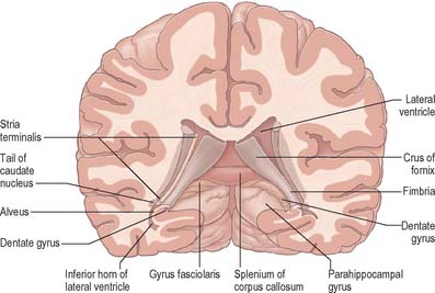
The hippocampus lies above the subiculum and medial parahippocampal gyrus, forming a curved elevation, approximately 5 cm long, along the floor of the inferior horn of the lateral ventricle (Fig. 23.15). Its anterior end is expanded, and here its margin may present two or three shallow grooves that give a paw-like appearance, the pes hippocampi. The ventricular aspect is convex. It is covered by ependyma, beneath which fibres of the alveus converge medially on a longitudinal bundle of fibres, the fimbria of the fornix. Passing medially from the collateral sulcus, the neocortex of the parahippocampal gyrus merges with the transitional juxtallocortex of the subiculum (Fig. 23.16). The latter curves superomedially to the inferior surface of the dentate gyrus, then laterally to the laminae of the hippocampus. This curvature continues, first superiorly, then medially above the dentate gyrus, and ends pointing towards the centre of the superior surface of the dentate gyrus. The dentate gyrus is a crenated strip of cortex related inferiorly to the subiculum, laterally to the hippocampus and, more medially, to the fimbria of the fornix (Fig. 23.16, Fig. 23.17). The form of the fimbria is quite variable, but medially it is separated from the crenated medial margin of the dentate gyrus by the fimbriodentate sulcus (Fig. 23.18). The hippocampal sulcus, of variable depth, lies between the dentate gyrus and the subicular extension of the parahippocampal gyrus. Posteriorly, the dentate gyrus is continuous with the gyrus fasciolaris and thus with the indusium griseum. Anteriorly, it is continued into the notch of the uncus, turning medially across its inferior surface, as the tail of the dentate gyrus (band of Giacomini), and vanishes on the medial aspect of the uncus (Fig. 23.17). The tail separates the inferior surface of the uncus into an anterior uncinate gyrus and posterior intralimbic gyrus.
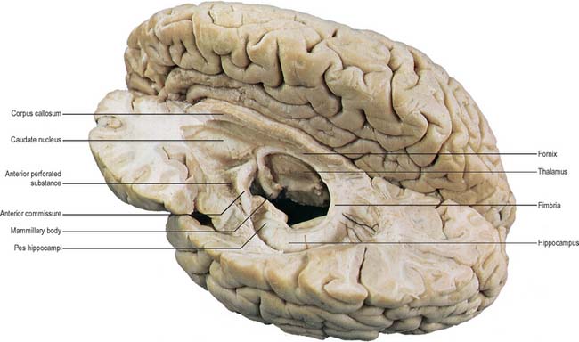
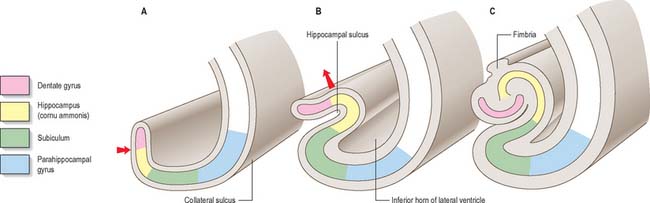
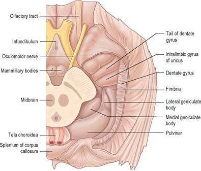
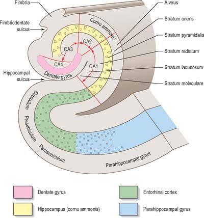
The trilaminar cortex of the dentate gyrus is the least complex of the hippocampal fields (Fig. 23.19). Its major cell type is the granule cell, found in the dense granule cell layer. Granule cells have unipolar dendrites that extend into the overlying molecular layer. The latter receives most of the afferent projections to the dentate gyrus, primarily from the entorhinal cortex (Fig. 23.20). The granule cell and molecular layers are sometimes referred to as the fascia dentata. The polymorphic layer, or hilus of the dentate gyrus, contains cells that give rise primarily to ipsilateral association fibres. They remain within the dentate gyrus and do not extend into other hippocampal fields.

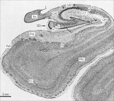
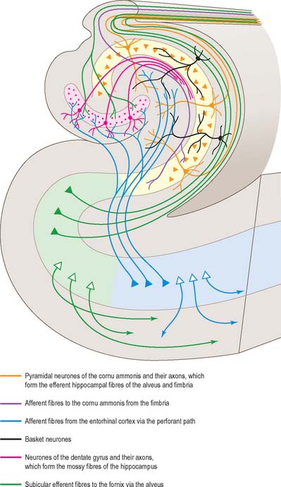
The hippocampus is trilaminar archicortex. It consists of a single pyramidal cell layer, with plexiform layers above and below it. It may best be divided into three distinct fields, CA1, CA2 and CA3 (Fig. 23.18, Fig. 23.19). Field CA3 borders the hilus of the dentate gyrus at one end, and field CA2 at the other. Field CA3 pyramidal cells are the largest in the hippocampus and receive the mossy fibre input from dentate granule cells on their proximal dendrites. The whole pyramidal cell layer in this field is about 10 cells thick. The border between CA3 and CA2 is not well marked. The CA2 field has the most compact layer of pyramidal cells. It completely lacks a mossy fibre input from dentate granule cells and receives a major input from the supramammillary region of the hypothalamus. Field CA1 is usually described as the most complex of the hippocampal subdivisions and its appearance varies along its transverse and rostrocaudal axes. The CA1/CA2 border is not sharp, and at its other end CA1 overlaps the subiculum for some distance. Approximately 10% of neurones in this field are interneurones.

It is common to describe several strata within the layers of the hippocampus. Starting from the ventricular aspect, these are the alveus (contains subicular and hippocampal pyramidal cell axons converging on the fimbria of the fornix); stratum oriens (mainly the basal dendrites of pyramidal cells and some interneurones); stratum pyramidalis; stratum lucidum (contains mossy fibres which make contact with the proximal dendrites of pyramidal cells in field CA3); and the stratum radiatum and stratum lacunosum-moleculare. The stratum lucidum is not as prominent in man as it is in other primates, and is not present in fields CA1 and CA2.

In the stratum radiatum and stratum oriens, CA3 and CA2 cells receive associational connections from other rostrocaudal levels of the hippocampus, as well as afferents from subcortical structures, e.g. the septal nuclei and supramammillary region. The projections from pyramidal cells of fields CA3 and CA2 to CA1, often called Schaffer collaterals, also terminate in the stratum radiatum and stratum oriens. The projections from the entorhinal cortex to the dentate gyrus (the perforant pathway) travel in the stratum lacunosum-moleculare, where they make synaptic contact en passant with the distal apical dendrites of hippocampal pyramidal cells.

The subicular complex is generally subdivided into subiculum, presubiculum and parasubiculum. The major subcortical projections of the hippocampal formation (to the septal nuclei, mammillary nuclei, nucleus accumbens and anterior thalamus), and to the entorhinal cortex, all arise from pyramidal neurones of the subicular complex. The subiculum consists of a superficial molecular layer containing apical dendrites of subicular pyramidal cells, a pyramidal cell layer, and a deep polymorphic layer. The presubiculum is medial to the subiculum and is distinguished by a densely packed superficial layer of pyramidal cells. It forms the boundary between the subicular complex as a whole and the entorhinal cortex. The cell layers deep to the parasubiculum are indistinguishable from the deep layers of the entorhinal cortex.

The entorhinal cortex (Brodmann’s area 28, Fig. 23.19) extends rostrally to the anterior limit of the amygdala. Caudally it overlaps a portion of the hippocampal fields. The more primitive levels of the entorhinal cortex (below the amygdala) receive projections from the olfactory bulb. More caudal regions do not generally receive primary olfactory inputs. The entorhinal cortex is divisible into six layers and is quite distinct from other neocortical regions. Layer I is acellular and plexiform. Layer II is a narrow cellular layer, which consists of islands of large pyramidal and stellate cells. These cell islands are a distinguishing feature of the entorhinal cortex. They form small bumps on the surface of the brain that can be seen by the naked eye (verrucae hippocampae), and provide an indication of the boundaries of the entorhinal cortex. Layer III consists of medium-sized pyramidal cells. There is no internal granular layer (another classic feature of entorhinal cortex); in its place is an acellular region of dense fibres called the lamina dissecans, which is sometimes called layer IV. Layers III and V are apposed in regions where the lamina dissecans is absent. Layer V consists of large pyramidal cells five or six deep. Layer VI is only readily distinguishable from layer V close to the border with the perirhinal cortex. Its cells continue around the angular bundle (subcortical white matter deep to the subicular complex made up largely of perforant path axons) to lie beneath the pre- and parasubiculum.

Glutamate and/or aspartate appears to be the major excitatory transmitter in three pathways in the hippocampal formation, namely the perforant pathway (which arises in the entorhinal cortex and terminates primarily in the dentate gyrus); the mossy fibres, which run from the dentate granule cells to the pyramidal cells of the CA3 field; and in the Schaffer collaterals of CA3 pyramidal cells, which terminate on CA1 pyramidal cells.

GABAergic neurones are found in the deep portions of the granule cell layer in the dentate gyrus (basket cells). The highest concentration of GABA receptors is found in the molecular layer of the dentate gyrus. In the hippocampus proper, GABAergic cells are found mostly in the stratum oriens, but also in the pyramidal cell layer and stratum radiatum.

There are many peptide-containing neurones in the hippocampal formation. Granule cells in the dentate gyrus appear to contain the opioid peptide dynorphin, which is also present in mossy fibres running to the CA3 field. Enkephalin, or a related peptide, may be present in fibres arising in the entorhinal cortex. There is a dense plexus of somatostatin-immunoreactive fibres in the molecular layer of the dentate gyrus and also in the stratum lacunosum-moleculare of the hippocampus. The polymorphic layer of the dentate gyrus, stratum oriens of the hippocampus and the deep layers of the entorhinal cortex, all contain somatostatin-immunoreactive neurones. Vasoactive intestinal polypeptide (VIP)-immunoreactive neurones are plentiful in many hippocampal fields, especially in the superficial layers of the entorhinal cortex. Cells containing cholecystokinin (CCK)-immunoreactivity are found in the hilar region of the dentate gyrus, in all layers of the hippocampus, especially in the pyramidal cell layer, and also throughout the subicular complex and entorhinal cortex. There are also substantial plexuses of CCK-immunoreactive fibres in the stratum lacunosum-moleculare, subicular complex and entorhinal cortex. Hippocampal CCK-immunoreactive cells may give rise to extrinsic projections, e.g. to the lateral septum and medial mammillary nucleus, because CCK-immunoreactive fibres are found in the fimbria/fornix.

The medial septal complex and the supramammillary area of the posterior hypothalamus are the two major sources of subcortical afferents to the hippocampal formation. There are also projections from the amygdaloid complex and claustrum (to the subicular complex and entorhinal cortex), as well as monoaminergic projections from the ventral tegmental area, the mesencephalic raphe nuclei and the locus coeruleus. The noradrenergic and serotoninergic projections reach all hippocampal fields, but are especially dense in the dentate gyrus.

The projections from the septal complex arise in the medial septal and vertical limb nuclei of the diagonal band. They travel via the dorsal fornix, fimbria, supracallosal striae and a ventral route through the amygdaloid complex. While these projections reach all hippocampal fields, the most prominent terminations are in the dentate gyrus, field CA3, presubiculum, parasubiculum and entorhinal cortex. Many of these medial septal/diagonal band neurones are GABAergic or cholinergic, and they form part of the topographically organized basal forebrain cholinergic system (cell groups Ch1 and Ch2).

Neurones in the supramammillary area also provide a significant innervation of the hippocampal formation. They arrive partly through the fornix and partly through a ventral route, and terminate most heavily in the dentate gyrus, and fields CA2 and CA3 of the cornu ammonis.

All divisions of the anterior thalamic nuclear complex and associated lateral dorsal nucleus project to the hippocampal formation, and are directed predominantly to the subicular complex. Some midline thalamic nuclei, particularly the parataenial, central medial and reuniens nuclei, also project to the hippocampal formation, especially to the entorhinal cortex.

The human fornix contains more than a million fibres. Cells in the CA3 field project bilaterally to the lateral nucleus of the septal complex, via the precommissural fornix. They give rise to the Schaffer collaterals to CA1 cells and to the commissural projections to the contralateral hippocampus. Neurones in the subicular complex and entorhinal cortex give rise to projections to the nucleus accumbens and to parts of the caudate nucleus and putamen. The subicular complex gives rise to the major, postcommissural fibre system of the fornix. The presubiculum, in particular, projects to the anterior thalamic nuclear complex (anteromedial, anteroventral and laterodorsal nuclei). Both the subiculum and presubiculum provide the major extrinsic input to the mammillary complex. Both the lateral and the medial mammillary nuclei receive afferents from the subicular complex.

Several fields in the temporal lobe neocortex, especially the parahippocampal gyrus, the dorsal bank of the superior temporal gyrus, the perirhinal cortex (Brodmann’s area 35) and the temporal polar cortex, together with the agranular insular cortex and posterior orbitofrontal cortex, project to the entorhinal cortex. Projections to the entorhinal cortex also arise from the dorsolateral prefrontal cortex (Brodmann’s areas 9, 10, 46), the medial frontal cortex (Brodmann’s areas 25, 32), the cingulate cortex (Brodmann’s areas 23, 24), and retrosplenial cortex. The subicular complex receives direct cortical inputs, e.g. from the temporal polar cortex, perirhinal cortex, parahippocampal gyrus, superior temporal gyrus and dorsolateral prefrontal cortex. The entorhinal cortex projects to the perirhinal cortex as well as to temporal polar cortex, caudal parahippocampal and cingulate gyri. In monkeys, the subicular complex also projects to a number of cortical areas, including perirhinal cortex, parahippocampal gyrus, caudal cingulate gyrus, medial frontal and medial orbitofrontal cortex.

Septum

The septum is a midline and paramedian structure (Fig. 23.13, Fig. 22.6). Its upper portion corresponds largely to the bilateral laminae of fibres, sparse grey matter and neuroglia, known as the septum pellucidum, which separates the lateral ventricles. Below this, the septal region is made up of four main nuclear groups: dorsal, ventral, medial and caudal. The dorsal group is essentially the dorsal septal nucleus, the ventral group consists of the lateral septal nucleus, the medial group contains the medial septal nucleus and the nucleus of the diagonal band of Broca, and the caudal group contains the fimbrial and triangular septal nuclei.

The major afferents to the region terminate primarily in the lateral septal nucleus. They include fibres carried in the fornix that arise from hippocampal fields CA3 and CA1 and the subiculum. There are also afferents arising from the preoptic area, anterior, paraventricular and ventromedial hypothalamic nuclei and the lateral hypothalamic area. The lateral septum receives a rich monaminergic innervation, including noradrenergic afferents from the locus coeruleus and medullary cell groups (A1, A2). It also receives serotoninergic afferents from the midbrain raphe nuclei and dopaminergic afferents from the ventral tegmental area (A10).

Projections from the lateral septum run to the medial and lateral preoptic areas, anterior hypothalamus, supramammillary and midbrain ventral tegmental area, via the medial forebrain bundle. There is also a projection to the medial habenular nucleus and some midline thalamic nuclei via the stria medullaris thalami, which runs on the dorsomedial wall of the third ventricle. The projections from the habenula via the fasciculus retroflexus to the interpeduncular nucleus and adjacent ventral tegmental area in the midbrain provide a route through which forebrain limbic structures can influence midbrain nuclear groups.

A large proportion of the medial septal/diagonal band neurones are cholinergic or GABAergic. They project to the hippocampal formation and cingulate cortex.

Amygdala

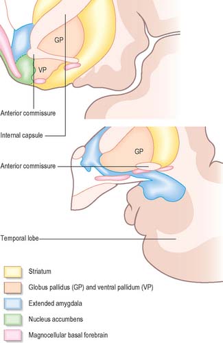
The amygdala (amygdaloid nuclear complex) consists of lateral, central and basal nuclei that lie in the dorsomedial temporal pole, anterior to the hippocampus, close to the tail of the caudate nucleus (Fig. 23.13) and partly deep to the gyrus semilunaris, gyrus ambiens and uncinate gyrus (Fig. 23.4). Collectively the nuclei form the ventral, superior and medial walls of the tip of the inferior horn of the lateral ventricle. The amygdala is partly continuous above with the inferomedial margin of the claustrum. Fibres of the external capsule and substriatal grey matter, including the cholinergic magnocellular nucleus basalis (of Meynert), incompletely separate it from the putamen and globus pallidus. Laterally, it is close to the optic tract.

The lateral nucleus has dorsomedial and ventrolateral subnuclei. The central nucleus has medial and lateral subdivisions. The basal nucleus is commonly divided into a dorsal magnocellular basal nucleus, an intermediate parvicellular basal nucleus, and a ventral band of darkly staining cells usually referred to as the paralaminar basal nucleus, because it borders the white matter ventral to the amygdaloid complex. The accessory basal nucleus lies medial to the basal nuclear divisions and is usually divided into dorsal, magnocellular, and ventral, parvicellular, parts. The lateral and basal nuclei are often referred to collectively as the basolateral area (nuclear group) of the amygdaloid complex.

It has been suggested that the basolateral complex of nuclei (lateral, basal, accessory basal) shares several characteristics with the cortex, and that it may be considered as a quasi-cortical structure. Although it lacks a laminar structure, it has direct, often reciprocal, connections with adjacent temporal and other areas of cortex, and it projects to the motor or premotor cortex. It receives a direct cholinergic and non-cholinergic input from the magnocellular corticopetal system in the basal forebrain, and has reciprocal connections with the mediodorsal thalamus. The distribution of small peptidergic neurones in the basolateral nuclear complex, e.g. those containing neuropeptide-Y (NY), somatostatin (SOM) and CCK, are also similar in form and density to those found in the adjacent temporal lobe cortex. Projection neurones from this part of the amygdala appear to utilize, at least in part, the excitatory amino acids glutamate or aspartate as a transmitter. Moreover, they project to the ventral striatum rather than to hypothalamic and brain stem sites. Thus, it may be appropriate to consider this part of the amygdaloid complex as a polymodal cortex-like area, separated from the cerebral cortex by fibres of the external capsule.

The central nucleus is present through the caudal half of the amygdaloid complex. It lies dorsomedial to the basal nucleus and is divided into medial and lateral parts. The medial part, which contains larger cells than the lateral part, resembles the adjacent putamen. The medial and central nuclei appear to have an extension across the basal forebrain, as well as within the stria terminalis, which merges with the bed nucleus of the stria terminalis. This extensive nuclear complex is sometimes referred to as the ‘extended amygdala’ (Fig. 23.21). It can be considered as a macrostructure formed by the centromedial amygdaloid complex (medial nucleus, medial and lateral parts of the central nucleus), the medial bed nucleus of the stria terminalis, and the cell columns that traverse the sublenticular substantia innominata, which lies between them. It has been suggested that portions of the medial nucleus accumbens may be included in the extended amygdala.

A consistent feature of the intrinsic connections among amygdaloid nuclei is that they arise primarily in lateral and basal nuclei, and terminate in the central and medial nuclei, which suggests a largely unidirectional flow of information. In brief, the lateral nucleus projects to all divisions of the basal nucleus, accessory basal nucleus, paralaminar and anterior cortical nuclei, and less heavily to the central nucleus: it receives few afferents from other nuclei. The magnocellular, parvicellular and intermediate parts of the basal nucleus project to the accessory basal, central (especially the medial part) and medial nuclei, as well as to the periamygdaloid cortex and the amygdalohippocampal area. The accessory basal nucleus projects densely to the central nucleus, especially its medial division, as well as to the medial and cortical nuclei. Its major intra-amygdaloid afferents arise from the lateral nucleus. The medial nucleus projects to the accessory basal, anterior cortical and central nuclei as well as to the periamygdaloid cortex and amygdalohippocampal area, while afferents arise especially from the lateral nucleus. The intrinsic connections of the cortical nucleus are not very well understood. The posterior part of the cortical nucleus projects to the medial nucleus, but it has been difficult to differentiate this projection from that arising in the amygdalohippocampal area. The central nucleus projects to the anterior cortical nucleus and the various cortical transition zones. It forms an important focus for afferents from many of the amygdaloid nuclei, especially the basal and accessory basal nuclei, and it has major extrinsic connections.

The organization of the extensive subcortical and cortical connections of the amygdala are consistent with a role in emotional behaviour. It receives highly processed unimodal and multimodal sensory information from the thalamus, sensory and association cortices; olfactory information from the olfactory bulb and piriform cortex; visceral and gustatory information relayed via brain stem structures and the thalamus. Its projections reach widespread areas of the brain, including the endocrine and autonomic domains of the hypothalamus and brain stem.

Afferent connections

The heaviest brain stem projection to the amygdala arises in the peripeduncular nucleus. The parabrachial nuclei also project to the central nucleus. The amygdala receives a rich monoaminergic innervation. The noradrenergic projection arises primarily from the locus coeruleus, serotonergic fibres arise from the dorsal and, to some extent the median, raphe nuclei, and the dopaminergic innervation arises primarily in the midbrain ventral tegmental area (A10). The basal and parvicellular accessory basal nuclei, the amygdalohippocampal area and nucleus of the lateral olfactory tract receive a very dense cholinergic innervation arising from the magnocellular nucleus basalis of Meynert.

The amygdala has rich interconnections with allocortical, juxtallocortical and, especially, neocortical areas. In addition to direct projections from the olfactory bulb to the nucleus of the lateral olfactory tract, anterior cortical nucleus and the periamygdaloid cortex (piriform cortex), there are also associational connections between all parts of the primary olfactory cortex and these same superficial amygdaloid structures. The amygdaloid complex has particularly extensive and rich connections with many areas of the neocortex in unimodal and polymodal regions of the frontal, cingulate, insular and temporal neocortices.

The anterior temporal lobe provides the largest proportion of the cortical input to the amygdala, predominantly to the lateral nucleus. Rostral parts of the superior temporal gyrus, which may represent unimodal auditory association cortex, project to the lateral nucleus. There are also projections from polymodal sensory association cortices of the temporal lobe, including perirhinal cortex (areas 35 and 36), the caudal half of the parahippocampal gyrus, the dorsal bank of the superior temporal sulcus, and both the medial and lateral areas of the cortex of the temporal pole.

The CA1 field of the hippocampus and adjacent subiculum, and possibly the entorhinal cortex, project to the amygdala, mainly to the parvicellular basal nucleus.

The rostral insula projects heavily to the lateral, parvicellular basal and medial nuclei. The caudal insula, which is reciprocally connected with the second somatosensory cortex, also projects to the lateral nucleus, thus providing a route by which somatosensory information reaches the amygdala. The caudal orbital cortex projects to the basal, magnocellular accessory basal and lateral nuclei. The medial prefrontal cortex projects to the magnocellular divisions of the accessory and basal nuclei.

Efferent connections

The central nucleus provides the major relay for projections from the amygdala to the brain stem and receives many of the return projections. It projects to the periaqueductal grey matter, ventral tegmental area, substantia nigra pars compacta, peripeduncular nucleus and tegmental reticular formation (midbrain); the parabrachial nuclei (pons); the nucleus of the solitary tract and the dorsal motor nucleus of the vagus (medulla).

The central nucleus is the major relay for amygdaloid projections to the hypothalamus. Amygdaloid fibres reach the bed nucleus of the stria terminalis primarily via the stria terminalis, but also via the ventral amygdalofugal pathway. In general, central and basal nuclei project to the lateral part of the bed nucleus, while medial and posterior cortical nuclei project to the medial bed nucleus. Anterior cortical and medial nuclei project largely to the medial preoptic area and anterior medial hypothalamus, including the paraventricular and supraoptic nuclei. There is a particularly prominent projection to the ventromedial and premammillary nuclei. The amygdala projects to the rostrocaudal extent of the lateral hypothalamus. The majority of the fibres originate in the central nucleus and run principally in the ventral amygdalofugal pathway and medial forebrain bundle.

There is a rich projection to the mediodorsal nucleus of the thalamus, which gives access to the prefrontal cortex, and also complements direct projections from the amygdala to the same cortical domain. The projection to the mediodorsal nucleus arises from most amygdaloid nuclei, but particularly from the lateral, basal and accessory basal nuclei and the periamygdaloid cortex. The major termination of amygdaloid afferents is in the medial, magnocellular part of the mediodorsal nucleus, especially rostrally. This part of the mediodorsal nucleus projects to the identical medial and orbital prefrontal cortical areas that receive amygdaloid afferents directly. However, this projection to the mediodorsal nucleus is not reciprocated. The central and medial nuclei do not project to the mediodorsal nucleus, but to the midline nuclei, especially the nucleus centralis and nucleus reuniens.

The parvicellular division of the basal nucleus, magnocellular accessory basal nucleus (but not the magnocellular basal nucleus), and the central nucleus all project to basal forebrain cholinergic cell groups, notably the nucleus basalis of Meynert and the horizontal limb nucleus of the diagonal band.

The striatum, and particularly the nucleus accumbens, receives prominent projections from the amygdaloid complex. The basal and accessory basal nuclei are the most important contributors to this projection. The ventral striatum sends many fibres to the ventral pallidum, which in its turn projects to the mediodorsal nucleus of the thalamus. Thus, the ventral striatopallidal system provides a second route through which the amygdala can influence mediodorsal thalamic–prefrontal cortical processes.

The lateral, magnocellular accessory basal and parvicellular basal nuclei contribute the largest proportion of efferents to the hippocampal formation. The main projection is from the lateral nucleus to the rostral entorhinal cortex, but many fibres also terminate in the hippocampus proper and the subiculum. There appears to be marked polarity in amygdalohippocampal connections – the amygdala has a greater influence on hippocampal processes than vice versa.

The amgygdaloid nuclear complex projects to widely dispersed neocortical fields. Amygdalocortical projections principally originate in the basal nucleus, and to a smaller extent in the lateral and cortical nuclei.

The amygdala projects to virtually all levels of the visual cortex in both temporal and occipital lobes. The largest component of these projections arises from the magnocellular basal nucleus. The amygdala also reciprocates projections to the auditory cortex in the rostral half of the superior temporal gyrus. Projections to the polymodal sensory areas of the temporal lobe generally reciprocate the amygdalopetal projections. Efferents from the lateral and accessory basal nuclei are directed to the temporal pole, particularly the medial perirhinal area.

The insular cortex is heavily innervated by the amygdaloid medial and anterior cortical nuclei. The orbital cortex and medial frontal cortical areas 24, 25 and 32, including parts of the anterior cingulate gyrus, also receive a heavy projection. Areas 8, 9, 45 and 46 of the dorsolateral prefrontal cortex, as well as the premotor cortex (area 6), receive a patchy innervation. The basal nucleus is an important source of these projections, which are augmented by contributions from the accessory basal (magnocellular and parvicellular divisions) and lateral nuclei.

WHITE MATTER OF CEREBRAL HEMISPHERE

The nerve fibres which make up the white matter of the cerebral hemispheres are categorized on the basis of their course and connections. They are either association fibres, which link different cortical areas in the same hemisphere; commissural fibres, which link corresponding cortical areas in the two hemispheres; or projection fibres, which connect the cerebral cortex with the corpus striatum, diencephalon, brain stem and the spinal cord.

ASSOCIATION FIBRES

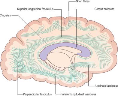
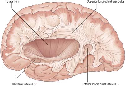
Association fibres (Fig. 23.22, Fig. 23.23) may be either short association (arcuate or ‘U’) fibres, which link adjacent gyri, or long association fibres, which connect more widely separated gyri.

Short association fibres may be entirely intracortical. Many pass subcortically between adjacent gyri, some merely pass from one wall of a sulcus to the other.

Long association fibres are grouped into bundles, e.g. uncinate fasciculus, cingulum, superior longitudinal fasciculus, inferior longitudinal fasciculus and fronto-occipital fasciculus.

The uncinate fasciculus connects the motor speech (Broca’s) area and orbital gyri of the frontal lobe with the cortex in the temporal pole. The fibres follow a sharply curved course across the stem of the lateral sulcus, near the anteroinferior part of the insula. The cingulum is a long, curved fasciculus, which lies deep to the cingulate gyrus. It starts in the medial cortex below the rostrum of the corpus callosum, follows the curve of the cingulate gyrus, enters the parahippocampal gyrus and spreads into the adjoining temporal lobe. The superior longitudinal fasciculus is the largest of the long association fasciculi. It starts in the anterior frontal region and arches back, above the insular area, contributing fibres to the occipital cortex (areas 18 and 19). It curves down and forwards, behind the insular area, to spread out in the temporal lobe. The inferior longitudinal fasciculus starts near the occipital pole. Its fibres, probably derived mostly from areas 18 and 19, sweep forwards, separated from the posterior horn of the lateral ventricle by the optic radiation and tapetal commissural fibres, and are distributed throughout the temporal lobe. The fronto-occipital fasciculus starts at the frontal pole. It passes back deep to the superior longitudinal fasciculus, separated from it by the projection fibres in the corona radiata. It lies lateral to the caudate nucleus near the central part of the lateral ventricle. Posteriorly, it fans out into the occipital and temporal lobes, lateral to the posterior and inferior horns of the lateral ventricle.

COMMISSURAL FIBRES

Commissural fibres cross the midline, many linking corresponding areas in the two cerebral hemispheres. By far the largest commissure is the corpus callosum. Others include the anterior, posterior and habenular commissures, and the commissure of the fornix.

Corpus callosum

The corpus callosum (Fig. 23.2, Fig. 23.24, Fig. 23.25; see Fig. 22.2) is the largest fibre pathway of the brain. It links the cerebral cortex of the two cerebral hemispheres, and it roofs much of the lateral ventricles. It forms an arch approximately 10 cm long, with an anterior end approximately 4 cm from the frontal poles and a posterior end approximately 6 cm from the occipital poles. Its anterior portion is known as the genu. This recurves posteroinferiorly in front of the septum pellucidum, then diminishes rapidly in thickness and is prolonged to the upper end of the lamina terminalis as the rostrum. The trunk of the corpus callosum arches back and is convex above. It ends posteriorly in the expanded splenium, which is its thickest part.
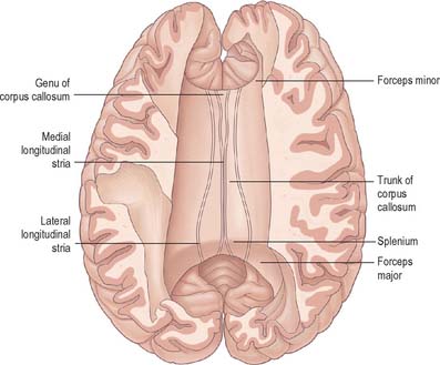
The median region of the trunk of the corpus callosum forms the floor of the great longitudinal fissure. Here, it lies close to the anterior cerebral arteries and the lower border of the falx cerebri, which may contact it behind. On each side, the trunk is overlapped by the cingulate gyrus, from which it is separated by the callosal sulcus. The inferior surface of the corpus callosum is concave in its long axis: it is attached to the septum pellucidum anteriorly and fused with the fornix and its commissure posteriorly.

The superior surface of the callosal trunk (Fig. 23.24) is covered by a thin layer of grey matter, the indusium griseum. This extends anteriorly around the genu and then continues into the paraterminal gyrus on the inferior aspect of the rostrum. It contains narrow longitudinal bundles of fibres on each side, the medial and lateral longitudinal striae. Posteriorly, the indusium griseum is continuous with the dentate gyrus and hippocampus through the gyrus fasciolaris (Fig. 23.25).

The splenium of the corpus callosum overhangs the posterior ends of the thalami, the pineal gland and tectum, but is separated from them by several structures. On each side the crus of the fornix and gyrus fasciolaris curve up to the splenium. The crus continues forwards on the inferior surface of the callosal trunk, but the gyrus fasciolaris skirts above the splenium, then rapidly diminishes into the indusium griseum. The tela choroidea of the third ventricle advances below the splenium through the transverse fissure, and the internal cerebral veins emerge between its two layers to form the great cerebral vein. Posteriorly the splenium is near the tentorium cerebelli, great cerebral vein and the start of the straight sinus.

Nerve fibres of the corpus callosum radiate into the white matter core of each hemisphere, thereafter dispersing to the cerebral cortex. Commissural fibres forming the rostrum extend laterally, below the anterior horn of the lateral ventricle, connecting the orbital surfaces of the frontal lobes. Fibres in the genu curve forwards, as the forceps minor, to connect the lateral and medial surfaces of the frontal lobes. Fibres of the trunk pass laterally, intersecting with the projection fibres of the corona radiata to connect wide neocortical areas of the hemispheres. Fibres of the trunk and splenium, which form the roof and lateral wall of the posterior horn and the lateral wall of the inferior horn of the lateral ventricle, constitute the tapetum. The remaining fibres of the splenium curve back into the occipital lobes as the forceps major.

Interhemispheric connections through the corpus callosum do not all represent a simple linking of loci in one hemisphere with the same loci in the other. In areas containing a clear representation of a contralateral sensorium (e.g. body surface, visual field), only those areas that are functionally related to midline representation are linked to the contralateral hemisphere. This is most clearly seen for the visual areas, where the cortex containing the representation of each midline retinal zone is linked to its counterpart on the contralateral side. A similar arrangement is seen in somatic areas, where the trunk representation is callosally linked, but the peripheral limb areas (hand and foot) are not.

Connections that link the same, or similar, areas on each side are termed homotopic connections. The corpus callosum also interconnects heterogeneous cortical areas on the two sides (heterotopic connections). These may serve to connect functionally similar, but anatomically different, loci in the two hemispheres, and/or to connect functional areas in one hemisphere with regions that are specialized for a unilaterally confined function in the other.

Anterior commissure

The anterior commissure is a compact bundle of myelinated nerve fibres which crosses anterior to the columns of the fornix and is embedded in the lamina terminalis, where it is part of the anterior wall of the third ventricle (Fig. 23.13). In sagittal section it is oval, its long (vertical) diameter is approximately 1.5 mm. Laterally it splits into anterior and posterior bundles. The smaller anterior bundle curves forwards on each side to the anterior perforated substance and olfactory tract. The posterior bundle curves posterolaterally on each side in a deep groove on the anteroinferior aspect of the lentiform complex, and subsequently fans out into the anterior part of the temporal lobe, including the parahippocampal gyrus. Areas thought to be connected via commissural fibres include: the olfactory bulb and anterior olfactory nucleus; the anterior perforated substance, olfactory tubercle and diagonal band of Broca; the prepiriform cortex; the entorhinal area and adjacent parts of the parahippocampal gyrus; part of the amygdaloid complex (especially the nucleus of the lateral olfactory stria); the bed nucleus of the stria terminalis and the nucleus accumbens; the anterior regions of the middle and inferior temporal gyri.

PROJECTION FIBRES

Projection fibres connect the cerebral cortex with lower levels in the brain and spinal cord. They include large numbers of both corticofugal and corticopetal projections. Corticofugal projection fibres converge from all directions to form the dense subcortical white matter mass of the corona radiata (Fig. 23.26). Large numbers of fibres pass to the corpus striatum and the thalamus, intersecting commissural fibres of the corpus callosum en route. The corona radiata is continuous with the internal capsule, which contains the majority of the cortical projection fibres.
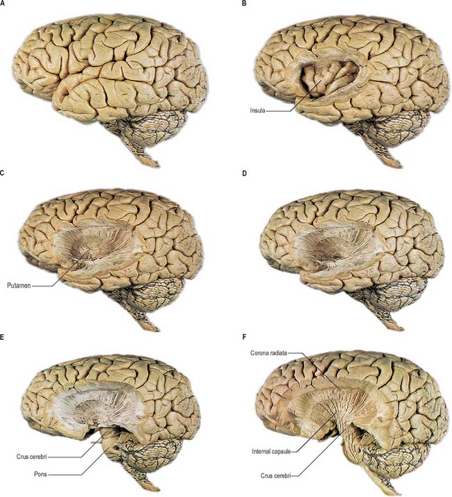
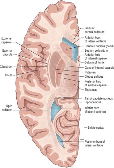
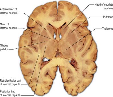
Internal capsule

In horizontal cerebral sections the internal capsule appears as a broad white band, with a lateral concavity, which accommodates the lentiform complex (Fig. 23.27, Fig. 23.28, Fig. 23.29). It has an anterior limb, genu, posterior limb, and retrolenticular (retrolentiform) and sublenticular (sublentiform) parts. Both anterior and posterior limbs are medial to the lentiform complex. The head of the caudate nucleus is medial to the anterior limb, and the thalamus is medial to the posterior limb. Cortical efferent fibres of the internal capsule continue to converge as they descend. Fibres derived from the frontal lobe tend to pass posteromedially, while temporal and occipital fibres pass anterolaterally. Many, but not all, corticofugal fibres pass into the crus cerebri of the ventral midbrain. Here, corticospinal and corticobulbar fibres are located in the middle half of the crus. Frontopontine fibres are located medially, whereas corticopontine fibres from temporal, parietal and occipital cortices are found laterally.

The anterior limb of the internal capsule contains frontopontine fibres, which arise from the cortex in the frontal lobe. They synapse with cells in the pontine nuclei. Axons of these cells enter the opposite cerebellar hemisphere through the middle cerebellar peduncle. Anterior thalamic radiations interconnect the medial and anterior thalamic nuclei and various hypothalamic nuclei and limbic structures with the frontal cortex.

The genu of the internal capsule is usually regarded as containing corticobulbar fibres, which are mainly derived from area 4 and terminate mostly in the contralateral motor nuclei of cranial nerves. Anterior fibres of the superior thalamic radiation, between the thalamus and cortex, also extend into the genu.

The posterior limb of the internal capsule includes the corticospinal tract. The fibres concerned with the upper limb are anterior. More posterior regions contain fibres representing the trunk and lower limbs. Other descending axons include frontopontine fibres, particularly from areas 4 and 6, and corticorubral fibres, which connect the frontal lobe to the red nucleus. Most of the posterior limb also contains fibres of the superior thalamic radiation (the somaesthetic radiation) ascending to the postcentral gyrus.

The retrolenticular part of the internal capsule contains parietopontine, occipitopontine, and occipitotectal fibres. It also includes the posterior thalamic radiation and the optic radiation, and interconnections between the occipital and parietal lobes and caudal parts of the thalamus, especially the pulvinar.

The optic radiation arises in the lateral geniculate body. It sweeps backwards, intimately related to the superolateral aspect of the inferior horn and the lateral aspect of the posterior horn of the lateral ventricle, from which it is separated by the tapetum.

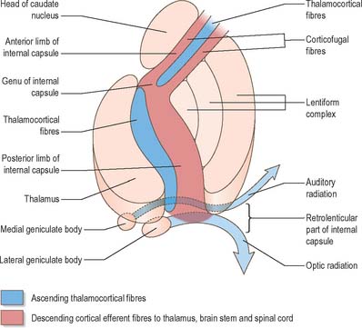
The sublenticular part of the internal capsule contains temporopontine and some parietopontine fibres, the auditory radiation from the medial geniculate body to the superior temporal and transverse temporal gyri (areas 41 and 42), and a few fibres that connect the thalamus with the temporal lobe and insula. Fibres of the auditory radiation sweep anterolaterally below and behind the lentiform complex to reach the cortex.

CEREBRAL ASYMMETRY

The two human cerebral hemispheres are not simply mirror images of each other. Much information on the lateralization of cerebral function has come from studying patients in whom the corpus callosum had been divided (commissurotomy) as a treatment for intractable epilepsy, and from those rare individuals who lack part, or all, of their corpus callosum. Commissurotomy produces the ‘split-brain’ syndrome, which has provided evidence supporting the notion that abilities or functions are predominantly associated with one or other hemisphere. Knowledge of such lateralization of function has been advanced more recently by functional brain imaging techniques such as positron emission tomography (PET).

The left hemisphere usually prevails for verbal and linguistic functions, for mathematical skills and for analytical thinking. The right hemisphere is mostly non-verbal. It is more involved in spatial and holistic or ‘Gestalt’ thinking, in many aspects of musical appreciation, and in some emotions. Memory also shows lateralization. Thus, verbal memory is primarily a left hemisphere function, while non-verbal memory is represented in the right hemisphere. These asymmetries are relative, not absolute, and vary in degree according to the function and individual concerned. Moreover, they apply primarily to right-handed men. Those men with left-hand preference, or mixed handedness, make up a heterogeneous group, which (as an approximation) shows reduced or anomalous lateralization, rather than a simple reversal of the situation in right-handers. For example, speech representation can occur in either or both hemispheres. Women show less functional asymmetry, on average, than men.

Certain cerebral anatomical asymmetries are apparent at both the macroscopic and histological levels. One of the most notable is in the planum temporale, which is usually larger on the left than the right side. Subtle asymmetries in the superior temporal lobe have been demonstrated in terms of overall size and shape, sulcal pattern, cytoarchitecture and at the neuronal level. It seems reasonable to assume that these differences underlie some of the functional asymmetry for language representation.

Asymmetries in areal size, cytoarchitecture or neurocytology occur elsewhere in the cerebral cortex as well as subcortically. For example, many brains have a wider right frontal pole and a wider left occipital pole. Brodmann’s area 45 in the inferior frontal lobe, corresponding to Broca’s area, contains a population of large pyramidal neurones that are found only on the left side. The cortical surface surrounding the central sulcus is larger in the left hemisphere, especially in the areas containing the primary somatosensory and motor maps of the arm, suggesting that one cerebral manifestation of hand preference is a larger amount of neural circuitry in the relevant parts of the cortex. Histological asymmetries are also found in areas that are not usually considered to be closely related either to language or handedness. The left entorhinal cortex has significantly more neurones than the right.

NEUROPSYCHOLOGICAL FUNCTIONS

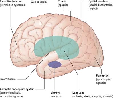
The neuropsychological functions of perception, spatial analysis, learned skilled movement, language, memory, problem solving (all executive functions) and emotion, are organized within the cerebral hemispheres (Fig. 23.30).

The organization of neuropsychological functions is highly localized and involves the association areas of the neocortex and the limbic system. The parietal association areas are concerned with the perceptual recognition of objects by kinaesthetic and visual stimuli, and the visuo-spatial orientation of the body and its parts in space. Large focal lesions (particularly of the right cerebral hemisphere) and bilateral lesions (e.g. in Alzheimer’s disease), lead to visual disorientation in space, with an inability to navigate the environment, locate objects, and dress in relationship to body parts. Acute focal lesions (especially of the right cerebral hemisphere) lead to neglect of the body and space in the opposite side of corporeal and visual space (neglect). Failure of recognition of objects by touch or vision, represents tactile or visual agnosia. The parietal-temporal cortical connections are particularly important for visual object recognition and lesions lead to an inability to identify, copy, or match, objects (apperceptive visual agnosia). The anterior superior parietal lobes and the related connections with the pre-motor areas, including the supplementary motor areas, are concerned in the execution of skilled movements through development and experience (praxis). Lesions of these areas lead to contralateral loss of skilled movements of the face, mouth and limbs (ideomotor apraxia).

The anterior temporal neocortex (middle and inferior temporal gyri) is concerned in ascribing meaning to perceptual stimuli. Bilateral lesions of these areas (e.g. in semantic dementia) leads to loss of recognition of words and percepts. Unilateral lesions in the left hemisphere particularly affect word meaning and naming (semantic or trans-cortical aphasia), whereas lesions of the right hemisphere lead primarily to loss of recognition of visual percepts (associative visual agnosia) and faces (prosopagnosia), and objects can be copied or matched but not identified for meaning or name.

The pre-motor areas (frontal association cortex, parietal and temporal association cortex) combine to form the ‘language area’ in the cortex that surrounds the lateral fissure in the left dominant hemisphere. Focal lesions of the language area lead to breakdown in verbal communication (aphasia) and loss of the ability to read (alexia), write (agraphia), and calculate (acalculia). The aphasic syndromes of Broca, conduction and Wernicke, correspond to lesions within the frontal, parietal, and temporal cortical areas, respectively. Lesions of the parietal cortex immediately posterior to the language area (angular gyrus) can lead to alexia, agraphia and acalculia, in the absence of aphasia.

The pre-frontal association cortex and its connections with the limbic system, and in particular, with the amygdala, are essential for problem solving behaviour (executive functions) and the affective motivational aspects of behaviour. Bilateral lesions of these areas (e.g. in frontotemporal dementia) lead to the ‘frontal lobe syndrome’, in which there is a radical change in personality, with loss of reason, judgement, and insight, together with loss of personal and social feelings (sympathy and empathy). The restricted involvement of the orbital frontal areas and limbic connections leads to over-activity and disinhibition (pseudo psychopathic behaviour). Spread of lesions into the dorsolateral surface of the pre-frontal area leads to an inert, apathetic, state (pseudo depression). Unilateral focal lesions of the pre-frontal areas are not usually associated with obvious cognitive or behavioural change.

The limbic allocortex and its connections comprising the Papez circuit (hippocampus, mammillary body, anterior nucleus of the thalamus, and cingulate gyrus) are responsible for the laying down of autobiographical (episodic) memory. Bilateral lesions (e.g. in Alzheimer’s disease) or following alcoholic encephalopathy (Korsakoff psychosis) lead to the loss of ability to learn new information (anterograde amnesia) or remember experiences in the relatively recent past (retrograde amnesia). However, perceptual information about the world and language (semantic memory) are preserved, because the temporal neocortical association areas are spared.

Patients with chronic epilepsy, who have undergone surgical section of the corpus callosum in order to relieve their seizures, portray few difficulties under normal circumstances. However, when these ‘split brain’ patients undergo psychological testing, the two halves of the brain appear to behave relatively autonomously, e.g. visual information directed to the right cerebral hemisphere alone does not evoke a verbal response, and consequently individuals cannot name objects or read words solely presented to the left visual field. Destruction of the splenium of the corpus callosum and its connections to the left occipital cortex, either by stroke or tumour, leads to the posterior disconnection syndrome of ‘alexia without agraphia’. Such individuals speak and write without difficulty but cannot understand written material (alexia). Disconnection of visual processes in the right hemisphere from the verbal processes of the dominant left cerebral hemisphere is thought to explain the syndrome.

REFERENCES

Alheid GF, Heimer L. New perspectives in basal forebrain organization of special relevance for neuropsychiatric disorders: the striatopallidal, amygdaloid and corticopetal components of the substantia innominata. Neuroscience. 1988;27:1-39.

Provides evidence that the region of the ‘substantia innominata’ in the basal forebrain is composed of parts of three forebrain structures: the ventral striatopallidal system; the extended amygdala; and the magnocellular corticopetal system..

Derbyshire SW, Jones AK, Gyulai F, Clark S, Townsend D, Firestone LL. Pain processing during three levels of noxious stimulation produces differential patterns of central activity. Pain. 1997;73:431-445.

Papez JW. A proposed mechanism of emotion. Arch Neurol Psychiat. 1937;38:725-743.

The classic description of the mechanism of emotion in man..

Passingham RE. The Frontal Lobes and Voluntary Action. Oxford: Oxford University Press, 1993.

Penfield W, Rasmussen T. The Cerebral Cortex of Man. New York: Macmillan, 1950.

The classic description of the organization of sensory and motor homunculi in the human cerebral cortex..

Peyron R, Laurent B, Garcia-Larrea L. Functional imaging of brain responses to pain. A review and meta-analysis. Neurophysiol Clin. 2000;30:263-288.

Sperry RW. Lateral specialization in the surgically separated hemisphere. In: Schmidt FO, Worden FG. The Neurosciences. Third study program. Cambridge, Mass: MIT Press; 1974:5-19.

Sperry RW. Consciousness, personal identity and the divided brain. Neuropsychologia. 1984;17:153-166.

Toga AW, Thompson PM, Mori S, Amunts K, Zilles K. Towards multimodal atlases of the human brain. Nature Rev Neurosci. 2006;7:952-966.