Cerebellum

Published on 18/03/2015 by admin

Filed under Basic Science

Last modified 22/04/2025

Print this page

rate 1 star rate 2 star rate 3 star rate 4 star rate 5 star
Your rating: none, Average: 3 (1 votes)

This article have been viewed 5850 times

CHAPTER 20 Cerebellum

The cerebellum occupies the posterior cranial fossa, separated from the occipital lobes of the cerebral hemispheres by the tentorium cerebelli. It is the largest part of the hindbrain; in adults the weight ratio of cerebellum to cerebrum is approximately 1 : 10 and in infants 1 : 20. The cerebellum lies dorsal to the pons and medulla, from which it is separated by the fourth ventricle. It is joined to the brain stem by three bilaterally paired cerebellar peduncles, and these contain all the afferent and efferent fibres associated with the cerebellum.

The basic internal organization of the cerebellum is of a superficial cortex overlying a dense core of white matter. The cortex is highly convoluted, forming narrow ridges, or folia, and intervening sulci and fissures. Aggregations of neuronal cell bodies embedded within the white matter constitute the fastigial, globose, emboliform and dentate nuclei – collectively known as the (deep) cerebellar nuclei. In general terms, the majority of afferent input to the cerebellum terminates in the cortex. The output from the cortex is carried by the axons of cortical Purkinje cells; most of these axons terminate in the deep cerebellar nuclei which are themselves the major origin of cerebellar efferent projections.

The cerebellum is an important part of the circuitry that links sensory to motor areas of the brain, and it functions to coordinate movement. It provides corrections during movement, which are the basis for precision and accuracy, and it is critically involved in motor learning and reflex modification. It receives information from peripheral receptors through spinal, trigeminal and vestibulocerebellar pathways and from the cerebral cortex and the tectum, via the intermediary of pontine nuclei. Cerebellar output is directed predominantly to the thalamus and thence to the motor cortex, and also to brain stem centres such as the red nucleus, vestibular nuclei and reticular nuclei that themselves give rise to descending spinal pathways.

EXTERNAL FEATURES AND RELATIONS

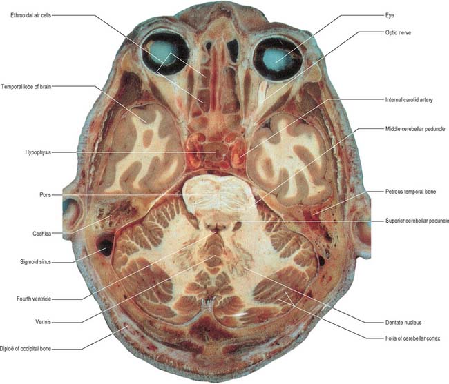
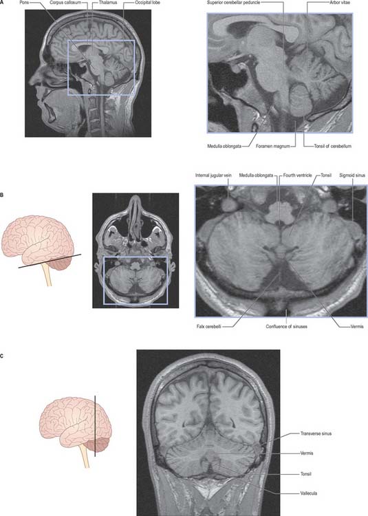
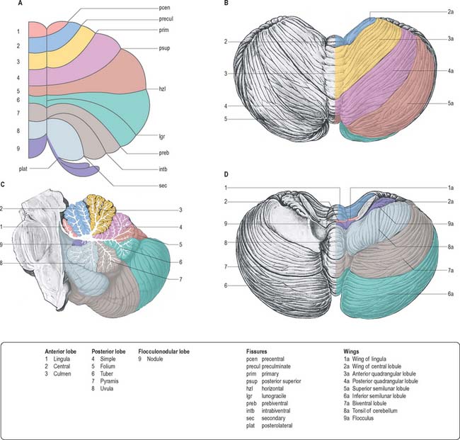
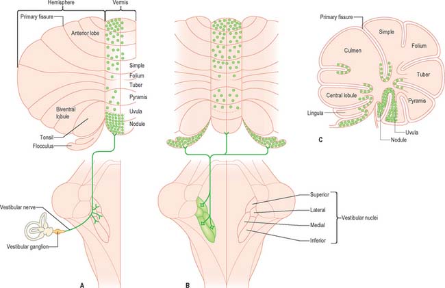
The cerebellum consists of two large, laterally located hemispheres which are united by a midline vermis (Figs 20.1, 20.2, 20.3). The superior surface of the cerebellum, which would constitute the anterior part of the unrolled cerebellar cortex, has a flat profile (Fig. 20.3). The paramedian sulci are shallow and the borders between vermis and hemispheres are indicated by kinks in the transverse fissures. The superior surface adjoins the tentorium cerebelli and projects beyond its free edge. The transverse sinus borders the cerebellum at the point where the superior and inferior surfaces meet. The inferior surface is characterized by a massive enlargement of the cerebellar hemispheres, which extend medially to overlie some of the vermis. Deep paramedian sulci demarcate the vermis from the hemispheres. Posteriorly the hemispheres are separated by a deep vallecula, which contains the dural falx cerebelli. The inferior cerebellar surface lies against the occipital squama. The shape of the surface facing the brain stem is irregular and forms the roof of the fourth ventricle and the lateral recesses on each side of it, while the cerebellar peduncles define the diamond shape of the ventricle when viewed from behind. Anterolaterally the cerebellum lies against the posterior surface of the petrous part of the temporal bone.

image

Fig. 20.2 Magnetic resonance images of the cerebellum of a 16-year-old female. A, sagittal B, axial. C, coronal.

(By courtesy of Drs JP Finn and T Parrish, Northwestern University School of Medicine, Chicago.)

The cerebellar surface is divided by numerous curved transverse fissures which separate its folia and give it a laminated appearance. The deepest fissures divide it into lobes and lobules. One conspicuous fissure, the horizontal fissure, extends around the dorsolateral border of each hemisphere from the middle cerebellar peduncle to the vallecula, separating the superior and inferior surfaces. Although the horizontal fissure is prominent, it appears relatively late in embryological development and does not mark the boundary between major functional subdivisions of the cortex. The deepest fissure in the vermis is the primary fissure, which curves ventrolaterally around the superior surface of the cerebellum to meet the horizontal fissures; it appears early in embryological development and marks the boundary between the anterior and posterior lobes.

Because the cerebellar cortex has a roughly spherical shape, the true relationships between its parts is somewhat obscure. Thus, the most anterior lobule of the cerebellar vermis, the lingula, lies very close to the most posterior lobule, the nodule. The lobules of the superior vermis that belong to the anterior lobe are the lingula, central lobule and culmen. The lingula is a single lamina of four or five shallow folia. Its white core is continuous with the superior medullary velum, and it is separated from the central lobule by the precentral fissure. The central lobule and culmen are continuous bilaterally with an adjoining lateral extension, or wing, in each hemisphere. The central lobule is separated from the culmen by the preculminary fissure. The culmen (with attached anterior quadrangular lobules) lies between the preculminary and primary fissures.

The simple lobule (with attached posterior quadrangular lobules) and the folium (with attached superior semilunar lobules) lie between the primary and the horizontal fissures: the two lobule sets are separated by the posterior superior fissure.

From the back forward, the inferior vermis is divided into the tuber, pyramis, uvula and nodule, in that order. The tuber is continuous laterally with the inferior semilunar lobules and separated from the pyramis by the lunogracile fissure. The pyramis and attached biventral lobules (containing an intrabiventral fissure) are separated from the uvula and attached cerebellar tonsils by the secondary fissure. Behind the uvula, and separated from it by the median part of the posterolateral fissure, is the nodule. The tonsils are roughly spherical and overhang the foramen magnum on each side of the medulla oblongata.

The nodule and attached flocculi constitute a separate flocculonodular lobe, which is separated from the uvula and tonsils by the deep posterolateral fissure. The flocculonodular lobe is richly interconnected with the vestibular nuclei, which lie at the lateral margin of the fourth ventricle.

FUNCTIONAL DIVISIONS OF THE CEREBELLUM

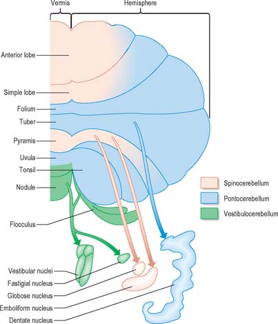
Functionally speaking, the cerebellum can be divided into a body, with inputs mainly from the spinal cord and pontine nuclei, and a flocculonodular lobe, which has strong afferent and efferent connections with the vestibular nuclei (Fig. 20.4). The body is subdivided into a series of regions dominated by their spinal or pontine inputs. The anterior lobe, simple lobule, pyramis and biventral lobules are the main recipients of spinal and trigeminal cerebellar afferents. Pontocerebellar input dominates in the folium, tuber and uvula, and throughout the entire hemisphere, including those regions that receive afferents from the spinal cord.

The mediolateral subdivision of the cerebellum into vermis and hemispheres represents a functional subdivision that is closely related to its output. In mammals, the increase in the size of the cerebellar hemispheres parallels the development of the cerebral cortex, and reflects the importance of the corticopontocerebellar input and of the efferent projections of the cerebellar hemispheres (through the dentate, emboliform and globose nuclei) to the thalamus and thence to the cerebral cortex.

INTERNAL STRUCTURE

The vast majority of cerebellar neuronal cell bodies are located within the outer, highly convoluted cortical layer. Beneath the cortex, the cerebellar white matter forms an extensive central core from which a characteristic branching pattern of nerve fibres (arbor vitae) extends towards the cortical surface. The white matter consists of afferent and efferent fibres travelling to and from the cerebellar cortex. Fibres crossing the midline in the white core of the cerebellum and the anterior medullary velum effectively constitute a cerebellar ‘commissure’; the afferent portion contains fibres of the restiform body and the middle cerebellar peduncle, and the efferent portion contains decussating fibres from the fastigial nucleus.

CEREBELLAR NUCLEI

Laterally, on either side within the white matter core, are four cerebellar nuclei. These are the dentate, emboliform, globose and fastigial nuclei. The dentate nucleus, which is located most laterally and is by far the largest, is the only nucleus easily visible to the naked eye (Fig. 20.1). It has the form of an irregularly folded sheet of neuronal cell bodies, with a medially directed hilum through which pass a mass of fibres mainly derived from dentate neurones and which form the bulk of the superior cerebellar peduncle. The emboliform and globose nuclei lie medial to the dentate and are equated to the nucleus interpositus (interposed nucleus) in lower species; the emboliform and globose nuclei may sometimes be referred to as the anterior and posterior interposed nuclei, respectively. Their efferent fibres join the superior cerebellar peduncle. The fastigial nucleus lies next to the midline, bordering on the roof of the fourth ventricle. A large proportion of the efferent fibres that leave this nucleus decussate within the cerebellar white matter and subsequently constitute the uncinate fasciculus, which passes dorsal to the superior cerebellar peduncle to enter the contralateral vestibular nuclei. Uncrossed fastigiobulbar fibres enter the vestibular nuclei by passing along the lateral angle of the fourth ventricle, and some fibres of the fastigial nucleus ascend in the superior cerebellar peduncle.

CEREBELLAR PEDUNCLES

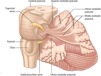
Three pairs of peduncles connect the cerebellum with the brain stem (see Ch. 19; Fig. 20.5).

The middle cerebellar peduncle is the most lateral and by far the largest of the three. It passes obliquely from the basal pons to the cerebellum and is composed almost entirely of fibres that arise from the contralateral basal pontine nuclei, with a small addition from nuclei in the pontine tegmentum.

The inferior cerebellar peduncle is located medial to the middle peduncle. It consists of an outer, compact fibre tract, the restiform body, and a medial, juxtarestiform body. The restiform body is a purely afferent system; it receives the posterior spinocerebellar tract from the spinal cord and the trigeminocerebellar, cuneocerebellar, reticulocerebellar and olivocerebellar tracts from the medulla oblongata. The juxtarestiform body is mainly an efferent system; it is made up almost entirely of efferent Purkinje cell axons from the vestibulocerebellum, on their way to the vestibular nuclei, and uncrossed fibres from the fastigial nucleus. It also contains primary afferent fibres travelling in the vestibular nerve and secondary afferent fibres from the vestibular nuclei. The crossed fibres from the fastigial nucleus pass dorsal to the superior cerebellar peduncle and enter the brain stem as the uncinate fasciculus at the border of the juxtarestiform and restiform bodies.

The superior cerebellar peduncle contains all of the efferent fibres from the dentate, emboliform and globose nuclei and a small fascicle from the fastigial nucleus. Its fibres decussate in the caudal mesencephalon, on their way to synapse in the contralateral red nucleus and thalamus. The anterior spinocerebellar tract reaches the upper part of the pontine tegmentum before looping down within this peduncle to join the spinocerebellar fibres entering through the restiform body.

CEREBELLAR CORTEX

Although the human cerebellum makes up approximately one-tenth of the entire brain by weight, the surface area of the cerebellar cortex, if unfolded, would be about half that of the cerebral cortex. The great majority of cerebellar neurones are small granule cells, so densely packed that the cerebellar cortex contains many more neurones than the cerebral cortex. Unlike the cerebral cortex, where a large number of diverse cell types are arranged differently in different regions, the cerebellar cortex contains a relatively small number of different cell types, which are interconnected in a highly stereotyped way. As a consequence, all regions of the cerebellar cortex look similar in histological sections.

The elements of the cerebellar cortex possess a precise geometric order, which is arrayed relative to the tangential, longitudinal and transverse planes in individual folia. The cortex contains the terminations of afferent ‘climbing’ and ‘mossy’ fibres, five varieties of neurone (granular, stellate, basket, Golgi and Purkinje), neuroglia and blood vessels.

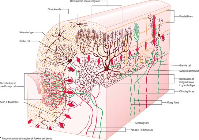
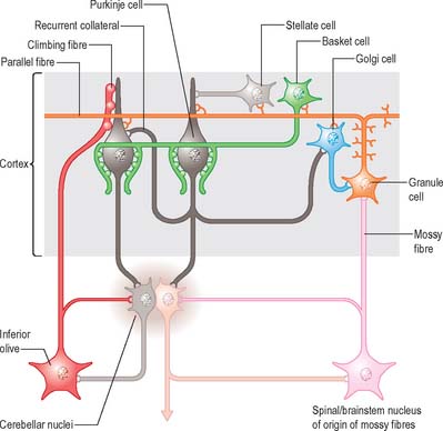
There are three main layers: molecular, Purkinje cell and granular (Fig. 20.6). The main circuit of the cerebellum involves granule cells, Purkinje cells and neurones in the cerebellar nuclei. Granule cells receive the terminals of the mossy fibre afferents (i.e. all afferent systems except the olivocerebellar fibres). The axons of the granule cells ascend to the molecular layer, where they bifurcate into parallel fibres, which are so called because they are oriented parallel to the transverse fissures and perpendicular to the dendritic trees of the Purkinje cells on which they terminate. Purkinje neurones are large and are the sole output cells of the cerebellar cortex. Their axons terminate in the cerebellar nuclei and in the vestibular nuclei. In addition to the dense array of parallel fibres, the dendritic trees of Purkinje cells receive terminals from climbing fibres which originate from neurones in the inferior olivary nucleus. The cerebellar cortex thus receives two distinct types of input: olivocerebellar climbing fibres, which synapse directly on Purkinje neurones, and mossy fibres, which connect to the Purkinje cells via granular neurones, whose axons are the parallel fibres.

Both parallel and climbing fibres excite the Purkinje cells, but they differ greatly in their firing characteristics and their effect on them. Purkinje cell axons inhibit their target neurones in the cerebellar nuclei; the latter project to motor control centres in the brain stem and cerebrum. The stellate, basket and Golgi cells are inhibitory interneurones and connect the cortical elements in complex geometrical patterns.

The molecular layer is approximately 300–400 μm thick. It contains a sparse population of neurones, dendritic arborizations, non-myelinated axons and radial fibres of neuroglial cells. Purkinje cell dendritic trees extend towards the surface and spread out in a plane perpendicular to the long axis of the cerebellar folia. Purkinje cell dendrites are flattened. The lateral extent of the Purkinje cell dendrites is some 30 times greater in the transverse plane than it is in a plane parallel to the cerebellar folia. Parallel fibres are the axons of granule cells, the stems of which ascend into the molecular layer where they bifurcate at T-shaped branches. The two branches extend in opposite directions as parallel fibres along the axis of a folium. Parallel fibres terminate on the dendrites of the Purkinje cells and Golgi cells, which they pass on their way, and on the basket and stellate cells of the molecular layer. Dendritic trees of Golgi neurones reach towards the surface. Unlike the flattened dendritic tree of the Purkinje cell, Golgi cell dendrites span the territory of many Purkinje neurones longitudinally as well as transversely. These dendrites receive synapses from parallel fibres. Some Golgi cell dendrites enter the granular layer, where they contact mossy fibre terminals. The cell bodies of Golgi neurones lie below, in the superficial part of the granular layer. The molecular layer also contains the somata, dendrites and axons of stellate neurones (which are located superficially within the molecular layer) and of basket cells (whose somata lie deeper within the molecular layer). Climbing fibres, the terminals of olivocerebellar fibres, ascend through the granular layer to contact Purkinje dendrites in the molecular layer. Radiating branches from large epithelial (Bergmann) glial cells give off processes that surround all neuronal elements, except at the synapses. Their conical expansions join to form an external limiting membrane at the surface of the cerebellum.

The Purkinje cell layer contains the large, pear-shaped somata of the Purkinje cells and the smaller somata of Bergmann glia. Clumps of granule cells and occasional Golgi cells penetrate between the Purkinje cell somata.

The granular layer (Fig. 20.6) is about 100 μm thick in the fissures and 400–500 μm on foliar summits. There are approximately 2.7 million granular neurones per cubic millimetre. It has been estimated that the human cerebellum contains a total of 4.6 × 10¹⁰ granule cells, and that there are 3000 granule cells for each Purkinje cell.

In summary, the granular layer consists of the somata of granule cells and the initial segment of their axons; dendrites of granule cells; branching terminal axons of afferent mossy fibres; climbing fibres passing through the granular layer en route to the molecular layer; and the somata, basal dendrites and complex axonal ramifications of Golgi neurones. Cerebellar glomeruli are synaptic rosettes consisting of a mossy fibre terminal that forms excitatory synapses upon the dendrites of both granule cells and Golgi cells.

Of the five cell types to be described, the first four are inhibitory, liberating γ-aminobutyric acid (GABA), and the fifth is excitatory, liberating L-glutamate. Figure 20.7 summarizes their main connections.

Purkinje cells have a specific geometry, which is conserved in all vertebrate classes (Fig. 20.6). They are arranged in a single layer between the molecular and granular layers. Individual Purkinje cells are separated by about 50 μm transversely and 50–100 μm longitudinally. Their somata measure approximately 50–70 μm vertically and 30–35 μm transversely. The subcellular structure of the Purkinje cell is similar to that of other neurones. One distinguishing feature is subsurface cisterns, often associated with mitochondria, which are present below the plasmalemma of somata and dendrites and may penetrate into the spines. The cisterns are intracellular calcium stores which are important links in the second messenger systems of the cell.

One, sometimes two, large primary dendrites arise from the outer pole of a Purkinje cell. From these an abundant arborization, with several orders of subdivision, extends towards the surface. Branches of each neurone are confined to a narrow sheet in a plane transverse to the long axis of the folium. Proximal first- and second-order dendrites have smooth surfaces with short, stubby spines, and are contacted by climbing fibres. Distal branches show a dense array of dendritic spines, which receive synapses from the terminals of parallel fibres. Inhibitory synapses are received from basket and stellate cells and from the recurrent collaterals of Purkinje cell axons, which contact the shafts of the proximal dendrites. The total number of dendritic spines per Purkinje neurone is in the order of 180,000.

The axon of a Purkinje cell leaves the inner pole of the soma and crosses the granular layer to enter the subjacent white matter. The initial axon segment receives axo-axonic synaptic contacts from distal branches of basket cell axons. Beyond the initial segment, the axon enlarges, becomes myelinated, and gives off collateral branches. The main axon ultimately forms a plexus in one of the cerebellar or vestibular nuclei. The recurrent collateral branches end on other Purkinje cells and on basket and Golgi neurones.

Basket and stellate cells are the neurones of the molecular layer. Their sparsely branched dendritic trees and the ramifications of their axons lie in a plane approximately perpendicular to the long axis of the folium, i.e. in the same plane as the Purkinje cell dendritic tree. Stellate cells are located in the superficial molecular layer and their axons synapse with the shafts of Purkinje cell dendrites. Both stellate and basket cells receive excitatory synapses from parallel fibres passing through their dendritic tree. Basket cells lie in the lower third of the molecular layer. Their somata receive synapses from Purkinje cell recurrent collaterals, climbing and mossy fibres as well as from the parallel fibres. Basket cell axons increase in size away from their somata and run deep in the molecular layer just above the Purkinje cells. Continuing for about 1 mm, each covers the territories of 10 to 12 Purkinje neurones. Collaterals of the basket cell axons ascend along Purkinje cell dendrites, and descend towards Purkinje cell somata and initial axonal segments forming pericellular networks, or ‘baskets’, around them. Branches from each basket cell axon also extend in the direction of the long axis of the folium to a further 3 to 6 rows of Purkinje neurones, flanking the axon. It follows that around 70 Purkinje cell neurones may receive synapses from a single basket neurone.

Most Golgi cell somata occupy the superficial zone of the granular layer, adjoining the Purkinje cell somata. Their dendrites radiate into the molecular layer. Unlike Purkinje cells, the dendritic trees of Golgi cells are not flattened, appearing much the same in transverse and longitudinal foliar section. In both planes they overlap the territories of several neighbouring Purkinje and Golgi cells. Some Golgi dendrites, however, divide in the granular layer and join cerebellar glomeruli, where they receive excitatory synaptic contacts from mossy fibres. The axon of the Golgi cell arises from the base of the cell body or proximal dendrite and immediately divides into a profuse arborization which extends through the entire thickness of the granular layer. The territory occupied by the axonal ramifications is of a volume that corresponds approximately to its dendritic tree in the molecular layer, and which overlaps with the axonal arborizations of adjacent Golgi cells. The main synaptic input to Golgi cell dendrites is from parallel fibres in the molecular layer. Purkinje cell recurrent collaterals and mossy and climbing fibres also terminate on their proximal dendrites and, more sparsely, on their somata.

Each granule cell has a spherical nucleus, 5–8 μm in diameter, with a mere shell of cytoplasm containing a few small mitochondria, ribosomes and a diminutive Golgi complex. Granule cells give rise to 3 to 5 short dendrites, which end in claw-like terminals within the synaptic glomeruli. The fine axons of granule cells enter the molecular layer and branch at a T-junction to form parallel fibres passing in opposite directions over a distance of several millimetres. Terminals located along the parallel fibres give them a beaded appearance and are sites of synapses upon the dendrites of Purkinje, stellate, basket and Golgi cells in the molecular layer. Most numerous are the synapses with Purkinje dendritic spines. It had been estimated that about 250,000 parallel fibres cross a single Purkinje dendritic tree, although every parallel fibre may not synapse with each dendritic tree that it crosses.

Two very different excitatory inputs innervate the cerebellar cortex, namely climbing fibres and mossy fibres.

Climbing fibres arise only from the inferior olivary nucleus. Olivocerebellar fibres cross the white matter and enter the granular layer where they branch to form climbing fibres. Each climbing fibre innervates a single Purkinje cell. There are about ten times as many Purkinje cells as there are cells in the inferior olive and so each olivocerebellar fibre branches into about 10 climbing fibres. Individual climbing fibres pass alongside the soma of a Purkinje cell, and then branch to make numerous synapses on the short, stubby spines that protrude from the proximal segments of Purkinje cell dendrites.

Mossy fibres take their origin from the spinal cord, the trigeminal, dorsal column, and reticular nuclei of the medulla, and the pontine tegmentum and basal pons. Like climbing fibres they are excitatory, but they contrast sharply in their anatomical distribution and physiological properties. As each mossy fibre traverses the white matter its branches diverge to enter several adjacent folia. Within each folium these branches expand into grape-like synaptic terminals (mossy fibre rosettes), which occupy the centre of cerebellar glomeruli.

Noradrenergic and serotoninergic fibres form a rich plexus in all layers of the cerebellar cortex. The aminergic fibres are fine, varicose and form extensive cortical plexuses: their release of noradrenaline and serotonin is assumed to be non-synaptic, and their effects paracrine, involving volumes of tissue. The serotoninergic afferents of the cerebellum take their origin from neurones in the medullary reticular formation, other than the raphe nuclei. The noradrenergic, coeruleocerebellar projection, when active, inhibits Purkinje cell firing not by direct action but via β-adrenergic-receptor-mediated inhibition of adenylate cyclase in the Purkinje cells. Cerebellar afferents have been traced from dopaminergic cells in the ventral tegmental area, and dopamine D2 and D3 receptors are present in the molecular layer. A plexus of thin, ChAT-containing fibres is centred on the Purkinje cell layer. The cells of origin of these cholinergic fibres are not known.

The connections of the cerebellum are organized in two perpendicular planes, corresponding to the planar organization of the cerebellar cortex. Efferent connections of the cortex are disposed in parasagittal sheets or bundles, which connect longitudinal strips of Purkinje cells with specific cerebellar or vestibular nuclei. The climbing fibre afferents to a Purkinje cell zone from the inferior olive display a similar zonal disposition. Cerebellar output is organized in modules, where a module consists of one or more Purkinje cell zones, their cerebellar or vestibular target nucleus, and their olivocerebellar climbing fibre input. Modular function is determined by the brain stem projections of the cerebellar or vestibular target nucleus. A general feature of the modular organization of the cerebellum is that GABAergic neurones in the cerebellar nuclei project to the subnuclei of the contralateral inferior olive, which give rise to their respective climbing fibre afferents. These recurrent connections are known as nucleo-olivary pathways.

Mossy fibre afferent systems from precerebellar nuclei in the spinal cord and the brain stem terminate in the granular layer of certain lobules in transversely oriented terminal fields. The transverse lobular arrangement of the mossy fibre afferents is enforced by the transverse orientation of the parallel fibres, which are axons of the granule cells and constitute the second link in the mossy fibre–parallel fibre input of the Purkinje cells. Parallel fibres cross and terminate on Purkinje cells belonging to several successive modules as they course through the molecular layer.

Purkinje cells can be activated in two different ways. Granule cell activity generates simple spikes, which resemble the response of other neurones in the brain, whereas activation by a climbing fibre produces a prolonged depolarization upon which several spike-like waves are superimposed. The rate of firing of single and complex spikes also differs markedly. While the Purkinje cell may fire simple spikes at a rate of hundreds per second, complex spikes occur at low frequencies, seldom more than three or four per second.

Purkinje cell activity is regulated by local Golgi, basket and stellate cells. Like Purkinje cells, Golgi cells have a rich dendritic tree that extends through the molecular layer. Unlike Purkinje cells, the Golgi cell dendrites are not restricted to a plane transverse to the folia and their axons do not leave the cerebellar cortex. Golgi cells regulate firing by presynaptic inhibition of the mossy fibre afferents, and so act as a governor, or rate limiter, of Purkinje cell activity. Stellate and basket cells synapse directly on Purkinje cells and are powerful inhibitors of their activity.

Structural and functional cerebellar localization

Since the cerebellar cortex is largely uniform in microstructure and microcircuitry, it seems likely that its basic mode of operation is also uniform. The most obvious input for this operation is provided by the mossy fibre afferents, which carry information from all levels of the spinal cord, and specialized sensory and motor information relayed from the cerebral cortex and subcortical motor centres. The most obvious output from the cerebellum is directed at motor systems. Purkinje cells are organized in modules, i.e. discrete, parallel zones that converge upon different cerebellar output nuclei coupled to different motor systems in the brain stem, spinal cord and cerebral cortex. Cerebellar function is therefore determined by temporal and spatial factors, e.g. inhibitory interneurones of the cerebellar cortex, which regulate the access of a particular combination of mossy fibre–parallel fibre inputs to an appropriate output. Plastic changes in the response properties of Purkinje cells, in the form of long-term depression of the parallel fibre–Purkinje cell synapses, may also contribute. Short-term and long-term changes in the response properties of Purkinje cells are under the influence of the climbing fibres.

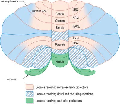
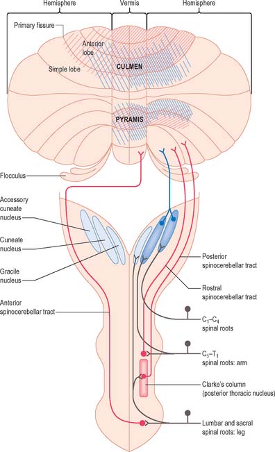
A double, mirrored, localization exists in the anterior and posterior cerebellum (Fig. 20.8). The anterior lobe, simple lobule, pyramis and the adjoining lobules of the hemisphere of the posterior lobe all receive branches from the same mossy and climbing fibres and project to the same cerebellar nuclei. The efferent pathways of these regions monitor the activity in the corticospinal tract and in the subcortical motor systems descending from the vestibular nuclei and reticular formation. The inputs to the cerebellum and the outputs from it are organized according to the same somatotopical patterns, but the orientation of these patterns is reversed. The representation of the head is found principally in the simple lobule, and caudally in a corresponding region of the posterior lobe. The double representation of the body follows in rough somatotopic order. Vestibular connections of the cerebellum display a similar double representation in the most rostral lobules of the anterior lobe and far caudally in the vestibulocerebellum (Fig. 20.9).

The folium, tuber, uvula, tonsil and posterior biventral lobule all receive an almost pure pontine mossy fibre input. Climbing fibres from the inferior olive and mossy fibres from the basilar and tegmental pontine nuclei relay visual and acoustic information from the respective cerebral association areas and midbrain tectum to the folium and tuber thought to represent a vermal visual/acoustic area (Fig. 20.8). The efferent connections of this area travel via the fastigial nucleus to gaze centres in the pons and midbrain.

AFFERENT CONNECTIONS OF THE CEREBELLUM

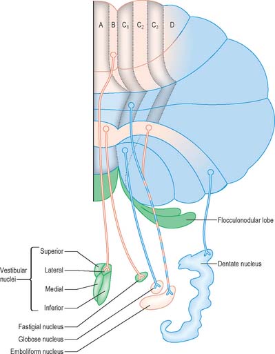
Mossy fibres and climbing fibres carry the afferent connections of the cerebellum. Mossy fibre systems terminate bilaterally in transversely oriented ‘lobular’ areas; there is considerable overlap amongst the terminations. Climbing fibres from different subnuclei of the inferior olive terminate contralaterally, on discrete longitudinal strips of Purkinje cells. This longitudinal pattern closely corresponds with the zonal arrangement in the corticonuclear projection (Fig. 20.10).

Spinocerebellar and trigeminocerebellar fibres

The spinal cord is connected to the cerebellum through the spinocerebellar and cuneocerebellar tracts and through indirect mossy fibre pathways relayed by the lateral reticular nucleus in the medulla oblongata. These pathways are all excitatory in nature and give collaterals to the interposed and fastigial nuclei before ending on cortical granule cells.

The posterior spinocerebellar tract takes its origin from the cells of Clarke’s column at the base of the dorsal horn in all thoracic segments of the spinal cord (Fig. 20.11). It enters the inferior cerebellar peduncle, gives collaterals to the cerebellar nuclei, and terminates, mainly ipsilaterally, in the vermis and adjoining regions of the anterior lobe and in the pyramis and adjoining lobules of the posterior lobe. Clarke’s column receives primary afferents of all kinds from the muscles and joints of the lower limbs, which reaches the nucleus via the fasciculus gracilis. It also receives collaterals from cutaneous sensory neurones. Accordingly, the tract transmits proprioceptive and exteroceptive information about the ipsilateral lower limb. Very fast conduction is required to keep the cerebellum informed about ongoing movements. The axons in the posterior spinocerebellar tract are the largest in the CNS, measuring 20 μm in diameter. The upper limb equivalent of the posterior spinocerebellar tract is the cuneocerebellar tract.

The anterior spinocerebellar tract is a composite pathway. It informs the cerebellum about the state of activity of spinal reflex arcs related to the lower limb and lower trunk. Its fibres originate in the intermediate grey matter of the lumbar and sacral segments of the spinal cord (Fig. 20.11). They cross near their origin, and ascend close to the surface as far as the lower midbrain before looping down in the superior cerebellar peduncle. Most fibres cross again in the cerebellar white matter. Thus their distributions to the cerebellar nuclei and cortex appear to be the same as those of the posterior tract.

The rostral spinocerebellar tract originates from cell groups of the intermediate zone and horn of the cervical enlargement. Although considered to be the upper limb and upper trunk counterpart of the anterior spinocerebellar tract, most of its fibres remain ipsilateral throughout their course. It enters the inferior cerebellar peduncle and terminates in the same cerebellar nuclei and folia as the cuneocerebellar tract.

The cuneocerebellar tract contains exteroceptive and proprioceptive components which originate from the cuneate and external cuneate nuclei respectively. The primary afferents travel in the fasciculus cuneatus. The tract itself is predominantly uncrossed and ends in the posterior half of the anterior lobe. Exteroceptive and proprioceptive mossy fibre components of the tract terminate differentially in the apical and basal part of the folia. The exteroceptive component overlaps the pontocerebellar mossy fibre projection in the apices of the folia of the anterior lobe.

Comparable sets of ipsilateral proprioceptive and interceptive cerebellar projections exist for the extensive territory of the trigeminal brain stem nuclei. These nuclei also project to the ipsilateral inferior olive, relaying from there to the contralateral cerebellar cortex and deep nuclei. The cortical representation of the head is directly behind the primary fissure.

Olivocerebellar fibres

Olivocerebellar climbing fibre connections

The inferior olivary complex can be subdivided into a convoluted principal olivary nucleus, and posterior and medial accessory olivary nuclei. Olivary fibres form the olivocerebellar projection to the contralateral cerebellar cortex, and give off collaterals to the lateral vestibular nucleus and to the cerebellar nuclei. Climbing fibres terminate on longitudinal strips of Purkinje cells. The zonal patterns of the olivocerebellar and Purkinje–nuclear projections correspond precisely. The accessory olivary nuclei project to the vermis and the adjacent hemispheres. The caudal halves of the posterior and medial accessory nuclei innervate the vermis. The caudal part of the posterior accessory nucleus projects to Deiters’ nucleus and to the B zone of the anterior vermis. The caudal half of the medial accessory olive gives rise to a projection to the fastigial nucleus and provides climbing fibres to the A zone. The rostral halves of the accessory olives project to the pars intermedia. Climbing fibres from the rostral dorsal accessory olive give collateral projections to the emboliform nucleus and terminate in zones C1 and C3. Zone C2 receives terminals from the rostral medial accessory olive, which provides a collateral projection to the globose nucleus. The principal nucleus projects to the contralateral hemisphere (D zone), and gives collaterals to the dentate nucleus.

The inferior olivary complex receives afferent connections from the spinal cord and from sensory relay nuclei in the brain stem, including the posterior column and sensory trigeminal nuclei. It also receives descending connections from the superior colliculus, parvicellular red nucleus and related nuclei in the midbrain and a GABAergic projection, mainly crossed, from the cerebellar nuclei and certain vestibular nuclei. The latter two connections are topically organized. Thus, the dentate nucleus projects to the principal nucleus, the emboliform nucleus to the rostral posterior accessory nucleus, and the globose nucleus to the rostral medial accessory nucleus. The fastigial nucleus is connected with the caudal medial accessory olive, but the fibres are less numerous. The caudal posterior accessory olive receives a projection from the lateral vestibular nucleus.

The posterior accessory olive and the caudal half of the medial accessory olive receive an input from the spinal cord and sensory relay nuclei. The middle region of the medial accessory olive receives a projection from the superior colliculus and projects to folium and vermis. The parvocellular red nucleus and related nuclei project to the olive through the ipsilateral descending central tegmental tract, which terminates in the rostral half of the medial accessory olive and the principal olive. The parvocellular red nucleus receives converging projections from the cerebellar nuclei and from the motor and premotor cortex. Direct pathways from the cerebral cortex to the inferior olive are sparse. The indirect pathways via the parvicellular red nucleus are much larger.

Climbing fibres that terminate in the vestibulocerebellum (flocculus and nodule) are derived from neurones of the medial accessory olive which receive a strong descending afferent connection from optokinetic centres in the midbrain. Optokinetic information is used by the flocculus in long-term adaptation of compensatory eye movements. Neighbouring neurones are under vestibular control and project to the nodule and the adjoining uvula.

Reticulocerebellar fibres

The lateral reticular nucleus of the medulla oblongata, and the paramedian reticular and tegmental reticular nuclei of the pons, give rise to mossy fibres. The latter nuclei also supply major collateral projections to the cerebellar nuclei. Spinoreticular fibres terminate in a somatotopical pattern within the entire lateral reticular nucleus, where they overlap with collaterals from the rubrospinal and lateral vestibulospinal tracts and a projection from the cerebral cortex.

The lateral reticular nucleus projects bilaterally to the vermis and hemispheres of the cerebellum. The projection from the dorsal part of the nucleus, which receives collaterals from the rubrospinal tract in addition to spinal afferents, is centred on the ipsilateral hemisphere. The ventral part of the nucleus, which receives a strong projection from the spinal cord and a collateral projection from the lateral vestibulospinal tract, projects bilaterally, mainly to the vermis. The lateral reticular nucleus provides a strong projection to the superior fastigial nucleus, the emboliform nucleus and the medial pole of the globose nucleus.

The paramedian reticular nucleus consists of cell groups at the lateral border of the medial longitudinal fasciculus. It receives fibres from the vestibular nuclei and the interstitiospinal and tectospinal tracts (which descend in the medial longitudinal fasciculus), and from the spinal cord and the cerebral cortex. It projects to the entire cerebellum.

The tegmental reticular nucleus of the pons is located next to the midline in the caudal half of the tegmentum. It receives afferent connections from the cerebral cortex, tectum, nucleus of the optic tract and cerebellar nuclei via the crossed descending branch of the superior cerebellar peduncle. Efferents from the tegmental reticular nucleus reach the cerebellum through the middle cerebellar peduncle. Some terminate superficially in the cortex of the anterior lobe, but many more end in the simple lobule, folium, tuber, vermis and adjoining flocculus. Additional efferents terminate in the caudal fastigial nucleus, dentate nucleus and lateral parts of the globose nucleus.

Pontocerebellar fibres

The cerebral cortex is the largest single source of fibres that project to the pontine nuclei. Fibres from the pontine nuclei access the cerebellum via the middle cerebellar peduncle, which is the largest afferent system of the human cerebellum. Many corticopontine fibres are collaterals of axons that project to other targets in the brain or spinal cord, e.g. it is likely that all corticospinal fibres give off collaterals to the pontine nuclei. Although corticopontine axons arise from lamina V pyramidal cells, the projection from different areas of the cerebral cortex is highly uneven. The areas of cerebral cortex that project to the pontine nuclei are those that are particularly involved in the control of movement. For example, in the case of visual areas, the input arises from extrastriate visual areas in the parietal lobe whose cells are responsive to movement, and function as important links in the visual guidance of movement. Dorsal pontine nuclei receive collateral branches from corticotectal fibres that project to the superior and inferior colliculi from the parietal, temporal and frontal areas of the cerebral cortex, and from tectopontine relays. The onward pontocerebellar projections are to the simple lobule and to the folium and tuber of the vermis.

Fibres of the pontine reticular nuclei are distributed bilaterally, with ipsilateral predominance, to all lobules of the cerebellum other than the lingula and nodule.

More than 90% of fibres in the middle cerebellar peduncle belong to the cortico-ponto-cerebellar pathway. Corticopontine fibres travel in the cerebral peduncle. Fibres from the frontal lobe occupy the medial part of the peduncle, and fibres from the parietal, occipital and temporal lobes occupy the lateral part. They synapse on some 20 million neurones in corresponding regions of the basilar pons. The onward pontocerebellar mossy fibre projection is predominantly to the lateral regions of the posterior and anterior lobes (Fig. 20.4) but collaterals are given off to the dentate nucleus.

EFFERENT CONNECTIONS OF THE CEREBELLUM

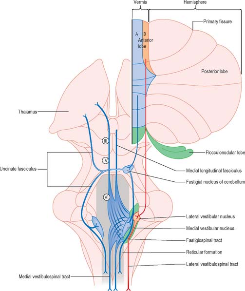
The output of the cerebellum consists of the inhibitory projections of the Purkinje cells to the cerebellar and vestibular nuclei, and the efferent connections of the cerebellar nuclei to motor centres in the brain stem and, through the thalamus, the motor cortex. Their effects on movement are always indirect, because there are no direct projections from the cerebellar nuclei to motor neurones. Disynaptic connections of the Purkinje cells in the anterior vermis and vestibulocerebellum with motor neurones controlling oculogyric and proximal limb muscles are mediated by the vestibular nuclei. The vermis also influences these motor neurones bilaterally through multisynaptic pathways that involve the fastigial and vestibular nuclei and the reticular formation (Fig. 20.12). The vermis cannot be considered as a single module. Each half of the vermis is composed of several modules (each made up of a longitudinal Purkinje cell zone and a target nucleus) and their supporting climbing fibre afferent projections.

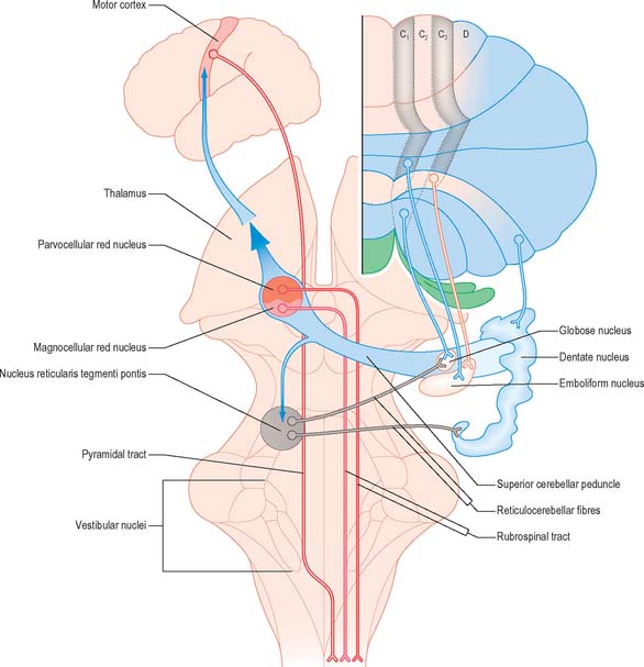
Each cerebellar hemisphere influences movements of the ipsilateral extremities by way of projections to the dentate and interposed (emboliform and globose) nuclei, which in turn project to the contralateral red nucleus, thalamus and motor cortex (Fig. 20.13).

Corticonuclear and corticovestibular fibres

Purkinje cells of each hemivermis project to the ipsilateral fastigial and vestibular nuclei. Purkinje cells of the hemisphere project to the interposed and dentate nuclei. Although the cerebellar cortex is organized in strips of Purkinje cell zones, which project to different cerebellar and vestibular nuclei, the borders between these strips are not apparent in the structure of the cortex when it is examined histologically using conventional staining methods. The vermis of the anterior lobe and simple lobule consist of two parallel strips, A and B, of Purkinje cells (Figs 20.10, 20.12). The medial strip (A zone) projects to the rostral pole of the fastigial nucleus, and the lateral strip (B zone) projects to the lateral vestibular nucleus. The B zone does not continue beyond the simple lobule. The cortex of the entire caudal vermis, which projects to the fastigial nucleus, is included in the A zone. The folium and tuber, which represent a region of the cerebellum that receives a visual input and which are involved in the accurate calibration of saccades, project to the caudal pole of the fastigial nucleus. The pyramis, uvula and nodule can be subdivided into several Purkinje cell zones. However, the significance of their connections with the cerebellar and vestibular nuclei is not well understood. Corticovestibular projections to the superior, medial and inferior vestibular nuclei, but not to the lateral nucleus, take their origin from the nodule and the adjacent region of the uvula.

The intermediate region consists of two strips of Purkinje cells (C1 and C3 zones), which project to the emboliform (anterior interposed) nucleus. They flank a single zone (C2) that projects to the globose (posterior interposed) nucleus (Figs 20.10, 20.13). The rest of the hemisphere projects to the dentate nucleus. There are indications for a subdivision of the hemisphere into two zones that project to the caudolateral and rostromedial parts of the dentate nucleus, respectively. The neurones of the caudolateral dentate are generally smaller than those of the rostromedial portion and the convolutions are broader. The efferent connections of the flocculus are mainly with the superior, medial and inferior vestibular nuclei and resemble those from the nodule and uvula.

Cerebellovestibular and cerebelloreticular fibres

Cerebellovestibular connections

The relationship between the cerebellum and the vestibular nuclei is complex (Figs 20.9, 20.12). In addition to the vestibulocerebellum (nodule, adjacent folia of the uvula and flocculus), the main vermis and the fastigial nucleus also project to the vestibular nuclei.

The vestibulocerebellum projects to the superior, medial and inferior vestibular nuclei. Neurones of these nuclei, which receive an input from the vestibular nerve and project to the nuclei controlling eye movements (vestibulo-ocular relay cells), are among the main targets of the Purkinje cells of the nodule and flocculus. Through these connections with vestibulo-ocular relay neurones, the flocculus is involved in the long-term adaptation of compensatory eye movements, the generation of smooth eye movements used to pursue an object, and the suppression of the vestibulo-ocular reflex during smooth pursuit. The function of the nodule in the control of eye movement is less well understood.

The vestibular nuclei are the main source of mossy fibre afferents to the nodule; their projection to the flocculus is relatively minor. Most mossy fibres which terminate in the flocculus arise from the reticular formation and relay optokinetic and visual information.

The lateral vestibular nucleus, which lacks an input from the labyrinth and receives Purkinje cell axons from the B zone of the anterior vermis, can be regarded as a displaced cerebellar nucleus. It gives rise to the lateral vestibulospinal tract, which descends to all levels of the spinal cord. It is avoided by the efferent pathways from the fastigial nucleus, which terminate more ventrally on large neurones in the magnocellular part of the medial vestibular nucleus and in the medial reticular formation. The medial and inferior vestibular nuclei receive a major input from the vestibular nerve and give rise to bilaterally ascending and descending tracts that course in the medial longitudinal fasciculus. The ascending tract is composed predominantly of the axons of vestibulo-ocular relay cells. The descending fibres form the medial vestibulospinal tract, which is particularly involved in head righting reflexes when the trunk is tilted.

Fastigial fibres, which terminate in the reticular formation, stimulate the bilaterally descending medullary reticulospinal tracts. The A zone of the vermis exerts a bilateral influence on ventromedially located spinal interneurones and motor neurones that innervate axial, truncal and proximal limb muscles. Some fibres of the uncinate tract descend as far as the cervical cord, where they terminate on the same motor neurones. The B zone exerts an influence on ipsilateral interneurones and motor neurones of the same system through its projection to the lateral vestibular nucleus and the lateral vestibulospinal tract.

The projections of the fastigial nucleus to the thalamus are relatively minor. They are bilateral, because the crossed ascending fibres of the uncinate fasciculus subsequently recross. Their targets include parts of the ventrolateral nucleus and the intralaminar, centrolateral and parafascicular nuclei. Fibres that terminate in the ventrolateral nucleus lie medial to the terminations of fibres from the dentate and interposed nuclei. This region of the ventrolateral nucleus projects to the motor cortex but also sends collaterals to the medullary reticular formation, which influences ventromedial interneurones and motor neurones in lumbar and sacral segments of the spinal cord via the medullary reticulospinal tracts.

The caudal region of the fastigial nucleus receives Purkinje cell axons from the folium and tuber, an area of the vermis that receives visual inputs. It projects to the contralateral horizontal gaze centre (paramedian pontine reticular formation, PPRF), and the vertical gaze centre (rostral interstitial nucleus of the medial longitudinal fasciculus, riMLF), and bilaterally, to deep layers of the superior colliculus. These projections probably mediate the adaptation of saccades by the vermal visual area.

The cerebellum influences visceromotor systems via the projections of the fastigial nucleus to the parasolitary nucleus (a region bordering the viscerosensory nuclei of the solitary tract), the dorsal motor nucleus of the vagus, the central grey matter, the serotoninergic raphe nuclei of the pons and medulla, and the noradrenergic locus coeruleus.

Other pathways from the cerebellar nuclei terminate on precerebellar relay nuclei that give rise to mossy or climbing fibres. Recurrent circuits involving the fastigial nucleus include the nucleus reticularis tegmenti pontis and a projection from the fastigial nucleus to the medial accessory olive. Nucleo-olivary projections arise from all the cerebellar nuclei, are crossed, and contain GABA as a neurotransmitter. The connections of the fastigial nucleus with the reticular nuclei are excitatory.

Cerebellar nuclei also project to the contralateral interstitial nucleus of Darkschewitsch, which lies at the boundary between the midbrain and diencephalon. This nucleus projects to the medial accessory olive via the central tegmental tract. The fastigial nucleus controls the climbing fibre output of the medial accessory olive, both via its nucleo-olivary projection and by its connection to the nucleus of Darkschewitsch.

Cerebellorubral and cerebellothalamic fibres

The axons of neurones in the dentate and interposed nuclei leave the cerebellum in the superior cerebellar peduncle. The superior peduncles, including their nucleo-olivary component, decussate in the caudal midbrain (Fig. 20.13) and each then gives off a small descending branch carrying fibres that terminate in the medial reticular formation of the pons and medulla and the pontine tegmental reticular nucleus. The nucleo-olivary fibres join this descending branch and terminate in the inferior olive in a strictly ordered manner. The ascending branch is distributed to the midbrain and diencephalon, mainly to the red nucleus and thalamus. The emboliform (anterior interposed) nucleus projects to the magnocellular part of the red nucleus. In humans this projection is very small and the target neurones give rise to a relatively trivial rubrospinal tract, which crosses in the caudal midbrain and terminates on lateral medullary interneurones and a small number of motor neurones in the upper cervical spinal cord.

The emboliform (anterior interposed) nucleus projects to lateral parts of the ventrolateral nucleus of the thalamus, which are connected with elements of the motor cortex projecting to axial and proximal limb muscles, and to the reticular formation of the pons and medulla. It also projects to the pontine tegmental reticular nucleus and to basal pontine nuclei, both of which give rise to mossy fibres. Its nucleo-olivary efferents terminate in the rostral half of the dorsal accessory olive.

The projections of the globose (posterior interposed) nucleus are very similar to those of the fastigial nucleus. The two nuclei share projections to the cord, the superior colliculus, the central grey matter and the raphe nuclei. Nucleo-olivary projections from the globose nucleus and the recurrent globose nucleus–interstitial nucleus–inferior olivary nucleus pathway converge upon the rostral half of the medial accessory olive. The thalamic projections overlap those from the fastigial and emboliform nuclei.

The dentate nucleus projects to the contralateral parvicellular red nucleus and the thalamus. The central tegmental tract takes its origin from the parvocellular red nucleus and terminates on the principal nucleus of the olive. The thalamic projection to the ventrolateral nucleus overlaps those of the other cerebellar nuclei. The inferior and lateral parts of the dentate nucleus project into the most medial region of the ventrolateral nucleus, which in turn projects to the premotor area of the frontal lobe.

The thalamus receives a massive input from other major motor systems, in addition to the input it receives from the cerebellum. In particular, the output of the basal ganglia is relayed to the thalamus by a projection from the globus pallidus (Ch. 22). Available evidence suggests that these two great subcortical motor systems terminate on different regions in the ventral thalamus and project to different targets in the motor and premotor cortex.

CEREBELLAR FUNCTIONS

The vermis of the cerebellum is involved in taking anticipatory action in order to maintain an upright posture when objects are picked up. For example, when a book is taken down from a shelf, the first muscle groups to be activated are not the flexors of shoulder, elbow or fingers, but the plantar flexors of the ankle. Contraction of the ankle flexors causes the forefeet to push the lower limbs and trunk backwards at the moment the hand grasps the book. Once the lift gets under way, the erector spinae muscles correct for the combined weights of the book and the reaching arm, in order to prevent forward sway of the head and trunk. Labyrinthine receptors simultaneously inform the cerebellum of any forward movement of the head, and appropriate antigravity thrust is exerted via one or both lateral vestibulospinal tracts.

The posterolateral region of the cerebellum has a role in postural fixation to prevent oscillation of distal limb parts caused by the viscoelastic properties of the muscles in response to sudden movements. If a volunteer is instructed to exert rapid wrist extension and to maintain the extended posture for two seconds, electromyographic records taken from the prime movers and antagonists reveal that the antagonists begin to contract prior to completion of the movement, and that they continue to contract and relax several times in alternating fashion with the prime movers, although with much less force, during the measured fixation period.

The cerebellum has a role in motor learning. Experimental studies have shown that when a novel motor skill is being learned, the olivocerebellar climbing fibre system becomes active when errors are made. The inferior olivary complex appears to be involved in correction, based on receipt of a copy of the intended movement from collateral branches of the corticospinal tract. Cerebellar output via the superior cerebellar peduncle is also copied on to the parvicellular red nucleus and projected from there to the inferior olive, where it can be compared with the original. Short bursts of climbing fibre activity depress the Purkinje cells responsible for producing the errors.

Many motor skills require precise timing, which involves an extreme degree of cooperation between prime movers and their antagonists. For example, reading a printed page requires that the scanning eyes snap back to the beginning of a line, time after time: even small errors may result in dyslexia, where slight incoordination of eye movements may cause the letters of a word to appear jumbled. Clinical testing of timing can be performed easily by checking the ability to perform rhythmic movements such as repetitive pronation/supination (Fig. 20.14).

image

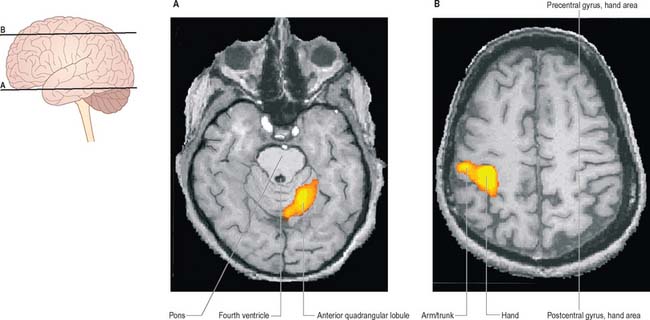
Fig. 20.14 fMRI (functional magnetic resonance imaging) of a volunteer executing repetitive finger movements of the right hand.

(By courtesy of Drs JP Finn and T Parrish, Northwestern University School of Medicine, Chicago.)

CEREBELLAR LESIONS

Midline cerebellar lesions, e.g. tumours such as medulloblastoma and secondary carcinoma, lead to abnormalities of posture, so that the patient cannot sit or stand, without toppling, usually backwards.

Unilateral lesions in the cerebellar hemispheres, e.g. stroke, produce ipsilateral symptoms and signs. Inco-ordination of eye movements leads to nystagmus, which is most marked when gaze is directed to the side of the lesion. Incoordination of the ipsilateral upper limb is manifested in the finger-nose test. The movements are inaccurate (dysmetria); the limb oscillates to and fro, the amplitude being greater as the target is reached (intention tremor); there is a breakdown of smooth co-operation between muscles controlling the joints of the upper limb (asynergia); alternating hand movements are clumsy (dysdiadochokinesia). When the arm is outstretched, there is a tendency for the limb to hyper-pronate and rise, and when pressure is applied to the extended arm and then released, there is a marked rebound of the limb. Unilateral incoordination of the lower limb is apparent because heel/shin testing is inaccurate.

Bilateral lesions of the cerebellar hemisphere lead to nystagmus and a form of dysarthria in which the syllables are extended and prosody is lost (scanning speech). The gait is wide based, reeling, and incoordinate (ataxia): the ataxia is not made significantly worse on eye closure (negative Romberg’s sign). Bilateral lesions can be acute, as in the encephalitis that follows chicken pox, or chronic, as in alcohol abuse, and the inherited spinocerebellar ataxias. The co-occurrence of nystagmus, scanning speech and intention tremor were held by the 19th century neurologist Charcot to be pathognomonic of multiple sclerosis.

It has been speculated that cerebellar lesions may lead to disorders of higher cortical function and changes in affect, but this has not been substantiated.