Central Serous Chorioretinopathy
Clinical Features:
Patients usually present with a unilateral decrease and distortion of central vision. Examination reveals a macular, well-circumscribed neurosensory retinal detachment often with one or more retinal pigment epithelial detachments. Often signs of CSCR can also be found in the contralateral eye (Fig. 12.1.1).
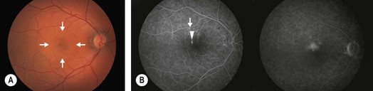
Figure 12.1.1 (A) Fundus photo showing a discrete, well-circumscribed elevation at the macula (arrows). (B) Fluorescein angiography in the early phase shows an area of hyperfluorescence (arrowhead) with leakage noted in the late phase. Note the adjacent areas of hyperfluorescence (arrow) indicative of RPE window defects characteristically seen in patients with central serous chorioretinopathy.
OCT Features
▶ Acute: the OCT reveals a well-circumscribed neurosensory retinal detachment seen as an elevation of the retinal layers with optically clear fluid occupying the space between the outer retina and the RPE layer (Fig. 12.1.2). Often (75%) these are also associated with a small pigment epithelial detachment, seen as elevation of the RPE layer with underlying shadowing. The retina may sometimes be thickened in the acute phase. Choroidal thickening is usually noted compared to normal as well as to fellow eyes in acute CSCR, and this may be better visualized using the EDI protocol on most commercial OCT scanners. This diffuse thickening may be seen to improve when the acute phase of the CSCR resolves.
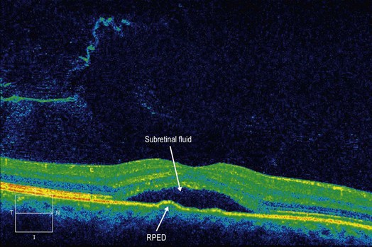
Figure 12.1.2 OCT scanning shows a neurosensory retinal detachment. A small pigment epithelial detachment can sometimes be visualized.
▶ Chronic CSCR may be accompanied by accumulation of hyper-reflective material in the subretinal space (Fig. 12.1.3). Cystic retinal changes and eventual retinal thinning has been reported overlying the areas of subretinal fluid in chronic CSCR. This may be accompanied by photoreceptor and RPE loss. The loss of photoreceptors on OCT may also be associated with decreased best corrected visual acuity even after resolution of subretinal fluid.
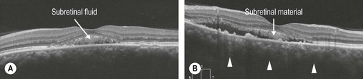
Figure 12.1.3 (A) Chronic central serous chorioretinopathy. Note the subretinal material accumulation and the change in reflectivity of the outer nuclear layer on OCT. (B) Enhanced depth imaging of chronic CSCR. Note that the bottom of the choroid cannot be visualized because of choroidal thickening (arrowheads). There is accumulation of subretinal material.
▶ Multifocal CSCR is characterized by multiple discrete areas of neurosensory detachments. As the CSCR resolves, the subretinal fluid is seen to decrease and then disappear. Quantitative OCT measurements of subretinal fluid are useful in monitoring for improvement and resolution.