Central Retinal Artery Occlusion
Clinical Features:
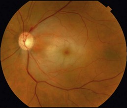
The classic finding is retinal whitening with a central cherry-red spot in the acute setting (Fig. 15.2.1). This corresponds to edema of the inner retina, which is most pronounced in the macula due to the prominent nerve fiber and ganglion cell layer. The central fovea lacks inner retina layers and therefore the underlying retinal pigment epithelium and choroidal pigment shows through giving the cherry-red spot.
OCT Features:
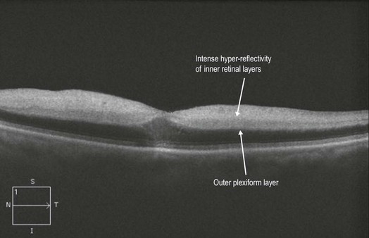
In the acute setting, there is intense hyper-reflectivity of the inner retinal layers (Fig. 15.2.2), corresponding to edema of the retinal layers supplied by the inner retinal vascular supply from the central retinal artery (watershed zone is between inner nuclear and outer plexiform layers). This hyper-reflectivity creates a shadowing effect, which degrades the normal signal from the outer retinal layers, therefore providing exaggerated contrast between them. Later, the edema resolves leaving attenuation and atrophy of the inner retinal layers (see branch retinal artery occlusion case, Chapter 15.1).