Causes mild to severe injury to upper GI tract


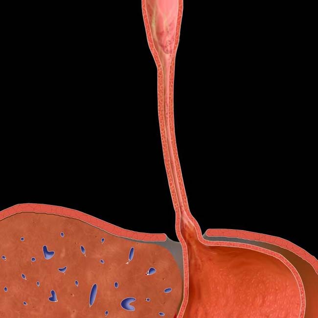
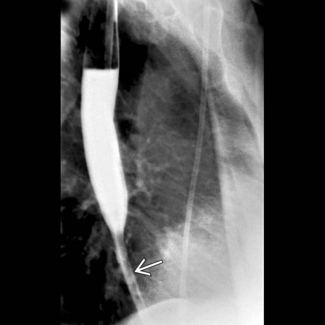
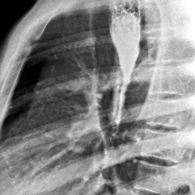
 from caustic ingestion shows a shortened and strictured esophagus, with the proximal stomach
  pulled into the chest. This stricture has been treated repeatedly by balloon dilation, and the patient has not required surgery.
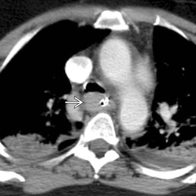
 and bilateral aspiration pneumonitis.
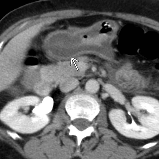
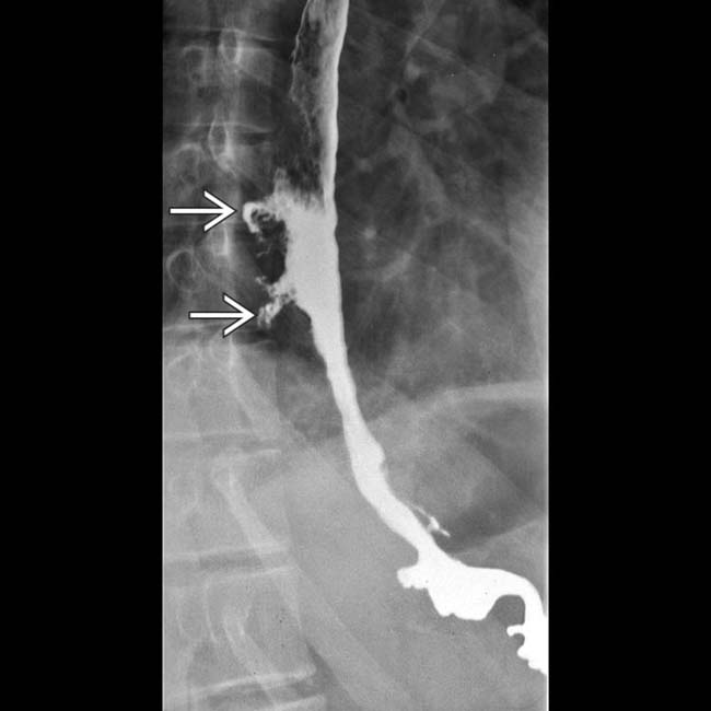
 , indicating corrosive gastritis.


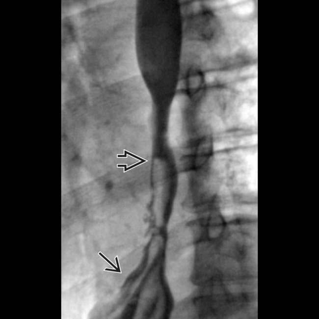
 of the distal half of the esophagus.
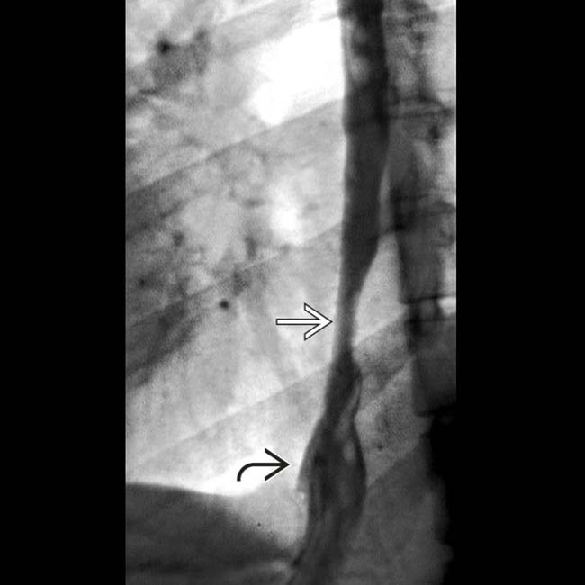
 and shortening of the distal esophagus with the stomach
  pulled into the lower chest.
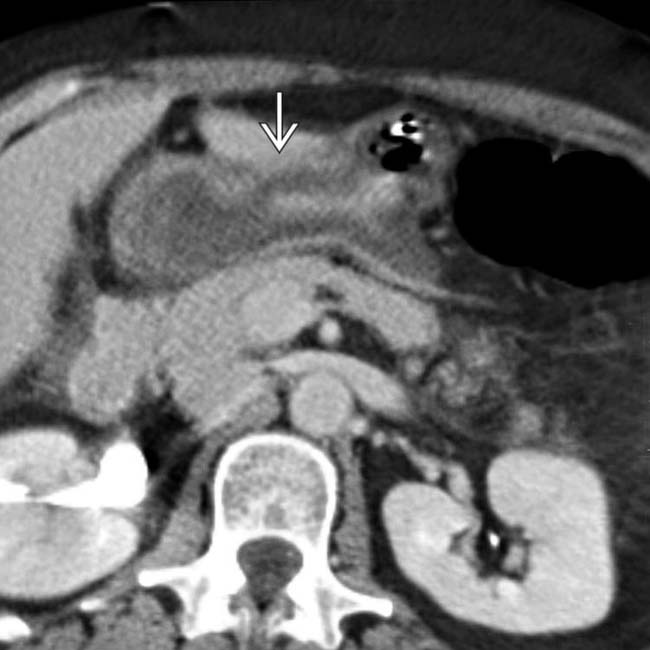
 . The patient subsequently developed esophageal perforation, respiratory failure, and sepsis before dying.
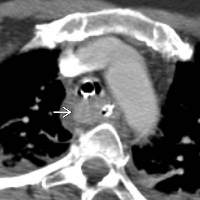
 .
.
 .
.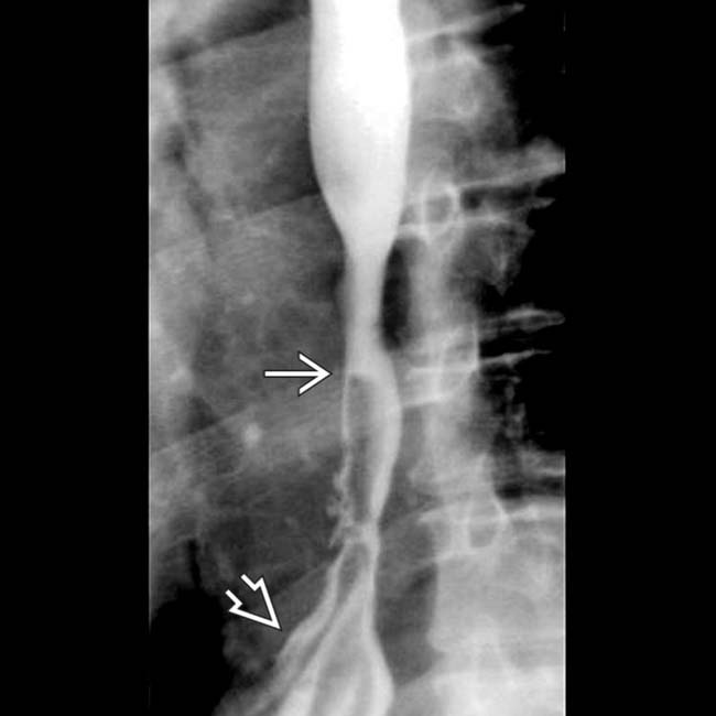
 , and a hiatal hernia
  . Reflux esophagitis may have a similar appearance.

