Caring for the patient undergoing biological therapy

Published on 09/04/2015 by admin

Filed under Hematology, Oncology and Palliative Medicine

Last modified 22/04/2025

Print this page

rate 1 star rate 2 star rate 3 star rate 4 star rate 5 star
Your rating: none, Average: 0 (0 votes)

This article have been viewed 1198 times

12 Caring for the patient undergoing biological therapy

Introduction

One of the most exciting and most recent developments in cancer treatments are the biological therapies. These have been developed as a result of the expansion of our knowledge of cancer biology. Many of these treatments are still in the trial phase and are still being investigated to optimise their effectiveness and to identify what side effects they may cause. However NICE has approved many of these treatments for general use. During your cancer/palliative care placement, you may come across patients receiving biological therapies alongside other cancer treatments such as cytotoxic therapy or radiotherapy. If you are allocated to a cancer centre, you may be able to arrange an insight visit with the clinical research team who coordinate and run cancer clinical trials.

Many of the treatments are administered orally and patients who are physically able will self-administer the drugs at home, so you may encounter the treatments while on a community placement or in the outpatient setting. Alternatively, if you have a placement in an inpatient area, try to find out what biological treatments some of your patients are on. Patients often confuse these drugs with cytotoxic therapy, but they act very differently and the side effects of biological therapies are variable in terms of the range of toxicities as well as the severity.

Biological therapies are treatments that use natural substances from the body, or drugs made from these substances. Biological therapies stimulate, direct or boost the body’s own immune cells to:

image Activity

Read Waugh and Grant (2010) (see References) or a similar textbook to refresh your knowledge of the different parts and cells of the immune system. How does the immune system work to get rid of non-self cells?

NMC Domain 3: 3.2

There are numerous types of biological therapies. Table 12.1 identifies some of the main groups and provides examples of drugs/agents. Each group is discussed in turn.

Table 12.1 Types of biological therapies

Groups of biological therapies Examples of agents
Cytokines Interferon, interleukins, tumour necrosis factor, colony-stimulating factors (G-CSF: pegfilgrastim/filgrastim and epoetin alfa)
Monoclonal antibodies (MoAbs) Trastuzumab (Herceptin), rituximab, bevacizumab, cetuximab
Cancer growth blockers Tyrosine kinase inhibitors: erotinib (Tarceva), imatinib (Glivec), getitinib (Iressa), sunitinib, dasatinib, lapatinib
Proteasome inhibitors: bortezomib (Velcade)
Anti-angiogenic agents Thalidomide
Cancer vaccines Bacillus Calmette–Guérin (BCG)
Gene therapies In development

Cytokines

Cytokines are natural protein messengers secreted from blood cells that coordinate the immune system. They attach to a receptor on the surface of white blood cells and trigger them to multiply, recognise and deal with anything that is foreign to the body, such as a cancer cell, bacteria, etc.

Interferon is a cytokine that is produced when white blood cells come in contact with a virus. It interferes with the ability of the virus to reproduce and stops other cells becoming infected with the virus. Interferon alpha is given subcutaneously. It interferes with or stops the cancer’s growth, makes cancer cells more vulnerable to being killed by white blood cells and reduces the number of blood vessels around the cancer. Interferon alpha is used to treat renal cell cancers, melanoma and chronic myeloid leukaemia. It can also be given topically to treat some types of skin cancer.

Another group of cytokines are the interleukins which are given intravenously (Batchelor 2006).

Yet another group is the haemopoietic growth factors. These stimulate production of white blood cells and help them mature. Rather than being used to eliminate cancer, these factors can be made synthetically, and boost recovery of the immune system when it has been damaged by cytotoxic therapy. Generally, granulocyte colony-stimulating factor (G-CSF) is used subcutaneously to stimulate neutrophil production and maturation, to prevent or minimise the severity and length of neutropenia and to lower the risk of infection when a patient is undergoing cytotoxic therapy and is likely to become immunocompromised. G-CSF is also used in haemopoietic transplant to increase the number of stems cells in the blood, before harvesting (discussed in Ch. 11).

Monoclonal antibodies

All cells have growth factor receptors on their cell surface. These are known as antigens. Antigens are activated when they come in contact with a specific growth factor, which enables the cell to divide. Antigens are also used by the immune system to recognise non-self cells that might cause harm. When B lymphocytes come into contact with something unfamiliar like a cancer cell or bacteria, they make B memory cells that remember and recognise it more easily next time. The B cells also produce specific antibodies (proteins) in the plasma which lock on to the specific antigen to activate an immune complex. This then aids other white cells to phagocytose (engulf) and destroy the foreign cell.

Some cancer cells have too many antigens on the surface which signal for increased cell division. To stop the cancer cells dividing, more copies of the specific antibody can be manufactured in the form of monoclonal antibodies (MoAbs). MoAbs inhibit cancer cellular growth by blocking the specific growth factor that tells the cell to divide. They also make these cancer cells more recognisable, so the white cells can kill them. For example, normal cells have two copies of human epidermal growth factor 2 (HER2) receptor protein. These cell receptors lock together with growth factors and trigger the cell to grow. Approximately 30–40% of patients with breast cancer have too many HER2 receptors as a result of an amplification of a gene which increases cell division. A MoAb known as trastuzumab (Herceptin) can be given intravenously and will attach to the receptors, blocking growth, slowing cell division and contributing to cell death. MoAbs are often given with cytotoxic drugs to increase the effectiveness of treatment. Other MoAbs include rituximab used to treat non-Hodgkin’s lymphoma, bevacizumab (Avastin) and cetuximab used to control metastatic colorectal and breast cancer.

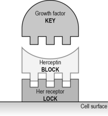
Not all patients (even with cancer cells with extra receptors) will respond. Cancer cells can learn to avoid binding to the antibodies, by downregulating (reducing) the number of receptors available. In addition, MoAbs are big molecules and cannot reach all the cells in a large cancer which has narrow blood vessels (Fig. 12.1).