Cardiopulmonary resuscitation

Published on 14/03/2015 by admin

Filed under Emergency Medicine

Last modified 22/04/2025

Print this page

rate 1 star rate 2 star rate 3 star rate 4 star rate 5 star
Your rating: none, Average: 0 (0 votes)

This article have been viewed 1128 times

Chapter 1 Cardiopulmonary resuscitation

Cardiopulmonary resuscitation (CPR) is one of the most difficult areas in clinical medicine and outcomes are often said to be extremely poor, especially in patients with unwitnessed out-of-hospital cardiac arrest. While many accepted practices do not yet have a strong evidence base, a number of principles can now reasonably be supported. Any attempt at resuscitation is worthwhile and, although some recommendations have changed, it is acceptable and appropriate that staff or bystanders trained in using previous practices use their known skills.

Early intervention is essential and, in general terms, every minute which passes without cardiac output leads to a dramatic worsening of prognosis. Basic life support (BLS) needs to be effective and initiated early and should be supplemented by advanced life support (ALS) as soon as possible.

In particular, direct current (DC) cardioversion should be performed urgently in those patients with a rhythm disturbance that is likely to be responsive. Immediate cardioversion of an arrhythmia associated with no (or poor) cardiac output should take priority over all else.

Resuscitation should never be withheld or delayed, except when there is no doubt that it is not in the patient’s best interests, and a full discussion with the patient and the patient’s family has already taken place. This needs to be documented with a specific instruction that resuscitation should not be performed (‘not for resuscitation’ or ‘allow natural death’).

CPR includes basic life support and advanced life support. The aim of CPR is to provide cardiac output and ventilation while facilitating the return of spontaneous circulation. A standardised approach allows teaching of the required skills and improves the likelihood that appropriate steps are taken under the stressful circumstances of a life-threatening emergency, even if those involved rarely perform CPR. Clearly defined roles allow an efficient and effective team approach.

Despite the difficulties in performing research in this area, the body of evidence is growing and new concepts such as circulatory support alone (chest compression without active ventilation), devices and techniques to provide more effective external chest compression, devices for minimally invasive internal cardiac massage and improved technology for electrical therapy (biphasic cardioversion) are under investigation. Recent work suggests controlled hypothermia may improve neurological outcome post-arrest in some patients. Broader public education, improved telecommunications and increased availability of defibrillators have also had considerable impact.

The daunting logistic task of teaching and maintaining competency in cardiopulmonary resuscitation, not only for healthcare professionals but also for the general public, has meant that changes in practice have appropriately been adopted slowly and cautiously.

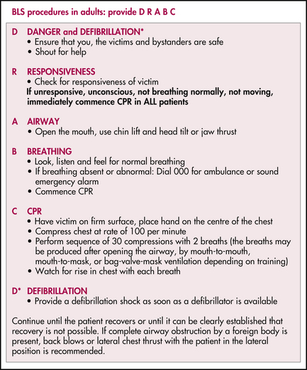
BASIC LIFE SUPPORT (BLS)

The general principles of basic life support (BLS) are the same in neonates, children and adults; however, there are some differences in their application because of the physiological and pathophysiological differences between these groups.

First assess safety and call for help, then rapidly initiate BLS. If there is life-threatening external bleeding, the immediate priority is to control blood loss by applying direct pressure and elevating the wound above the level of the heart. Protect from the environment (including maintaining body temperature), constantly reassess, handle gently and provide reassurance. In trauma patients be aware of the possibility of spinal injury and be careful to avoid neck movement whenever possible.

In all age groups, begin BLS if no sign of life is present, i.e. the patient is unconscious and unresponsive, not breathing and not moving. A pulse check is not required to initiate BLS and it is important not to delay commencing chest compressions. Give two initial rescue breaths and then commence chest compressions. Apply compression to the lower half of the sternum to one-third of the depth of the chest at a rate of 100 per minute. The compression-to-ventilation ratio is 30:2 with one or more rescuers, and the general principle is that compressions should be fast, hard, early and uninterrupted.

Continue until the patient recovers or until it can be clearly established that recovery is not possible. If significant hypothermia is present (< 32°C), perform resuscitation in conjunction with active warming of the patient.

Complications of CPR may include trauma to the ribs, sternum, lungs, liver, spleen or heart. Pneumothorax, haemothorax or fat embolism may all occur. Gastric distension (with air) and aspiration are also likely.

When performing CPR, be aware of the risks of sharps injury. Take appropriate precautions if exposure to poisons (e.g. skin contact with organophosphates) or to infective agents (e.g. meningococcus) is possible.

RESUSCITATION OF THE NEWBORN

A small percentage of newborn babies require some resuscitation at birth, usually initial breathing assistance. Deliveries in the emergency department are unexpected or precipitous and, therefore, carry a higher risk of neonatal complications. Prepare for full neonatal resuscitation with the formation of an experienced resuscitation team that is separate from the staff assisting the mother.

Newborn babies can rapidly develop hypothermia. Have pre-warmed towels and baby blankets ready and a radiant heater above the neonatal resuscitation area.

Initial assessment occurs while the newborn is being dried, the cord clamped and the baby placed on dry bedding under a radiant heater, or placed on the mother’s chest and covered with a dry blanket if it is obvious that no resuscitation is required.

Assess response to stimulation, colour, tone, respiratory rate/effort and heart rate (which should be 110–160 bpm). If the baby is not moving or breathing, place on the neonatal resuscitation bed under a radiant heater and provide stimulation by drying with a soft towel.

Routine suctioning is not recommended. Suctioning the pharynx of meconium-stained liquor before delivery of the shoulders is not recommended. If there are secretions causing obstruction, these can be very gently and briefly suctioned. If there has been meconium-stained liquor with subsequent respiratory compromise, brief gentle suctioning under direct vision (laryngoscopy) may be performed.

If the baby has not started breathing or the heart rate is < 100 bpm, provide positive pressure ventilation with a self-inflating 240-mL neonatal resuscitation bag. Oxygen can be added if there is no improvement after the first few minutes. Use a soft-rimmed mask that covers the mouth and nose, but not the eyes. Keep the head in a neutral position as extension or flexion can kink and obstruct the airway.

Deliver breaths at a rate of 40–60/min with just enough volume to see the chest wall rise. The heart rate should rise above 100 bpm and the baby should become pink. Ongoing bradycardia is usually due to hypoxia and inadequate ventilation.

If the heart rate is < 60 bpm after 30 seconds of positive pressure ventilation with oxygen, commence chest compressions. Use two fingers on the lower half of the sternum, or both hands encircling the chest with both thumbs on the lower half of the sternum. Compress the chest to one-third of its depth at a rate of 100/min. Perform three compressions before pausing to deliver one ventilation.

Intubation can then be performed if there is an experienced operator available.

Drugs and IV fluids are rarely required. If the heart rate remains below 60 bpm despite adequate ventilation, oxygenation and chest compressions, drugs may be administered but must not detract from the above measures.

Intravenous access is usually readily available through the umbilical vein. Peripheral veins are an often difficult option, but preferred to the intraosseous route in the neonate.

Table 1.1 Drugs used in paediatric resuscitation

Drug Strength IV/IO dose
Adrenaline 1:10,000 0.1 mL/kg (10 μg/kg)
Sodium bicarbonate 8.4% 1 mL/kg (1 mmol/kg)
Dextrose 10% 2 mL/kg
Fluid/volume Normal saline (or blood if indicated) 10 mL/kg
Naloxone (Only if indicated and note the short half-life) 400 μg/mL 0.25 mL/kg (100 μg/kg)

ADVANCED LIFE SUPPORT (ALS) IN CHILDREN

Commence advanced life support (ALS) as soon as staff and equipment become available. This involves drug and electrical therapy and more advanced airway management in addition to the BLS already being performed. Staff present should take up clearly defined roles. The compression-to-ventilation ratio can now be reduced to 15 compressions to 2 breaths.

Parents or primary caregivers may wish to be present during the resuscitation process. This should be encouraged with the provision that there is a senior nursing or medical staff member purely allocated to explain events and answer questions.

The airway should be managed by the most experienced operator. Airway adjuncts can be used. An oropharyngeal airway is sized from the centre of the lips to the angle of the mandible; a nasopharyngeal airway is sized from the tip of the nose to the tragus of the ear. Unrelieved airway obstruction requires gentle inspection with a laryngoscope and suction or Magill’s forceps to clear any material. Secure an airway as soon as possible. Intubation is ideal; however, prolonged attempts may result in hypoxaemia and bag-mask ventilation is often adequate.

For a newborn, a size 3-mm (internal diameter) uncuffed endotracheal tube (ETT) is used. For children over 1 year, the size is approximately determined by the formula: size (mm) = [age (years) ÷ 4] + 4. Cuffed tubes are now also commonly used in children.

Administer drugs into the largest and most central intravenous route. Allow no more than 90 seconds in attempting intravenous access before intraosseous access is obtained. All drugs and fluids that can be given intravenously can be given via the intraosseous route at the same dosage. Adrenaline, atropine and lignocaine can be given via an ETT (requiring higher doses), but this is the third-line option.

A heart rate of less than 60 bpm in an infant (< 1 year) or less than 40 bpm in a child (< 8 years) should be regarded as cardiac arrest. Treat asystole, low output/severe bradycardia or pulseless electrical activity with adrenaline 10 μg/kg IV/IO every 2–5 minutes. Seek and treat any reversible causes (refer to Box 1.2). Administer 20 mL/kg fluid bolus. Consider sodium bicarbonate 1 mmol/kg IV/IO and atropine 20 μg/kg IV/IO. Pacing may be of value in rare cases.

Defibrillate ventricular fibrillation (VF) or pulseless ventricular tachycardia (VT) immediately. If the arrest was witnessed, up to three (‘stacked’) shocks can be delivered in quick succession. The initial energy level is 2 J/kg but deliver all subsequent shocks at 4 J/kg, rounding up to the nearest energy level. Then initiate 2 minutes of CPR before analysing the rhythm again and delivering a single shock of 4 J/kg, if indicated. Adrenaline 10 μg/kg IV/IO can be administered every 2–5 minutes. Persistent or refractory VF or VT may also be treated with amiodarone 5 mg/kg IV/IO, which may be repeated. Also consider sodium bicarbonate 1 mmol/kg IV/IO, magnesium sulfate 0.05–0.1 mmol/kg IV/IO, potassium chloride 0.05 mmol/kg IV/IO. Seek and treat any reversible causes (refer to Box 1.2).

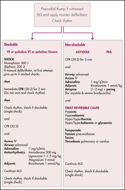
ADULT ADVANCED LIFE SUPPORT (ALS)

Begin advanced life support (ALS) as soon as staff and equipment become available. Uninterrupted effective chest compressions with prompt defibrillation remain the priority. Staff should take on clearly defined roles according to the resources available. These roles should include a team leader, airway person, chest compressor, defibrillator, drug administrator and a timekeeper/scribe. ALS includes electrical and pharmacological therapy and advanced airway management, in addition to the BLS already taking place.

A precordial thump may be administered to a patient if it is a witnessed cardiac arrest and cardiac monitoring shows a ‘shockable’ rhythm (i.e. VF or pulseless VT) and defibrillation is not immediately available. This should not delay defibrillation and is to be followed immediately by chest compressions.

Defibrillation electrode pads are now widely available. Place one electrode pad (or paddle) at the right parasternal area over the second intercostal space, and the other electrode pad (or paddle) in the left mid-axillary line over the sixth intercostal space. There must be no contact between the two pads (paddles). Paddles also require firm pressure and conductive gel pads. Avoid ECG electrodes, medications patches and pacemakers. Suspend physical contact with the patient during defibrillation.

Most defibrillators are now biphasic. Maximum joules are administered from the first shock and are used for every subsequent defibrillation attempt. This may be 200 J or even 360 J, depending on the defibrillator and institution. There is no proven benefit in delivering more than 200 J with a biphasic defibrillator.

In a witnessed arrest, three shocks in quick succession may be delivered initially. ‘Stacked’ shocks can only be performed with a manual defibrillator. Then analyse the rhythm every 2 minutes and deliver a further single shock, if indicated.

A defibrillated heart is ‘stunned’, even after successful defibrillation, so that cardiac output may still be very poor. For this reason, it is recommended to always resume chest compressions after defibrillation unless there are immediately obvious signs of life. Then assess cardiac output 2 minutes later at the next rhythm analysis point.

Administer adrenaline 1 mg every 2–5 minutes. The intravenous route is ideal. The intraosseous route is now preferable to the endotracheal route

image

Figure 1.2 Procedure for adult cardiac arrest

Adapted from the Australian Resuscitation Council adult cardiorespiratory arrest algorithm

even in adults. Intraosseous access devices for adult patients are being developed and will become more widely available in the near future.

Securing the airway with an endotracheal tube is ideal; however, it should not take priority. Chest compressions should not be interrupted for more than 20 seconds in order to intubate. Two-person bag-and-mask ventilation is also preferable to an oesophageal intubation of a traumatised and potentially obstructed airway.

Amiodarone 300 mg IV/IO may be administered for refractory VF/VT. Other drugs can be administered as part of addressing ‘reversible causes’.

Consider reversible causes in all arrests. These are easily remembered as the ‘four Hs and four Ts’, a list of causes and contributors to cardiac arrest that should each be addressed.

Return of spontaneous circulation may occur without the patient regaining consciousness. These patients may have a greater chance of a good neurological outcome if they are cooled in the first few hours to 32–34°C for 12–24 hours. They therefore require intubation and ventilation with sedation and paralysis and further management in the intensive care environment. They may also require urgent coronary revascularisation.

In cases where there is no return of spontaneous circulation, senior medical staff may decide to stop resuscitation efforts and declare the person deceased. This decision is made in the context of the duration and nature of the arrest, the premorbid state of the patient and the likelihood of significant neurological recovery.