Cardiology

Published on 24/06/2015 by admin

Filed under Internal Medicine

Last modified 22/04/2025

Print this page

rate 1 star rate 2 star rate 3 star rate 4 star rate 5 star
Your rating: none, Average: 0 (0 votes)

This article have been viewed 1768 times

Cardiac conditions are very commonly encountered in the long case setting. Given that cardiac disease is the most common morbidity in the world’s adult population, a cardiac condition could be encountered as the main pathology or an associated condition in the long case setting. Common cardiology long cases the candidate should be well versed in are: heart failure, ischaemic heart disease, arrhythmias and hypertension. Also important are valve disease and associated clinical decision-making processes, including timing of surgery if indicated in the patient with a cardiac valvular pathology.

HYPERTENSION

Case vignette

A 57-year-old man is admitted to hospital with severe chest pain. The pain is of sudden onset, tearing in character and radiating to the back. He is diaphoretic and anxious. On examination his pulse is regular at 110 bpm. His heart sounds are normal and the chest is clear. His blood pressure in the right arm is 190/100 mmHg and in the left arm 170/70 mmHg.

Approach to the patient

Examination

Check the blood pressure and look for a postural drop. If the blood pressure is very high, check the other arm and repeat the measurement at the end of the physical examination for confirmation.

Investigations

Creatinine clearance needs to be checked to clarify renal function but does not really help to distinguish between primary renal disease causing high blood pressure and renal impairment secondary to hypertensive nephrosclerosis. If there is no significant proteinuria or renal failure, a trial of effective blood pressure lowering for 6 months can be given. Deterioration of renal function at any stage is an indication for investigation. The additional investigations in this setting include a renal ultrasound (looking for renal shrinkage suggesting chronic renal failure or renovascular disease, or enlarged kidneys suggesting conditions such as polycystic kidney disease), a DTPA scan (to assess renal perfusion) and a renal arterial Doppler study (to exclude renal artery stenosis). If clinical evidence indicates renal artery stenosis, further study with computed tomography (CT) or magnetic resonance (MR) angiography is indicated.
The major endocrine investigation is the aldosterone/renin ratio, to detect primary hyperaldosteronism. This is now believed to account for 10–15% of people presenting with essential hypertension, but this does not mean that they all need adrenalectomy. Other endocrine investigations include: serum renin index (to exclude renin hypersecretion) and 24-hour urinary cortisol (to screen for Cushing’s syndrome).
If there is suspicion (tachycardia, palpitations, sweating, anxiety, postural hypotension) of phaeochromocytoma, perform serum catecholamine level, urinary metanephrines and urinary vanillylmandelic acid level.

Management

It is not wise to commence treatment at the first diagnosis itself unless there is malignant hypertension, end-organ damage (see box) or significant other vascular risk factors, or comorbidity. (Treat with antihypertensive agents if the diastolic pressure is > 100 mmHg, or systolic > 200 mmHg, or systolic pressure > 160 mmHg together with end-organ damage. The presence of other cardiovascular risk factors would be another indication for treatment.) Observation for 3–6 months with recommendation of non-pharmacological methods such as progressive muscle relaxation, weight reduction (if relevant), reduction of alcohol consumption, salt restriction and regular physical exercise would suffice initially. It is important to advise the patient against smoking. If present, hyperlipidaemia and diabetes should be treated. If the blood pressure remains elevated (>140/90 mmHg) despite adequate lifestyle modification (or due to failure of lifestyle modification), pharmacotherapy should be initiated.

Selection of the appropriate antihypertensive agent should be guided by several factors, including: the patient’s comorbidities, age, sex, ethnic background and drug allergies. Initially an attempt should be directed at monotherapy, and the commonly used agents are thiazide diuretics, beta-blockers, calcium channel blockers, angiotensin-converting enzyme (ACE) inhibitors and angiotensin II receptor blocker (ARB). If monotherapy is inadequate, combination therapy can be considered. An ACE inhibitor with a diuretic, or a beta-blocker with a diuretic, are two such combinations. There are combination pills containing an ACE inhibitor or an ARB together with a thiazide diuretic that can be prescribed. Hypertension that is not well controlled with conventional agents even with up titration and combination warrants further investigation and the addition of potent, less commonly used agents such alpha receptor blockers, centrally acting agents or arterial vasodilators.

Comorbidities that can influence the choice of therapy

Diabetes mellitus—ACE inhibitors are the most suitable agents. Where ACE inhibitors are not tolerated, the other options to consider include ARBs and calcium channel blockers. Beta-adrenergic receptor blockers and thiazide diuretics can interfere with glycaemic control. ACE inhibitors and ARBs have significant and useful synergy in severe high blood pressure and diabetic nephropathy.

Gout—beta-blockers, ACE inhibitors, calcium channel blockers and alpha-blockers are suitable. Thiazide diuretics can exacerbate gout.

Dyslipidaemia—ACE inhibitors, calcium channel blockers and alpha-blockers are recommended. Beta-blockers may be less desirable due to their adverse effects on the lipid profile.

Ischaemic heart disease—diuretics, beta-blockers, calcium channel blockers, ACE inhibitors and ARBs are suitable because of their protective properties in coronary vascular disease.

Congestive cardiac failure—ideal agents include beta-blockers, ACE inhibitors, ARBs and diuretics, which also have proven value in the management of cardiac failure.

Peripheral vascular disease—calcium channel blockers, alpha-adrenergic receptor blockers and diuretics are desirable agents. Beta-blockers are contraindicated.

Pregnancy—for mild hypertension in pregnant patients, methyldopa and the alpha- and beta-adrenergic receptor blocking agent labetolol are good choices. In preeclampsia, nifedipine is a suitable agent; however, urgent delivery of the baby is an absolute requirement. Severe hypertension in the pregnant patient can be managed with intravenous (IV) hydralazine.

Adverse effects of some antihypertensive agents

It is important to have a commanding knowledge of the properties and adverse effects of the commonly used antihypertensive agents. Below is a list of adverse effects seen with different classes of antihypertensive agents, together with some important properties of selected agents.

ISCHAEMIC HEART DISEASE AND ACUTE MYOCARDIAL INFARCTION

Sometimes examiners like to test candidates’ knowledge of the management of common acute medical conditions. No condition is more common than acute myocardial infarction in the physician trainee’s case repertoire, and the candidate is expected to be thoroughly familiar with the management of this condition.

Case vignette

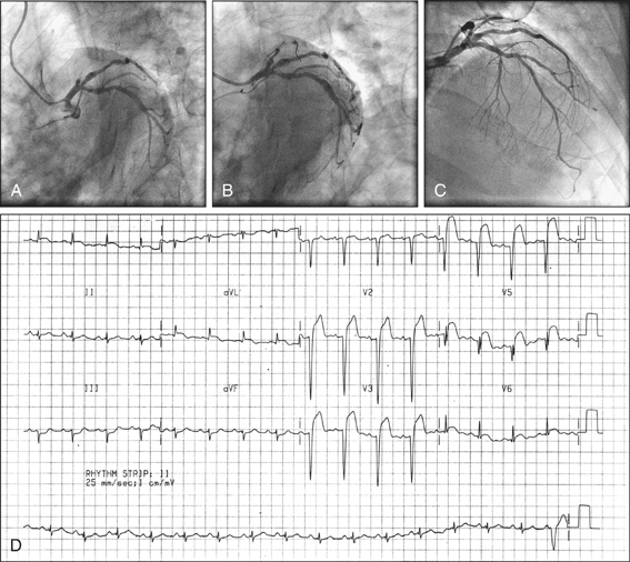
A 45-year-old man presents with retrosternal chest tightness of 3 hours’ duration. The pain is dull in nature, 7/10 in severity and radiating along the left arm. The onset was at rest. He also complains of progressive dyspnoea and associated nausea. He denies any cardiovascular risk factors apart from a strong family history. On examination his pulse rate is 100 bpm, low in volume and regular. His blood pressure is 90/60 mmHg. There are fine crepitations bibasally in the lung fields. The ECG shows deep T wave inversions in leads I, III and aVF.

Upon stabilisation the patient is admitted to the coronary care unit (CCU). His Killip class is 2 (see box). He is managed on aspirin, metoprolol 12.5 mg twice daily and IV heparin. While in CCU his blood pressure drops to 70/40 mmHg and pulse rate to 40 bpm. His pain is progressive and the ECG shows further deepening of the T wave inversions and new ST depression in the said leads, and also in leads aVR, V1 and V2.

He is commenced on an IV glycoprotein IIb/IIIa inhibitor and referred for early catheterisation. Catheterisation reveals occlusion of the posterior descending branch of the right coronary artery, which is successfully reopened by balloon angioplasty and stenting. On day 2 he develops fever and pleuritic chest pains.

On day 3 he develops acute pulmonary oedema and cardiogenic shock. Auscultation reveals a new harsh pansystolic murmur audible all over the precordium.

(Adapted from Killip T, Kimball J T 1967 Treatment of myocardial infarction in coronary care unit. A two-year experience with 250 patients. American Journal of Cardiology 20:457)

Approach to the patient

Examination

First and foremost, check the vital signs and establish haemodynamic stability. If the patient is having an acute episode of coronary ischaemia (or other emergency such as pneumothorax, pulmonary embolus) they may become rapidly unstable, with haemodynamic compromise or cardiopulmonary arrest.
Once the patient is stable, perform a detailed cardiovascular examination. Look in the fundus for hypertensive or diabetic changes. Listen to the heart for additional sounds such as the third and fourth heart sounds. Listen to the lung bases for crepitation of congestion.

Management

In any acute myocardial infarction the first priority is to assess the patient’s clinical stability and assess the requirement for, and urgency associated with, coronary revascularisation. Remember: patients presenting with an infarction could present with acute pulmonary oedema, cardiogenic shock, malignant ventricular tachyarrhythmias or severe bradycardia. Once the patient’s haemodynamic stability and cardiac rhythm stability are established, perform an urgent ECG to confirm the diagnosis and identify the nature of the infarction—whether it is an ST segment elevation infarction (STEMI) or a non-ST segment elevation infarction (non-STEMI).

STEMI

If the infarction is a STEMI, urgent reperfusion therapy is needed. Acute reperfusion therapy could be primary percutaneous transluminal coronary angioplasty (primary PTCA) with insertion of a stent or thrombolysis. If the centre offers a primary angioplasty service and the patient fulfils the criteria (see box), urgent transfer to the cardiac catheterisation laboratory should take place. Patients presenting in the first 4–6 hours of onset of chest pain are considered suitable for primary PTCA. Previous coronary artery bypass grafts, peripheral vascular disease, untreatable terminal illness and dementia are exclusion criteria for this procedure.
If primary PTCA is not an option, the patient should be thrombolysed with the relevant thrombolytic agent. If the patient presents within the first hour after the onset of chest pain, thrombolysis would be a preferred option (the ‘golden hour’ phenomenon). Usually a recombinant tissue plasminogen activator (rTPA) or an analogue is given to the patient immediately. Streptokinase is an alternative for patients over the age of 65 or those with evidence of an inferior myocardial infarction presenting after 4 hours of the onset of chest pain. Those who have been treated with streptokinase previously should not be given streptokinase again, due to the heightened risk of an allergic reaction.
The ECG criteria for primary PTCA and thrombolysis are similar (see box), but thrombolysis may be useful up to 12 hours or even 24 hours after the onset of chest pain.
If the decision is made to administer thrombolytics, the patient should not have any contraindications (e.g. risk of haemorrhage or allergy) to such therapy. If thrombolytic therapy has not been effective and the patient is a strong candidate for reperfusion therapy, attempts should be made to organise urgent rescue angioplasty.
image

Figure 3.3 Coronary angiogram of occluded artery (A) before primary PTCA (B) during primary PTCA (C) after primary PTCA; (D) ECG showing ST segment elevation in the antroseptal and lateral leads, indicating complete occlusion of the LAD artery

Non-STEMI

If a non-STEMI, the patient should be managed initially with anticoagulation and antiplatelet therapy. The patient should subsequently be referred for early coronary catheterisation.
Patients with acute coronary syndrome benefit from IV antiplatelet therapy in the way of platelet glycoprotein IIb/IIIa inhibitor agents such as tirofiban or eptifibatide in addition to heparin.
The patient should be admitted to the CCU for continuous ECG monitoring. Serial cardiac enzymes or troponin levels should monitored. Patients who sustain right ventricular damage may become profoundly hypotensive. They need IV volume infusion as the first line of therapy. Those in cardiogenic shock require inotropic therapy in the form of IV dobutamine or dopamine to support cardiac function and haemodynamic stability.

Post-infarct ventricular fibrillation

Ventricular fibrillation within the first 24 hours of an infarction indicates a more favourable prognosis than those occurring afterwards. Episodes of ventricular fibrillation after the first 24 hours signify a guarded prognosis, and consideration should be given to the implantation of a cardiac defibrillator. Inferior or posterior myocardial infarctions can damage the AV node or the cardiac conduction system, leading to heart block. These patients need urgent insertion of a temporary pacemaker. Some may recover their nodal and conduction function as the oedema and inflammation associated with the acute event settles, but others who sustain permanent damage need insertion of a permanent pacemaker (PPM). Complete heart block in a patient who suffers an anterior myocardial infarction signifies a bad prognosis due to the extensive area of myocardial damage.

Follow-up

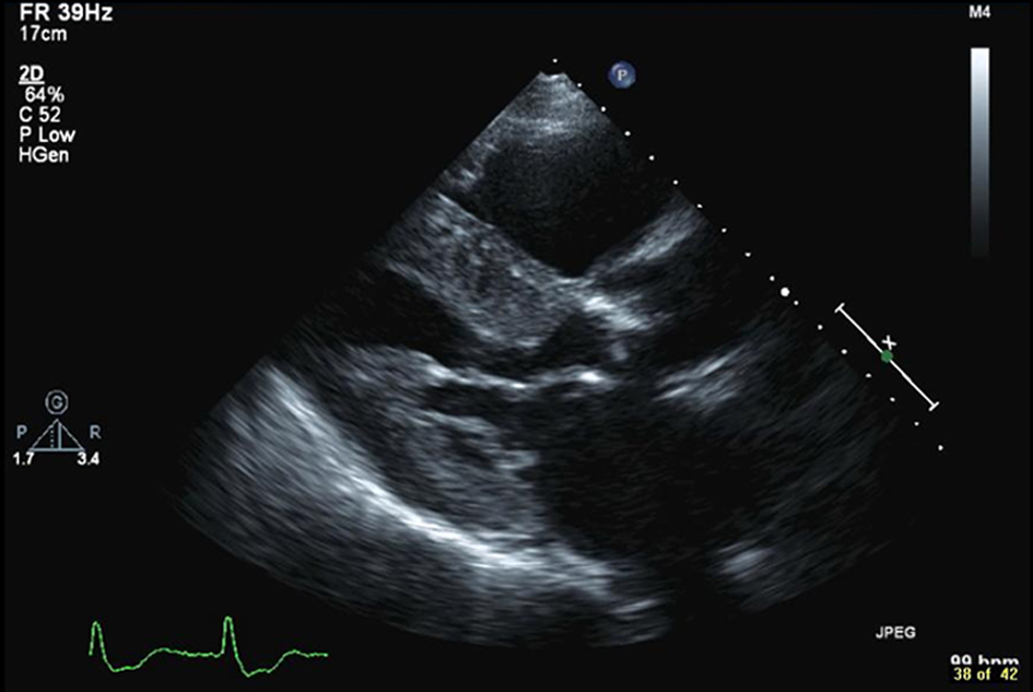
Follow-up management includes a transthoracic echocardiogram to assess the ejection fraction and to look for segmental left ventricular wall motion abnormalities, valvular defects, ventricular septal defect, ventricular thrombus and ventricular aneurysms. Ejection fraction can also be assessed by performing a nuclear gated heart pool scan. Patients who have an ejection fraction of less than 40% after an infarction are at high risk (30%) of sudden cardiac death over the next 5 years. These patients qualify to be treated with a prophylactic implanted cardiac defibrillator (ICD) usually 40 days after the infarction, according to current evidence.
Patients suffering from post-infarct angina need early coronary angiography to define the coronary anatomy before deciding on definitive therapy. Stable patients should have an exercise stress study, looking for reversible ischaemia. This may take the form of a stress echocardiography, stress ECG or nuclear medicine perfusion study. The presence of reversible ischaemia or ischaemia associated with stress are indications for coronary angiography. Definitive treatment with angioplasty, with or without stenting of the involved artery, or coronary artery bypass grafting, should be decided upon as guided by the coronary anatomy.
Prior to discharge, patients should be recruited to a post-myocardial infarction rehabilitation program and modification of cardiovascular risk factors should be encouraged. Some patients may need counselling and significant reassurance to help them recover from the acute event. Others may need advice on lifestyle and occupational issues, relevant information and education, counselling on sexual matters, dietary advice, help with giving up smoking and partner counselling.

CONGESTIVE CARDIAC FAILURE

Approach to the patient

History

The patient may complain of anorexia, which may be due to hepatic congestion. Ask about the various medications she or he has been treated with and any adverse effects associated with therapy.
Look for agents that can precipitate heart failure, such as NSAIDs, rosiglitazone,diltiazem and verapamil. Obtain a detailed social history, enquiring into the level of support available at home and how the patient copes with their physical limitations. Ask whether the patient has a scale at home and whether their body weight is monitored regularly. Enquire about the heart failure care plan and compliance thereof.

Examination

In the physical examination, look for the presence of tachypnoea and tachycardia. The patient may be hypertensive or hypotensive. Assess the JVP and quantify the elevation. Ascertain the character of the arterial pulse, which is usually thready and weak. Patients with severe cardiac failure may demonstrate Cheyne-Stokes respiration. Feel the apex beat, looking for a lateral shift or a heave. Auscultation may reveal an S3 gallop. Listen for murmurs that would suggest a valvular pathology such as aortic stenosis, aortic regurgitation and/or mitral regurgitation and tricuspid regurgitation. Left ventricular enlargement may lead to mitral annular dilatation, which may cause functional mitral regurgitation with a pansystolic murmur.
Listen to the lung fields for crepitations and percuss for stony dullness of an effusion. Examine the abdomen for tender hepatomegaly and ascites. Severe tricuspid regurgitation with congestion may cause pulsatile hepatomegaly. Check for peripheral oedema and define its distribution.
Investigations in the setting of acute and chronic cardiac failure and the many different therapeutic options available are popular topics used by examiners to test the candidate’s knowledge. Never forget the socioeconomic aspects of cardiac failure and the important management goals of improving the patient’s quality of life and preventing recurrent hospital admissions.

Pathogenic causes

One of the most common causes of cardiac failure is ischaemic heart disease. Cardiac failure in ischaemic heart disease could be due to a massive infarction and associated ventricular wall damage, severe mitral regurgitation, acute interventricular septal rupture or ischaemic cardiomyopathy secondary to multiple previous infarctions. Severe hypertension can lead to systolic heart failure as well as diastolic heart failure secondary to left ventricular hypertrophy. Valvular heart disease, particularly severe mitral and aortic valvular disease, can lead to congestive systolic or diastolic failure.
Acute bacterial endocarditis can cause damage to the valve leaflets and may lead to cardiac failure. Dilated cardiomyopathy, idiopathic or due to numerous causes (e.g. viral myocarditis, chronic alcohol abuse, cardiotoxic chemotherapeutic agents such as anthracyclines or other cardiotoxins), can cause cardiac failure. The causes of high-output cardiac failure are severe anaemia, beriberi, Paget’s disease, thyrotoxicosis and arteriovenous fistulae.

Investigations

Your battery of investigations should be aimed at confirming your clinical diagnosis, assessing the severity and identifying the underlying cause. The most important investigations in a patient with cardiac failure are:

1. Full blood count—looking for anaemia

2. Electrolyte profile and renal function indices—hyponatraemia is common in severe congestive cardiac failure. A low cardiac output can lead to inadequate renal perfusion and thus to prerenal renal failure. Renal effect of diuretic therapy and associated volume depletion/dehydration are important factors of concern.

3. Plasma B-type natriuretic peptide (BNP) levels

4. Levels of serum markers of cardiac damage—such as troponin I or T and the MB fraction of the enzyme creatinine kinase (CKMB)

5. Urine analysis—looking for protein, blood, white cells and organisms

6. Arterial blood gases—to assess the level of oxygenation and the acid–base status, particularly in the severely dyspnoeic and those in pulmonary oedema

7. Electrocardiogram—looking for evidence of acute ischaemia, previous ischaemic cardiac damage, arrhythmias and left ventricular hypertrophy. Severe left ventricular hypertrophy can cause diastolic cardiac failure. The most common cause of left ventricular hypertrophy is chronic hypertension. Left ventricular hypertrophy together with asymmetrical septal hypertrophy is seen in hypertrophic obstructive cardiomyopathy. Patients in significant cardiac failure have a propensity to develop arrhythmias, particularly ventricular tachycardia, which can progress to ventricular fibrillation.

8. Chest X-ray—looking for cardiomegaly as well as pulmonary congestion

9. Transthoracic echocardiogram—looking for wall motion abnormalities and valvular lesions. The echocardiogram will quantify the ejection fraction and the dynamic ventricular chamber and wall dimensions.

10. Thyroid function tests—hyperthyroidism can lead to high-output cardiac failure. Hypothyroidism causes low-output cardiac failure as well as diastolic cardiac failure due to pericardial effusion.

11. Serum vitamin B1 levels in potentially malnourished patients—to exclude beriberi (remember: this is very rare in our society!)

12. Renal arterial Doppler study—to exclude renal artery stenosis in the patient who suffers from recurrent episodes of unexplained pulmonary oedema (flash pulmonary oedema).

Management

Acute decompensated cardiac failure

1. Acute decompensated cardiac failure or pulmonary oedema is a medical emergency. Vital observations should be made first: respiratory rate, pulse rate, blood pressure and arterial oxygen saturations via pulse oximetry. The primary objectives are reestablishment and maintenance of haemodynamic stability and adequate tissue oxygenation.

2. Sit the patient bolt upright immediately and give supplementary oxygen via a facial mask at a rate of 6 L/min. If the patient is still desaturating, try giving 100% oxygen via a non-rebreather mask (great caution should be exercised when giving supplementary oxygen to patients with a history of chronic fixed airways disease). Those refractory to oxygen supplementation alone may respond to continuous positive airway pressure (CPAP) or bilevel positive airways pressure (BIPAP)—non-invasive ventilation.

3. Vascular access should be established with at least one wide-bore cannula in each arm. Give IV frusemide 80–120 mg, while monitoring the urine output. Administer subcutaneous or IV morphine at an initial dose of 2.5–5 mg. This has an anxiolytic effect as well as the capacity to reduce the preload by vasodilatation. Intravenous or topical nitrate therapy should also be given. This reduces the preload as well as the afterload in addition to facilitating coronary perfusion. Refractory fluid overload may require IV infusion of frusemide at the rate of 5–10 mg/h. Glyceryl trinitrate infusion helps reduce preload and thus improve symptoms. However, the patient should have a satisfactory blood pressure.

4. The patient needs insertion of an indwelling urinary catheter and close nursing supervision.

5. The following investigations should be performed immediately: a 12-lead ECG, mobile chest X-ray, arterial blood gases, full blood count, serum electrolyte profile, together with renal function indices and markers of myocardial damage. The patient needs an urgent echocardiogram to ascertain the cardiac anatomy, function and in particular the ejection fraction and cardiac filling pressures. Some patients may benefit from a right heart catheter (Swan-Ganz catheter) to continuously monitor right heart pressures. This information helps in the fluid balancing act.

6. Once clinically stable, the patient’s anti-failure therapy should be optimised, as discussed below. The patient should be placed on salt and fluid restriction (e.g. 1000–2000 mL/day) while maintaining a strict fluid balance, and should be weighed daily.

7. Nesiritide (a recombinant BNP) is an arterial and venous vasodilator. This agent has some proven benefit in the treatment of decompensated heart failure and fluid overload. Nesiritide should not be given if the patient is in shock or is very hypotensive.

9. Levosimenden is a calcium sensitiser that has shown benefit in the treatment of acute decompensated heart failure. It improves cardiac output and decreases pulmonary capillary wedge pressure. This is an expensive drug that has proven survival benefits to the patient.

Long-term management of cardiac failure

Below is an outline of the fundamental principles in the pharmacological management of systolic cardiac failure.

1. If the patient is haemodynamically stable, beta-blocker therapy should be considered first. Carvedilol has alpha- as well as beta-adrenoreceptor-blocking qualities. It is indicated in symptomatic cardiac failure with a severity consistent with Class II–III according to the NYHA classification (see box, p 35). This drug has to be started at a low dose and the dose gradually increased over a few weeks while observing the level of tolerance and efficacy. It can be commenced in hospital and followed up after discharge. Bisoprolol and metoprolol (long acting) have also shown benefit to patients in randomised controlled trials. Bisoprolol has cardiac beta receptor selectivity and as such can be given to patients with asthma or emphysema. It is better tolerated by patients with low blood pressure due to its lack of alpha receptor blocking activity.

2. ACE inhibitor therapy for symptomatic as well as asymptomatic congestive cardiac failure has been very beneficial, particularly in patients with an ejection fraction of less than 40%. When commencing therapy, short-acting ACE inhibitors such as captopril 6.25 mg given three times a day are preferred over long-acting, once-daily preparations such as perindopril or lisinopril. If short-acting preparations are used at the beginning, adverse effects of therapy can be better managed by stopping the drug promptly. When the patient demonstrates good tolerance, switch to a long-acting preparation for ease of administration and better compliance. All patients treated with ACE inhibitors should be monitored for hyperkalaemia and progression of renal failure.

3. Angiotensin II receptor inhibitors can be used for symptomatic cardiac failure where there is ACE inhibitor intolerance. Both agents demonstrate equal efficacy.

4. Diuretic therapy should be commenced. Loop diuretics are preferred for their strong diuretic action, which facilitates afterload reduction. These agents can also lead to vasodilatation and thus cause preload reduction and relieve pulmonary congestion.

6. Oral or transdermal nitrate therapy, for preload reduction and the relief of cardiac ischaemia.

7. Hydralazine combined with isosorbide dinitrate is a proven alternative for the ACE inhibitor-intolerant, symptomatic patient.

8. Digoxin is useful in persistent congestive cardiac failure despite ACE inhibitor therapy. This is of benefit to patients in sinus rhythm as well as those in atrial fibrillation.

9. Oral anticoagulation is indicated for patients with a history of previous thromboembolism, chronic or paroxysmal atrial fibrillation or a left ventricular thrombus.

10. Aldosterone receptor antagonists such as spironolactone (and eplerenone) administered long term have been shown to minimise the aldosterone-mediated myocardial fibrosis in patients with chronic congestive cardiac failure; a cardiac failure patient has high levels of aldosterone in the circulation as a compensatory response.

11. Intravenous diuretics and IV inotropic (dobutamine) therapy may be useful in refractory fluid retention and refractory cardiac failure.

12. Cardiac resynchronisation with biventricular pacing is beneficial to the patient in sinus rhythm, in particular those who have a prolonged QRS complex in the ECG (> 120 ms). The case for cardiac resynchronisation can be further proved if there is echo evidence of dyssynchrony.

13. An implanted cardiac defibrillator has been shown to be protective for patients who suffer from recurrent symptomatic sustained ventricular tachycardia and episodes of ventricular fibrillation.

14. Highly specialised centres offer left ventricular assist devices (LVADs) that can be implanted to assist the pump function of the left ventricle. These battery-operated devices can be used as bridging therapy prior to cardiac transplantation and in selected groups of patients as destination (definitive) therapy. In very refractory cases, cardiac transplantation should be considered.

Diastolic cardiac failure

Clinical assessment is paramount in these patients. Physical examination should show evidence of pulmonary congestion, elevated JVP and an S4 gallop on auscultation.

General management of cardiac failure

In all cardiac failure patients, consider vaccination against Pneumococcus as well as influenza virus. The candidate will be expected to formulate a working plan, in collaboration with the community resources available, to optimally manage and maintain the patient on discharge. Patient education, nutrition, physical activity, fluid balance, weight monitoring and early recognition of decompensation are important aspects of such a plan. Consideration should be given to a shared-care plan, in association with the patient’s general practitioner. Ambulatory care provision by a multidisciplinary heart failure team offers significant clinical and prognostic benefits to all heart failure patients.

ATRIAL FIBRILLATION

Case vignette

An 86-year-old woman is admitted with palpitations and dyspnoea on minimal exertion. She also complains of retrosternal heaviness. She has a history of hypertension and diverticular disease. She has had several previous admissions with falls and per rectal bleeding. She is managed on amlodipine 5 mg daily. She is an independently living widow. On examination her pulse rate is 120 bpm with an irregularly irregular rhythm. Her blood pressure is 150/95 mmHg. The ECG shows atrial fibrillation and voltage evidence of left ventricular hypertrophy.

Approach to the patient

Atrial fibrillation (AF) is the most common arrhythmia seen in clinical practice and is highly likely to be encountered in the long case setting. Studies have demonstrated a 1% prevalence of AF among adults. Prevalence increases with age and with structural heart disease. Atrial fibrillation can double mortality and increase the risk of stroke five-fold. Patients may present initially with palpitations, angina, presyncope and fatigue. Atrial fibrillation with a rapid ventricular response can cause clinical instability and the patient may develop coronary ischaemia or heart failure.

Examination

Physical examination may reveal an irregularly irregular pulse. Establish the quality of peripheral pulse and the rate. Check the blood pressure for hypertension. Look for signs of hyperthyroidism. Listen to the precordium for a mitral murmur. Look for signs of congestive cardiac failure. Check for evidence of amiodarone toxicity, such as slate-brown skin discolouration, pulmonary fibrosis and hypo- or hyperthyroidism.

Management

Management of AF has two primary objectives: rate control and/or rhythm control. The secondary objectives include symptom management and stroke prevention. The relative prognostic merit of rhythm control over rate control as definitive therapy is still being debated and pivotal trials thus far have failed to deliver a verdict.

Rate control

Those who are at high risk of embolic stroke due to left atrial thrombus are best treated with rate control and anticoagulation. The stroke risk is high if the patient is over the age of 65, in the presence of coronary risk factors, mitral disease or if AF has been present for more than 48 hours. The ideal agent in this setting is warfarin and the INR should be maintained at 2–3. Those below 65 years of age with no additional risk features may be managed with aspirin alone.

Rapid ventricular response in the setting of atrial fibrillation (rapid AF) contributes to most presenting symptoms. AF with rapid ventricular response can precipitate coronary ischaemia, congestive heart failure and pulmonary oedema. Chronic rapid AF may lead to tachycardia-induced cardiomyopathy and heart failure.
More rapid rate control is achieved with IV digoxin, verapamil, diltiazem or a beta-blocker such as propanolol. Intravenous amiodarone is also useful in combination for rate control; however, inadvertent pharmacological cardioversion could happen.
Oral agents are preferred over IV agents, given the side effects of hypotension and severe bradycardia that could be encountered with the latter. Long-term rate control is achieved by oral administration of the same agents as mentioned above. If the patient is experiencing severe symptoms or is haemodynamically unstable, consider urgent direct current (DC) cardioversion, preferably upon the exclusion of left atrial thrombus by TOE.
Amiodarone can increase serum digoxin levels and therefore caution should be exercised when using these two agents in combination. Those who are refractory to pharmacological rate control or intolerant of the same may benefit from radiofrequency ablation of the AV node with the implantation of a PPM.

Rhythm control

There is no convincing evidence to support the benefits of rhythm control over rate control in patients with AF. One study showed a worse outcome for mortality and stroke risk in patients who underwent cardioversion. This may be attributed to lower usage of warfarin in this patient population. However, rhythm control may help alleviate symptoms, and reduce the number of drugs the patient has to take and also the inconvenience of monitoring INR. It is reasonable to cardiovert AF without TOE or anticoagulation if done within 48 hours after the onset.
After 48 hours, safe practice guidelines dictate that the patient be anticoagulated (with INR maintained at 2–3) for about 3 weeks prior to cardioversion and for about 4 weeks after. However, if a TOE can be performed to exclude any suggestion of a thrombus, cardioversion can take place safely without preprocedure anticoagulation. Direct current cardioversion with a starting shock of 200 J is the most efficient and rapid means of achieving rhythm control. The shock should be synchronised to avoid precipiting ventricular fibrillation (VF).
Pharmacological rhythm control can be achieved and maintained with agents such as flecainide, sotalol, amiodarone, procainamide and quinidine. With their usage, proarrhythmia and QTc prolongation should be watched for. Amiodarone has very complex and unpredictable pharmacokinetics and a long list of adverse effects (see box). Flecainide should not be used to treat patients with known heart disease. Patients with ischaemic heart disease may benefit from the beta-blocker effect of sotalol.
Non-pharmacological rhythm control can be achieved with surgical ‘maze’ procedure or percutaneous isolation and ablation of pulmonary veins. Careful patient selection and operator expertise are essential.

HEART TRANSPLANTATION

Pre-transplantation work-up

Post-transplantation management

1. Endomyocardial biopsies
These are performed to assess for allograft rejection. They may be performed as frequently as every week for the first month, with the frequency decreasing over time. Commonly around 15 heart biopsies are performed in the first year after a heart transplant.

2. Surveillance and investigations
Follow-up visits are frequent for the first month because regulation of immunosuppression is being adjusted during this time. The frequency of visits gradually diminishes until the patient is generally seen on a 6-monthly or annual basis. Certain centres perform coronary angiography annually after transplantation, to monitor the patient for the development of coronary allograft vasculopathy (CAV). Alternatively, dobutamine stress echocardiography is performed to screen for CAV, particularly in the later years. This is particularly the case as often the patients have a degree of renal impairment precluding the use of contrast agents used during coronary angiography. However, intravascular ultrasound (IVUS) is the most accurate way to detect CAV, although this is not a routine test and is performed in only a few centres.

3. Immunosuppressant therapy
The cornerstone of immunosuppression is triple therapy with cyclosporin, mycophenolate mofetil and prednisolone. Alternative agents include azathioprine, sirolimus and tacrolimus. Basiliximab (Simulect®), an interleukin-2 receptor antibody, may be used early (days 1 and 4).
It is also important to recognise that there are numerous significant drug interactions to be aware of, especially with the immunosuppressant medications.

Complications

1. Sepsis
Infection is a significant problem in transplant patients. Preventive measures should be instituted. During the early post-transplant course, bacterial infections are of primary concern. Fungal infections can appear, particularly among inpatients, diabetics or those over-immunosuppressed. Prophylaxis for Pneumocystis carinii is universally administered, as is therapy for CMV infection. Maintain vigilance for other uncommon infectious processes including Listeria, Legionella, Chlamydia and Nocardia infections.

2. Rejection
Hyperacute rejection can occur immediately after blood flow is restored to the allograft. Thereafter, rejection can be classified as either acute cellular rejection (common form) or acute vascular/humoral rejection (less common) and chronic rejection.
Rejection is monitored for by regular endomyocardial biopsies. Endomyocardial biopsies can be classified as: Grade 0 (none), Grade 1R (mild), Grade 2R (moderate), or Grade 3R (severe) (International Society for Heart Lung Transplantation (ISHLT) Revised 2004 classification).
Depending on severity, rejection can usually be treated with pulsed IV methyl prednisolone for 3 days followed by a weaning dose of increased oral prednisolone (usually starting at 60 mg). Alternatively, anti-thymocyte globulin may be required in severe cases.

3. Late graft failure

4. Malignancy
Malignancy is also a significant problem in heart transplant patients, particularly in the later stages. Heart transplant patients are at increased risk of both solid organ tumours and lymphoproliferative tumours. Skin cancers are a frequent problem (squamous cell carcinomas especially but also basal cell carcinomas and melanomas).

5. Hypertension
This is a common problem after cardiac transplantation, both early and late. Prednisolone and cyclosporin are common early causes, with later renal impairment a common exacerbating factor. It is important to treat hypertension, as some transplant candidates may not have been exposed to significant blood pressure elevations for some time and it can result in hypertensive encephalopathy.

6. Dyslipidaemia
This is also a common problem following heart transplantation and exacerbates the development of transplant CAV. It is multifactorial in aetiology, with immunosuppressants, renal impairment and loop diuretics all contributing to its development. Heart transplant patients are usually treated with a statin on a routine basis to manage this condition.

7. Renal impairment
Renal dysfunction, both early and late, can be a significant cause of morbidity and mortality for heart transplant patients. Again it is multifactorial in aetiology. However, it is commonly due to calcineurin inhibitors (‘cyclosporin kidney’).

8. Diabetes
Up to one-third of post heart transplant patients may have or develop diabetes. This may be brought on or exacerbated by steroids and immunosuppressant therapy (cyclosporin and tacrolimus).

9. Osteoporosis
This is frequently a major longer-term cause of significant morbidity for the heart transplant patient. Long-term steroid use, renal impairment and calcineurin inhibitors are common causes. As such, these patients are often managed on calcium, vitamin D, weight-bearing exercises and bisphosphonates.

10. Arrhythmias
These are relatively common after heart transplantation. It is important to recognise that atrial arrhythmias (AF and atrial flutter) are often markers of acute rejection and usually dictate the performance of an endomyocardial biopsy.
Some patients may have a persistent bradycardia after a heart transplant (due to sinus node dysfunction) and may require a PPM (around 15% require a PPM).

11. Psychological disturbance

12. Vaccination precautions
Heart transplant patients are immunosuppressed and therefore should not be given live attenuated vaccinations, as these could lead to fulminant infection. Patients should only be given killed vaccines or toxoid vaccines.

Survival rates

Overall survival rates after heart transplantation are 80% at 1 year and 50% at 10 years (ISHLT data 1992–2001).