Cardiogenic Shock

Published on 15/05/2015 by admin

Filed under Internal Medicine

Last modified 22/04/2025

Print this page

rate 1 star rate 2 star rate 3 star rate 4 star rate 5 star
Your rating: none, Average: 0 (0 votes)

This article have been viewed 2123 times

Chapter 19

Cardiogenic Shock

1. Define cardiogenic shock.

    Cardiogenic shock is a state of end-organ hypoperfusion due to cardiac failure and the inability of the cardiovascular system to provide adequate blood flow to the extremities and vital organs. In general, patients with cardiogenic shock manifest persistent hypotension (systolic blood pressure less than 80 to 90 mm Hg or a mean arterial pressure 30 mm Hg below baseline) with severe reduction in cardiac index (less than 1.8 L/min/m2) in the presence of adequate or elevated filling pressure (left ventricular end-diastolic pressure more than 18 mm Hg or right ventricular end-diastolic pressure more than 10 to 15 mm Hg).

2. What are the various types of shock?

    Blood flow is determined by three entities: blood volume, vascular resistance, and pump function. There are three main types of shock: (1) hypovolemic, (2) vasogenic or distributive, and (3) cardiogenic. Examples of causes of hypovolemic shock include gastrointestinal bleeding, severe hemorrhage, and severe diabetic ketoacidosis (as a result of volume depletion). Examples of vasogenic shock include septic shock, anaphylactic shock, neurogenic shock, and shock from pharmacologic causes. There are many causes of cardiogenic shock, although acute myocardial infarction (MI) is the most common. Cardiogenic shock can be separated into true cardiac causes, such as MI, and extracardiac causes, such as obstruction to inflow (tension pneumothorax, cardiac tamponade) or outflow (pulmonary embolus).

3. Describe the clinical signs observed in cardiogenic shock and other types of shock?

    The medical history and clinical examination help in making the diagnosis of cardiogenic shock. Feeling the extremities and examining the jugular veins provide vital clues: warm skin is suggestive of a vasogenic cause; cool, clammy skin reflects enhanced reflex sympathoadrenal discharge leading to cutaneous vasoconstriction, suggesting hypovolemia or cardiogenic shock. Distended jugular veins, rales, and an S3 gallop suggest a cardiogenic cause rather than hypovolemia. Figure 19-1 presents an algorithm for the evaluation and treatment of cardiogenic shock.
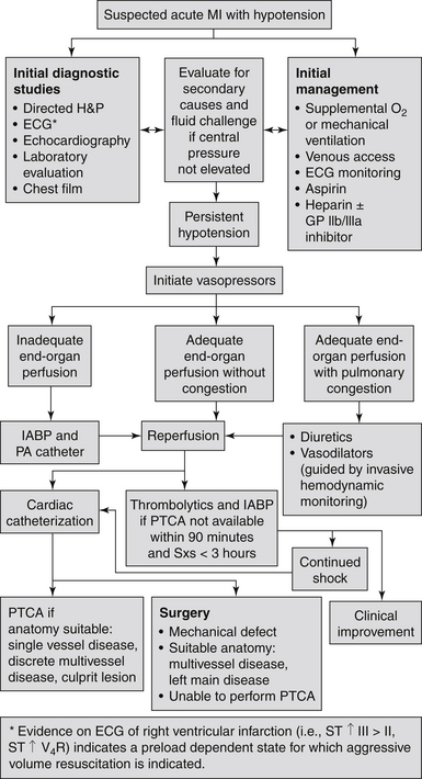
    It is important to note that the clinical examination and chest radiograph may not be reliable predictors of the pulmonary capillary wedge pressure (PCWP). Neither clinically reflect an elevated PCWP in up to 30% of cardiogenic shock patients. In addition, both cardiac tamponade and massive pulmonary embolism can present as cardiogenic shock without associated pulmonary congestion. Right-sided heart catheterization with intracardiac pressure and cardiac pressure measurements is important to confirm the diagnosis of cardiogenic shock.

4. Do all patients with cardiogenic shock have an increased heart rate?

    No. Patients with cardiogenic shock related to third-degree heart block or drug overdose (such as β-blockers and calcium channel antagonists overdose) can present with bradycardia and require temporary transvenous pacemaker implantation.

5. What are the determinants of central venous pressure (CVP)?

    The normal CVP is 5 to 12 cm H2O. Intravascular volume, intrathoracic pressure, right ventricular function, and venous tone all affect the CVP. To reduce variability caused by intrathoracic pressure, CVP should be measured at the end of expiration.

6. What is the significance of a loud holosystolic murmur in a patient with shock after acute myocardial infarction?

    New loud holosystolic murmurs with MI indicate either papillary muscle rupture or an acute ventricular septal defect (VSD). These may be indistinguishable, but acute VSD usually occurs with an anteroseptal MI and has an associated palpable thrill. Papillary rupture often does not have a thrill and is usually seen in inferior MI. These often cause shock on the basis of reduced forward blood flow and can be differentiated by echocardiography or pulmonary artery catheterization. Both require emergent cardiothoracic surgery for early repair. Note that in some patients, particularly those who develop acute mitral regurgitation, the murmur may be soft or inaudible (as a result of a small pressure gradient between the left ventricle and left atrium [or right ventricle]).

7. How can one differentiate cardiogenic from septic shock?

    In classic septic shock, the systemic vascular resistance (SVR) and the PCWP are reduced and the cardiac output is increased. These are usually opposite to the findings in cardiogenic shock. However, a significant decrease in cardiac output may occur in advanced and late stages of sepsis (cold septic shock, which carries a very high mortality rate). Many patients with cardiogenic shock may have normal SVR (i.e., relatively low), even while on vasopressor therapy. Patients with cardiogenic shock may also become dry (normal or low PCWP) with overzealous diuresis. Conversely, septic shock patients may become wet (high PCWP) with overzealous volume replacement. It is therefore ill advised to depend solely on the aforementioned hemodynamic criteria to differentiate cardiogenic shock from septic shock.

8. What is the most common cause of cardiogenic shock?

    Acute MI remains the leading cause of cardiogenic shock in the United States. In fact, despite the decline in its incidence with progressive use of timely primary percutaneous coronary intervention (PCI), cardiogenic shock still occurs in 5% to 8% of hospitalized patients with ST segment elevation myocardial infarction (STEMI). Unlike what is commonly believed, cardiogenic shock may also occur in up to 2% to 3% of patients with non–ST segment elevation myocardial infarction (NSTEMI). Overall, 40,000 to 50,000 cases of cardiogenic shock occur annually in the United States.

9. Describe the pathophysiology of cardiogenic shock among patients with acute myocardial infarction?

    Left ventricular (LV) pump failure is the primary insult in most forms of cardiogenic shock. The degree of myocardial dysfunction that initiates cardiogenic shock is often, but not always, severe. Hypoperfusion causes release of catecholamines, which increase contractility and peripheral blood flow, but this comes at the expense of increased myocardial oxygen demand and its proarrhythmic and cardiotoxic effects. The decrease in cardiac output also triggers the release of vasopressin and angiotensin II, which lead to improvement in coronary and peripheral perfusion at the cost of increased afterload. Neurohormonal activation also promotes salt and water retention, which may improve perfusion but exacerbates pulmonary edema.

    The reflex mechanism of increased SVR is in some cases not fully effective, and many patients have normal SVR even while on vasopressor therapy. Systemic inflammation, including expression of inducible nitric oxide synthase and generation of excess nitric oxide, is believed to contribute to the pathogenesis and inappropriate vasodilatation in some cases of cardiogenic shock.

10. Describe other mechanisms that cause or contribute to cardiogenic shock after myocardial infarction.

    It is critical to exclude mechanical complications after MI, which may cause or exacerbate cardiogenic shock in some patients. These mechanical complications include ventricular septal rupture, ventricular free wall rupture, and papillary muscle rupture. Two-dimensional (2-D) echocardiography is the preferred diagnostic modality and should be promptly performed when mechanical complications are suspected. Among STEMI patients with suspected mechanical complications in whom a 2-D echocardiogram is not available, diagnostic pulmonary artery catheterization should be performed (class I recommendation by the 2004 American College of Cardiology/American Heart Association [ACC/AHA] guidelines). The detection of mechanical complications before coronary angiography may help in dictating the revascularization strategy (surgical revascularization with mechanical repair of the complication rather than primary PCI of the infarct vessel without mechanical repair of the complication).

    Additional contributing factors to shock after MI include hemorrhage, infection, and bowel ischemia (the latter may be related to embolization after large anterior MI or persistent hypotension, or may be a complication of prolonged intraaortic balloon pump placement).

11. Can right ventricular (RV) dysfunction result in cardiogenic shock?

    RV dysfunction may cause or contribute to cardiogenic shock. Predominant RV shock represents 5% of all cardiogenic shock cases complicating MI. RV failure may limit LV filling via a decrease in the RV output, ventricular interdependence, or both. Treatment of patients with RV dysfunction and shock has traditionally focused on ensuring adequate right-sided filling pressures to maintain cardiac output and adequate left ventricular preload. However, caution should be undertaken not to overfill the RV and compromise LV filling (via ventricular interdependence).

    Shock caused by isolated RV dysfunction carries nearly as high a mortality as LV shock and benefits equally from revascularization.

12. List the other major causes of cardiogenic shock.

    Cardiovascular causes of cardiogenic shock are given in Table 19-1.

13. What is the mainstay therapy for patients with cardiogenic shock complicating myocardial infarction?

    Acute reperfusion and prompt revascularization for cardiogenic shock improves survival substantially and is considered the mainstay therapy for cardiogenic shock after acute MI. The Should We Emergently Revascularize Occluded Coronaries for Cardiogenic Shock (SHOCK) study was a landmark trial conducted in the 1990s enrolling patients with acute MI complicated by cardiogenic shock and randomizing patients to emergency revascularization or initial medical stabilization. Six-month mortality was lower in the early revascularization group than in the medical stabilization group (50% versus 63%, P = 0.03). At 1 year, survival was 47% for patients in the early revascularization group compared with 34% in the initial medical stabilization group (P < 0.03). The benefits of early revascularization persisted at long-term follow-up, and the strategy of early revascularization was associated with 67% relative improvement in 6-year survival compared with initial medical stabilization. Currently, the ACC/AHA guidelines recommend early revascularization in cardiogenic shock for those aged 75 years or younger.

14. Which is the best revascularization strategy in patients with cardiogenic shock complicating myocardial infarction?

    Among patients assigned to revascularization in the SHOCK trial, PCI accounted for 64% of initial revascularization attempts and coronary artery bypass grafting (CABG) for 36%. Although those treated with CABG had more diabetes and worse coronary disease, survival rates were similar with both revascularization strategies. Therefore, emergency CABG is an important treatment strategy in patients with cardiogenic shock and should be considered a complementary revascularization option to PCI in patients with extensive coronary disease.

    The ACC/AHA guidelines recommend CABG in cardiogenic shock patients with multivessel coronary artery disease. However, staged multivessel PCI may be performed if surgery is not an option. A single-stage procedure may be considered if the patient remains in shock after PCI of the infarct-related artery and if the other vessel has a critical flow-limiting lesion and supplies a large myocardium at risk.

15. Does the timing of revascularization matter in the treatment of cardiogenic shock?

    It is essential that revascularization and reperfusion are conducted promptly. Among patients with STEMI and cardiogenic shock, primary PCI should be done within a door-to-balloon time of less than 90 minutes. Although fibrinolytic therapy is less effective, it is indicated when timely PCI is unlikely to occur and when MI and cardiogenic shock onset are within 3 hours. Unlike what is commonly believed, prompt CABG is feasible. In the SHOCK trial, patients underwent CABG at a median time of 2.7 hours after randomization. It is also important to emphasize that approximately three-fourths of patients with cardiogenic shock after MI develop shock after hospital admission. Therefore, prompt revascularization and early reperfusion after acute MI may serve as a strategy to prevent the occurrence of cardiogenic shock.

16. Describe the common medical therapies for cardiogenic shock.

    Antiplatelet and antithrombotic therapies with aspirin and heparin should be administered to all patients with MI. Negative inotropes and vasodilators (including nitroglycerin) should be avoided. Optimal oxygenation and a low threshold to institute mechanical ventilation should be considered. Antiarrhythmic therapies (intravenous amiodarone) should be instituted when needed.

    Pharmacologic support with inotropic and vasopressor agents may be needed for short-term hemodynamic improvement. Although inotropic agents have a central role in cardiogenic shock treatment because the initiating event involves contractile failure, these agents increase myocardial adenosine triphosphate (ATP) consumption and hence their short-term hemodynamic benefits occur at the cost of increased oxygen demand. Higher vasopressor doses have also been associated with poorer survival. Therefore, these agents should be used in the lowest possible doses and for the shortest time possible. The ACC/AHA guidelines recommend norepinephrine for more severe hypotension because of its high potency. Often, dobutamine is needed as an additional inotropic agent.

17. What is the mainstay mechanical therapy for cardiogenic shock?

    Intraaortic balloon pump counterpulsation (IABP) has long been the mainstay of mechanical therapy for cardiogenic shock. IABP support should be instituted promptly, even before transfer for revascularization. IABP improves coronary and peripheral perfusion via diastolic balloon inflation (perfusion augmentation) and augments LV performance via systolic balloon deflation (afterload reduction). Thus, accurate timing of IABP inflation and deflation is important for the optimal hemodynamic support of the failing heart. IABP was performed in 86% of patients in the SHOCK trial. The overall and major complication rates were 7% and 3%, respectively, and were more evident among women and in patients with small body size and peripheral arterial disease.

18. What is the role of total circulatory support in cardiogenic shock?

    Temporary mechanical circulatory support with LV assist devices (LVADs) interrupts the vicious cycles of ischemia, hypotension, and myocardial dysfunction and allows the recovery of stunned and hibernating myocardium and reversal of neurohormonal derangements. LVAD support involves circulation of oxygenated blood through a device that drains blood from the left side of the heart and returns blood to the systemic arteries with pulsatile or continuous flow. These devices are advocated as a bridge to surgical revascularization, although they have been used as destination therapy for patients with end-stage cardiomyopathy and no alternative therapeutic options. Device-related complications and irreversible organ failure remain their major limitations. Surgically implanted LVADs remove blood through a cannula placed at the LV apex and return blood to the ascending aorta.

    Percutaneous LVADs are currently available and can be placed in the cardiac catheterization laboratory. The TandemHeart (CardiacAssist, Inc., Pittsburgh) removes blood from the left atrium using a cannula placed through the femoral vein and into the left atrium via transseptal puncture. Blood is then returned to a systemic artery, usually the femoral, with retrograde perfusion of the abdominal and thoracic aorta. Another percutaneous device, the Impella (Abiomed, Danvers, Mass.), is a novel intravascular microaxial blood pump, which can be introduced via the femoral artery and placed across the aortic valve into the left ventricle to unload the left ventricle and provide a short-term mechanical support for the failing heart (Fig. 19-2). Extracorporeal life support (ECLS) involves extracorporeal circulation of blood through a membrane oxygenator, which relieves both the right side and left side of the heart and the lungs of part of their workload.
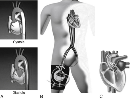
19. What are the mortality and morbidity rates of cardiogenic shock?

    In the modern era, the mortality rate of cardiogenic shock is approximately 50%. In the SHOCK trial, the 3- and 6-year survival rates among patients in the early revascularization group were 41% and 33%, respectively, with persistence of treatment benefits for up to 6 years. Importantly, 83% of 1-year survivors in the SHOCK trial were in New York Heart Association (NYHA) classes I or II and many of them enjoyed an acceptable quality of life at 6-year follow-up. Therefore, cardiogenic shock should be regarded as a very serious but treatable (and possibly preventable) condition that, when treated aggressively and in a timely manner, carries a reasonable chance for full recovery.

Bibliography, Suggested Readings, and Websites

1. Anderson, J.L., Adams, C.D., Antman, E.M., et al. ACC/AHA 2007 guidelines for the management of patients with unstable angina/non–ST-elevation myocardial infarction. J Am Coll Cardiol. 2007;50:e1–e157.

2. Hochman, J.S., Sleeper, L.A., Webb, J.G., et al. Early revascularization in acute myocardial infarction complicated by cardiogenic shock. SHOCK Investigators. Should we emergently revascularize occluded coronaries for cardiogenic shock. N Engl J Med. 1999;341:625–634.

3. Hochman, J.S., Sleeper, L.A., Webb, J.G., et al. Early revascularization and long-term survival in cardiogenic shock complicating acute myocardial infarction. JAMA. 2006;295:2511–2515.

4. Jneid, H., Anderson, J.L., Wright, R.S., et al. 2012 ACCF/AHA focused update of the guideline for the management of patients with unstable angina/non-ST-elevation myocardial infarction (updating the 2007 guideline and replacing the 2011 focused update): a report of the American College of Cardiology Foundation/American Heart Association Task Force on Practice Guidelines. J Am Coll Cardiol. 2012;60(7):648–681.

5. Levine, G.N., Bates, E.R., Blankenship, J.C., et al. 2011 ACCF/AHA/SCAI Guideline for Percutaneous Coronary Intervention: a report of the American College of Cardiology Foundation/American Heart Association Task Force on Practice Guidelines and the Society for Cardiovascular Angiography and Interventions. Circulation. 2011;124:e574–e651.

6. O’Gara, P.T., Kushner, F.G., Ascheim, D.D., et al. American College of Cardiology Foundation/American Heart Association Task Force on Practice Guidelines: 2013 ACCF/AHA guideline for the management of ST-elevation myocardial infarction: a report of the American College of Cardiology Foundation/American Heart Association Task Force on Practice Guidelines. Circulation. 2013;127(4):e362–e425.