CHAPTER 15 Capsular Shrinkage in the Treatment of Wrist Instability
Basic Science
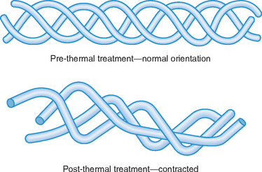
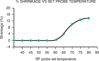
What is old is new again. Capsular shrinkage was used by Hippocrates 2,400 years ago to stabilize dislocated shoulders. Luckily, anesthetic techniques and thermal delivery methods have improved during the past two millennia. Recently (comparatively speaking), the biology of capsular shrinkage has been extensively studied in animal models. These studies have shown that the triple helix of collagen “unwinds” and “shrinks” when heated to 60° C, maximum shrinkage being achieved between 65 and 75° C (Figures 15.1 and 15.2). The hydrogen bonds that maintain the 3D configuration of the type I collagen triple–helix rupture as the collagen is heated beyond 60° C. The denatured collagen can potentially shorten to 50% of the resting length of the untreated collagen. The shortened denatured collagen acts as scaffolding onto which new collagen is deposited.1 The new collagen fibers maintain this shortened conformation, thus assuring the long–term maintenance of the shortening.
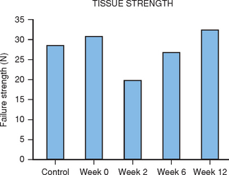
Biomechanical studies have demonstrated that the tensile strength of heated collagen decreases rapidly and does not return to normal values for 12 weeks.2 The tensile strength returns to nearly 80% normal by six weeks after heating (Figure 15.3). This transient loss of tensile strength would suggest that the application of stress to recently heated collagen is contraindicated. Premature loading of the shrunk collagen will lead to a lengthening of the collagen. This has been verified in an animal model.3,4 Based on this data, it would seem reasonable to recommend at least six to eight weeks of joint immobilization after capsular shrinkage. Clearly heavy loading of the joint should be avoided for 12 weeks.
Indications and Contraindications
Technique
Generalities
Shrinkage requires very low energy settings. The RF devices must be adjusted to heat the tissue to a temperature of between 65 and 75° C. It is wise to start at low energy and slowly increase the energy output until the desired shrinkage is observed. If a laser is used, it should be set to very low energy [i.e., 0.2 to 0.5 Joules at 15 pulses per second (3 to 7.5 Watts)]. The laser is held away from the target ligament and slowly advanced until the ligament is seen to shrink.
Once the shrinkage has stopped, continued heating will only further weaken the ligament without increasing the shrinkage. The color of the ligament changes from white to light yellow during the shrinkage. Lu et al. have suggested that a cross–hatching shrinkage pattern optimizes the in–growth of healthy tissue and hastens the recovery of the ligament.5 During the shrinkage, the traction on the wrist should be reduced as much as possible to permit optimal shrinkage.
Scapholunate Instability
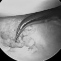
The question is what should or can be shrunk to stabilize the scapholunate axis. The SL interosseous ligament is a heterogenous structure. Its central portion is composed of fibrocartilage that is not shrinkable (Figure 15.4). The dorsal and palmar portions of the SL ligament are composed of type I collagen and are, however, shrinkable (Figure 15.5). The arthroscope and thermal wand can be placed in either the 3–4 or 4–5 portal. The 70–degree side–firing laser probe and 90–degree RF probe can be placed in the 4–5 portal, with the scope placed in the 3–4 portal. If the zero–degree probes are used, the scope should be placed in the 4–5 portal and the wand passed through the 3–4 portal. Extreme caution is exercised so as to avoid injury to normal hyalin cartilage. Good fluid inflow and outflow are critical because the arthroscopy fluid removes the excess thermal energy generated by the probes.
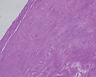
FIGURE 15.4 Histology of central fibrocartilaginous portion of the scapholunate ligament.
(Taken from Berger RA. [chapter title]. WP Cooney, RL Lilnscheid, JH Dobyns (eds.), The Wrist Diagnosis and Operative Treatment. St. Louis: Mosby 1998.)
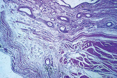
FIGURE 15.5 Histology of capsule demonstrating loose collagen (CF) in a fibrous stratum (FS).
(Taken from Berger RA. [chapter title]. WP Cooney, RL Lilnscheid, JH Dobyns (eds.), The Wrist Diagnosis and Operative Treatment. St. Louis: Mosby 1998.)
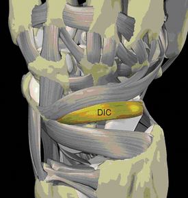
Capsular shrinkage of the dorsal intercarpal (DIC) ligament could potentially reinforce the stabilizing effect of SL ligament shrinkage. The DIC is attached to the distal dorsal aspect of the scaphoid and the dorsal triquetrum6,7 (Figure 15.6). Shrinkage of this ligament could simulate the tensioning of this ligament noted during open capsulodesis.8 To accomplish this, the scope and laser/RF angled probes would be placed in the radial and ulnar midcarpal portals. Alternatively, the scope can be placed in the midcarpal joint via the radial midcarpal volar portal described by Slutsky,9 with the thermal probes passed through the ulnar midcarpal portal.
Lunotriquetral and Ulnocarpal Instability
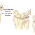
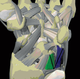
Mild forms of lunotriquetral instability can be treated with ulnocarpal ligament shrinkage. The author has applied this technique in a limited number of cases with satisfying results. This procedure takes advantage of the anatomy of the ulnotriquetral and ulnolunate ligaments. These ligaments form a V as they diverge from their origin on the palmar distal radioulnar ligament and insert on the palmar aspect of the lunate or triquetrum (Figure 15.7).
Results
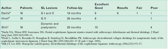
Few capsular shrinkage clinical trials have been reported. Table 15.1 lists three such trials. They all deal with the treatment of mild SL instability. They all suggest that capsular shrinkage has been very helpful for the majority of the patients treated. In addition to these results, Dr. Battistalla discusses his results in Chapter 10.
There are no clinical studies reporting the outcome of capsular shrinkage for lunotriquetral, ulnocarpal, or midcarpal instability. The author has found ulnocarpal ligament shrinkage for ulnocarpal ligament laxity, mild TFCC laxity, and mild lunotriquetral instability to be rewarding in the 12 patients so treated. He has used shrinkage to treat mild midcarpal instability in just two cases. Both patients are better, although one remains somewhat symptomatic when she applies heavy stress to the wrist.
1 Lopez MJ, Hayashi K, Vanderby RJr., Thabit GIII, Fanton GS, Markel MD. Effects of monopolar radiofrequency energy on ovine joint capsular mechanical properties. Clin Orthop. 2000;374:286-297.
2 Hecht P, Hayashi K, Lu Y, Fanton GS, Thabit GIII, Vanderby RJr., Markel MD. Monopolar radiofrequency energy effects on joint capsular tissue: Potential treatment for joint instability. An in vivo mechanical, morphological, and biochemical study using an ovine model. Am J Sports Med. 1999;27(6):761-771.
3 Naseef GSIII, Foster TE, Trauner K, Solhpour S, Anderson RR, Zarins B. The thermal properties of bovine joint capsule: The basic science of laser- and radiofrequency-induced capsular shrinkage. Am J Sports Med. 1997;25(5):670-674.
4 Hayashi K, Markel MD. Thermal capsulorrhaphy treatment of shoulder instability: Basic science. Clin Orthop. 2001;390:59-72.
5 Lu Y, Hayashi K, Edwards RBIII, Fanton GS, Thabit GIII, Markel MD. The effect of monopolar radiofrequency treatment pattern on joint capsular healing: In vitro and in vivo studies using an ovine model. Am J Sports Med. 2000;28(5):711-719.
6 Mitsuyasu H, Patterson RM, Shah MA, Buford WL, Iwamoto Y, Viegas SF. The role of the dorsal intercarpal ligament in dynamic and static scapholunate instability. J Hand Surg [Am]. 2004;29(2):279-288.
7 Viegas SF, Yamaguchi S, Boyd NL, Patterson RM. The dorsal ligaments of the wrist: Anatomy, mechanical properties, and function. J Hand Surg [Am]. 1999;24(3):456-468.
8 Szabo RM, Slater RRJr., Palumbo CF, Gerlach T. Dorsal intercarpal ligament capsulodesis for chronic, static scapholunate dissociation: Clinical results. J Hand Surg [Am]. 2002;27(6):978-984.
9 Slutsky DJ. Clinical applications of volar portals in wrist arthroscopy. Tech Hand Up Extrem Surg. 2004;8(4):229-238.