Ovarian Cancer (Epithelial)

Published on 10/03/2015 by admin

Filed under Obstetrics & Gynecology

Last modified 22/04/2025

Print this page

rate 1 star rate 2 star rate 3 star rate 4 star rate 5 star
Your rating: none, Average: 0 (0 votes)

This article have been viewed 1905 times

Ovarian Cancer (Epithelial)

Synonyms/Description

Ovarian carcinoma or adenocarcinoma

Etiology

Ovarian cancer is the second most common (endometrial is number one) genital tract malignancy (1% to 2% lifetime risk) and the most deadly (overall 5-year survival of 45%). Familial predisposition accounts for 5% to 10% of women who develop ovarian cancer and is typically associated with BRCA1 and BRCA2 gene mutations. Ovarian cancer is more prevalent with increasing age, usually occurring in postmenopausal women. Most cancers are of the epithelial type, the most common of these being serous cystadenocarcinoma (greater than 50%); the cell type comprising this tumor is very similar to that found in the fallopian tubes. Endometrioid adenocarcinoma (15% to 20% of epithelial ovarian cancers) and clear cell tumors of the ovary (6% of epithelial tumors) are associated with pelvic endometriosis and tend to occur in younger women. Mucinous adenocarcinoma (5% to 10% of epithelial tumors) is a mucin-producing tumor and resembles the intestinal or endocervical cell types. Other rarer cell types include carcinosarcoma (epithelial and mesenchymal mixed tumor), transitional cell, squamous cell carcinoma, and Brenner tumors (see related sections elsewhere in the book).
Traditionally, ovarian cancer was thought to arise de novo from the surface epithelium (mesothelium) of the ovary. However, there is recent evidence that the most common ovarian cancer originates from the fallopian tube. This precursor lesion, called serous intraepithelial tubal carcinoma (STIC), is very similar to high-grade ovarian serous carcinoma. The current theory is that most of these tumors arise from a STIC in the fimbriated end of the fallopian tube then spread to the ovary.
Even when the ovaries have been removed, a patient can still develop primary peritoneal carcinoma. This entity, known as serous surface papillary carcinoma of extra-ovarian origin, is considered a very aggressive cancer.

Ultrasound Findings

Pattern recognition and Doppler features of an ovarian mass are the most accurate method of differentiating a malignant from a benign tumor when performed by an experienced practitioner. In general, unilocular clear cysts with smooth borders, no nodularity, and absent internal blood flow are characteristic features of a benign mass. Papillary projections with abundant blood flow, large and irregular solid components, thick septations, and irregular walls are signs of malignancy. These features are characteristic of all epithelial ovarian tumors; hence, it is not possible to distinguish the exact tissue diagnosis sonographically. Once the tumor is advanced, ascites and peritoneal seeding are common and easily visualized sonographically.
Color flow Doppler is very important to evaluate the presence or absence of prominent vessels within the solid areas of the tumor. Prior reports suggested that spectral Doppler indices might be helpful to distinguish malignant from benign lesions. Although vessels in a malignant mass have a lower resistive index and abundant diastolic flow compared with those seen in benign masses, this finding has not been helpful in premenopausal women. The corpus luteum is very vascular with a low resistive pattern similar to tumor vessels, making the spectral Doppler waveform potentially useful only in postmenopausal patients. The mapping of blood vessels in the center of a mass is far more informative than the spectral Doppler waveform. More recently, some authors have suggested using 3-D ultrasound with power Doppler to quantify the blood flow within the solid portions of the tumor. Three-dimensional power Doppler vascular indices used to quantify blood flow include the vascularization index, flow index, and vascularization-flow index. Although evaluating the blood flow indices within a mass may be of some value, it does not seem to add substantially to the subjective sonographic assessment of the mass by experienced practitioners.
The International Ovarian Tumor Analysis (IOTA) multicenter group has devised and tested several approaches to determining whether an adnexal mass is malignant. Its five simple rules include five features suggestive of malignancy: M1, irregular contour; M2, ascites; M3, 4+ papillations; M4, irregular, multilocular, solid tumor with a largest diameter greater than or equal to 100 mm; M5, strong vascularity. The group also compiled five features suggestive of a benign mass: B1, unilocular; B2, solid components less than 7 mm in largest diameter; B3, acoustic shadows; B4, smooth, multilocular tumor; B5, no visible blood flow. The presence of one or more of the M features in the absence of any B feature conveys a diagnosis of malignancy. If one or more B features are present in the absence of any M feature, the diagnosis is benign. If both M features and B features exist in the same mass, the diagnosis is inconclusive. The group studied 1938 patients with an adnexal mass of which 1396 (72%) were benign, 373 (19.2%) had primary invasive cancers, 111 (5.7%) had borderline tumors, and 58 (3%) had metastatic tumors to the ovary. The M and B rules gave definitive results in 77% of masses. In the other 23% cases, pattern recognition by an expert was necessary to aid in the diagnosis. These simple rules ultimately resulted in a sensitivity and specificity for predicting a malignant mass of 92% and 96%, respectively. For comparison, the corresponding sensitivity and specificity of subjective assessment by a specialist were 91% and 96%, respectively. The IOTA group also reported that serum CA125 does not improve the diagnostic performance ultrasound models for diagnosis. There is recent evidence that serial CA125 readings with attention to trends for an individual patient may be a better way to use the test than a single cutoff value.
In summary, ultrasound scoring by morphologic criteria and subjective reading by an expert have a similar, but imperfect accuracy. Either way, about 15% of benign and 10% of malignant masses will be misdiagnosed. Furthermore there is no evidence that any other imaging modality performs better than ultrasound for the evaluation of ovarian masses.

Differential Diagnosis

The differential diagnosis of solid or complex ovarian masses is vast. The main goal of the ultrasound examination is to determine whether the mass is suspicious for malignancy or almost certainly benign. Once the mass is deemed to have malignant sonographic characteristics (thick septations, irregular walls, vascular nodularity, or solid areas with abundant blood flow), the differential diagnosis includes primary ovarian tumors versus metastatic lesions to the ovary. The sonographic appearance of primary and secondary ovarian malignancies is very similar. Furthermore a tubal cancer also has an appearance similar to an ovarian tumor, so the sonographic appearance does not help to differentiate between these types of malignancies. A mass associated with ascites is worrisome for a malignancy. A borderline ovarian tumor typically has some sonographic characteristics of malignancy, but the nodularity is often smaller and perhaps less vascular. A uterine tumor such as a sarcoma can be exophytic and mimic a malignant adnexal mass. If the mass has indeterminant characteristics such as some nodularity with limited blood flow, it could be a cystadenoma or cystadenofibroma. Although purely solid masses can be epithelial cancers, they are more likely to be fibromas or teratomas or dysgerminomas because epithelial malignancies tend to have some cystic component of varying sizes.

Clinical Aspects and Recommendations

The treatment of ovarian cancer usually includes surgical debulking and chemotherapy. Once the diagnosis is suspected, the patient is typically referred to a multidisciplinary team including gynecologic and medical oncologists.

Figures


image

Figure O2-1 Seven-centimeter ovarian cancer. Transvaginal view of the solid-appearing mass with abundant vascularity with irregular, jagged vessels.

image

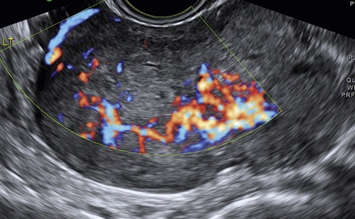
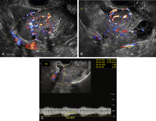
Figure O2-2 A to C, Ovarian cancer. Nine-centimeter ovarian cancer showing small cystic areas within a largely solid mass with extensive vascularity. C shows the low resistive index of 0.4 using spectral Doppler. This abundant diastolic flow is frequently seen in malignant tumors; however, the mere presence of disorganized blood vessels in the center of the mass is the most important Doppler finding.


image

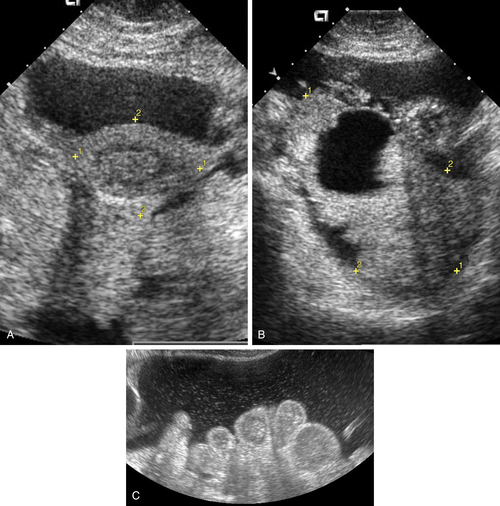
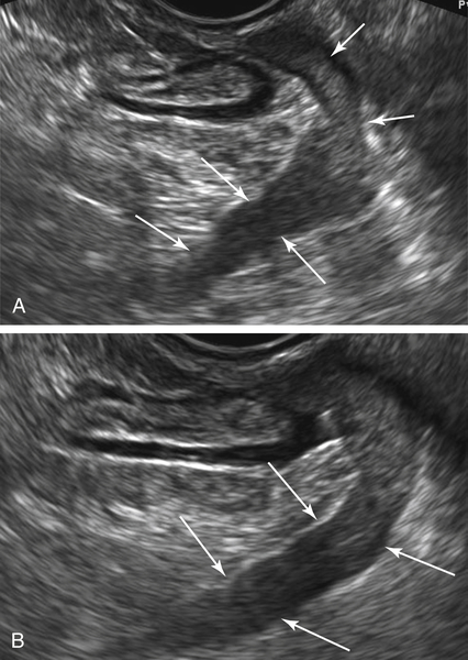
Figure O2-3 Advanced ovarian cancer. A shows a large amount of ascites anterior to the uterus (calipers) on a transverse view of the pelvis. B shows the large complex irregular mass (calipers), which was the ovarian tumor. Note the surrounding ascites. C shows the extensive ascites surrounding bowel loops, higher up in the abdomen.


image

Figure O2-4 Clear cell carcinoma. A shows a largely cystic tumor with areas of nodularity that are often associated with endometriosis. B shows that the solid nodules contain blood flow, which is a characteristic of malignancy.

image

Figure O2-5 Serous cystadenocarcinoma in a pregnant patient who presented for a routine 9-week obstetric scan. Note the typical deranged and irregular branching vascularity within the nodule.


image

Figure O2-6 A and B, Very large serous cystadenocarcinoma, almost completely solid with a few small internal cysts. C shows the intense vascularity with typical disorganized tumor vessels.


image

Figure O2-7 Bilateral papillary serous cystadenocarcinoma. A shows the right-sided lesion, which is predominantly cystic with several solid areas and a thick septum. B and C, The left-sided lesion is mostly solid with a small cystic component.


image

Figure O2-8 A to C, Typical serous cystadenocarcinoma with a mostly solid texture and extensive disorganized vascularity.


image

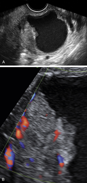
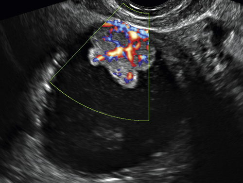
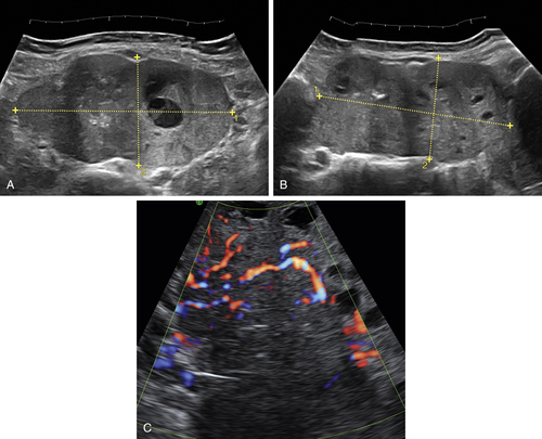
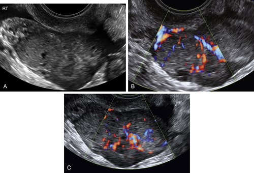
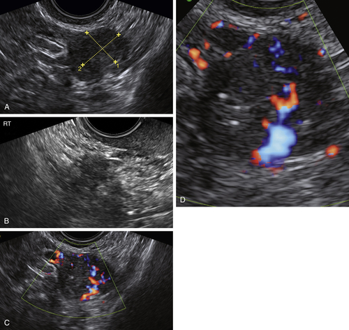
Figure O2-9 Stage 1 ovarian cancer in a 73-year-old patient. A shows an enlarged left ovary with a subtle 1.5-cm solid mass (calipers). B shows the normal (small) right ovary for comparison. C and D show the abundant blood flow to the small tumor. Typically it is difficult to demonstrate blood vessels within a normal postmenopausal ovary, making the flow pattern in the left ovary abnormal.


image

Figure O2-10 A and B, Primary extraovarian peritoneal carcinoma. Arrows show the linear solid mass coating the lateral side wall of the pelvis and peritoneal cavity.


Videos

Video 1 on ovarian cancer (epithelial) is available online.

Suggested Reading

Alcázar J.L., Rodriguez D. Three-dimensional power Doppler vascular sonographic sampling for predicting ovarian cancer in cystic-solid and solid vascularized masses. J Ultrasound Med. 2009;28:275–281.

Ameye L., Valentin L., Testa A.C., Van Holsbeke C., Domali E., Van Huffel S., Vergote I., Bourne T., Timmerman D. A scoring system to differentiate malignant from benign masses in specific ultrasound-based subgroups of adnexal tumors. Ultrasound Obstet Gynecol. 2009;33:92–101.

Campbell S. Ovarian cancer: role of ultrasound in preoperative diagnosis and population screening. Ultrasound Obstet Gynecol. 2012;40:245–254.

Kaijser J., Bourne T., Valentin L., Sayasneh A., Van Holsbeke C., Vergote I., Testa A.C., Franchi D., Van Calster B., Timmerman D. Improving strategies for diagnosing ovarian cancer: a summary of the International Ovarian Tumor Analysis (IOTA) studies. Ultrasound Obstet Gynecol. 2013;41:9–20.

Sharma A., Apostolidou S., Burnell M., Campbell S., Habib M., Gentry-Maharaj A., Amso N., Seif M.W., Fletcher G., Singh N., Benjamin E., Brunell C., Turner G., Rangar R., Godfrey K., Oram D., Herod J., Williamson K., Jenkins H., Mould T., Woolas R., Murdoch J., Dobbs S., Leeson S., Cruickshank D., Fourkala E.O., Ryan A., Parmar M., Jacobs I., Menon U. Risk of epithelial ovarian cancer in asymptomatic women with ultrasound-detected ovarian masses: a prospective cohort study within the UK collaborative trial of ovarian cancer screening (UKCTOCS). Ultrasound Obstet Gynecol. 2012;40:338–344.

Timmerman D., Ameye L., Fischerova D., Epstein E., Melis G.B., Guerriero S., Van Holsbeke C., Savelli L., Fruscio R., Lissoni A.A., Testa A.C., Veldman J., Vergote I., Van Huffel S., Bourne T., Valentin L. Simple ultrasound rules to distinguish between benign and malignant adnexal masses before surgery: prospective validation by IOTA group. BMJ. 2010;341:c6839.

Timmerman D., Valentin L., Bourne T.H., Collins W.P., Verrelst H., Vergote I. Terms, definitions and measurements to describe the sonographic features of adnexal tumors: a consensus opinion from the International Ovarian Tumor Analysis (IOTA) Group. Ultrasound Obstet Gynecol. 2000;16:500–505.

Vang R., Shih E.M., Kurman R.J. Fallopian tube precursors of ovarian low- and high-grade serous neoplasms. Histopathology. 2013;62:44–58.