Burns

Published on 23/06/2015 by admin

Filed under Emergency Medicine

Last modified 22/04/2025

Print this page

rate 1 star rate 2 star rate 3 star rate 4 star rate 5 star
Your rating: none, Average: 0 (0 votes)

This article have been viewed 1301 times

3.5 Burns

Introduction

Burns sustained by children are a common presentation to emergency departments and often cause significant distress to both the child and the parents. The mortality is increased in younger children. Deaths are generally related to flame burns, which may be complicated by inhalation of smoke and other toxic gases (e.g. in house fires). Early fatalities are related to respiratory complications, whereas late deaths are usually related to infection. The use of early debridement and skin grafting has led to an increased survival rate in patients who would have previously died because of infection.

Most burns are fortunately less serious, resulting mainly from scalds. This commonly occurs in pre–school-aged children due to their inquisitive nature precipitating accidents in the home. Flame burns occur in older children often experimenting with inflammables. Chemical and electrical burns are uncommon. One must be alert to the possibility of burns presenting as a manifestation of non-accidental injury in a young child.

Several preventive strategies can help decrease the risk and degree of burns sustained, especially with thermal burns. Lowering the temperature of hot water heaters to a maximum of 50°C significantly increases the contact time needed to produce deep or full-thickness burns. Flame-resistant clothing and smoke detectors in homes have saved many lives. Spill-proof mugs, guards around wood fire stoves, and child-resistant taps have all been shown to prevent burns. Further prevention strategies will have a far bigger impact on burns than advances in burn management.

Pathophysiology

The skin is the largest organ in the body, and its functions include:

Therefore children with extensive burns have difficulty retaining fluid and regulating temperature, and are at risk of infection.

The skin is composed of two main layers.

After a burn, injury to the deeper specialised epithelial cells prompts a change into stratified squamous epithelium. These cells proliferate, gradually covering the burn with a non-epithelial barrier. Therefore, if the dermal structures are damaged, skin grafting is the only means to cover the skin. When these deeper layers are involved, scarring results and contractures may occur.

The depth of the burn will depend on the temperature of the substance in contact with the skin, the length of time the substance is in contact with the skin, and the extent of subsequent cooling of the burned skin area. Hypothermia due to cooling occurs quickly in children due to their higher surface area to weight ratio, compared to adults. Also, children have thinner skin, which leads to deeper burns for a given contact temperature and duration.

Classification

Burns are generally classified into superficial, partial thickness or full thickness. Previous nomenclature (first, second, and third degree) has been replaced to give a more accurate description of the burn. In the emergency department setting, the definitive assessment of the ‘depth’ of the burn may be difficult, as the appearance can evolve during the first 24–48 hours. Likewise, the burn is not generally uniform in depth, and it may take time to delineate between superficial and deeper areas. Superficial and partial-thickness burns are the most common burns seen in children.

Examination

Note that children who are distressed may require the provision of immediate appropriate analgesia at the outset to aid examination.

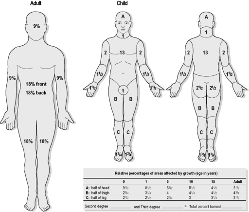
Evaluation of burn area

The extent and depth of the burn should be assessed after the patient has been stabilised. This is usually done from a body chart (Fig. 3.5.1), which can be useful to aid documentation of the burn. This chart is used because the surface area involved will alter depending on the age of the child and the parts of the body where the burn is located. A simple method, using the palmar surface of the child’s hand and fingers, can also be used to estimate the area of burn. This correlates to approximately 1% of the child’s total surface area. The adult formula using the ‘rule of nines’ can be used in adolescents older than 15 years.

Management

Prehospital

The main aims of prehospital care are stabilising ABCs, preventing ongoing burn injury, provision of analgesia, covering the area involved, and rapid transfer to an emergency department.

The first priority in any burn is to assess and stabilise the airway and breathing. Oxygen should be administered where there is suspected carbon monoxide poisoning with inhalation burns. Circulation is generally not a problem in the first hour after a major burn, and rapid transport to hospital should occur. If transfer time is greater than 1 hour, and the burn is greater than 20%, then intravenous fluid replacement should begin en route where possible.

The burns should be covered with water-soaked sterile cloth or the newer tea tree oil soaked pads. Excessive cooling of major burns causes hypothermia and worsens the patient’s outcome. Recently sustained minor burns should be cooled under running water for at least 20 minutes. This acts to minimise the extent of the burn and also affords some pain relief.

Analgesia is generally required early, and a single dose of a narcotic (e.g. fentanyl 1.5–2 mcg kg–1 intranasally) is a good choice in burns less than 20%. Greater than 20%, then intravenous access should be obtained and analgesia titrated to responses as well as administering intravenous fluids.

If electrical injury has been sustained, then cardiac monitoring should occur during transfer. Patients with chemical burns should undergo extensive washing of the affected area before transport.

Emergency department

The initial priority should focus on stabilisation of airway, breathing, and circulation, with concurrent provision of analgesia. If airway burns are suspected, then early intubation should be considered. Patients with obvious stridor due to upper airway compromise require urgent intubation. Supplemental oxygen should be instituted and oxygen saturation monitored.

Other potential indications for ventilation in major burns include:

The fluid losses due to the burn itself do not cause early circulatory failure, and other contributing injuries should be sought in the patient with early shock. It is also important to note the time the patient is evaluated, relative to the time of the burn. Children who have delay (e.g. a few hours) in presentation may arrive with circulatory compromise from skin fluid losses.

Intravenous access should be placed in all children with burns body surface area (BSA) of >20%. Fluid resuscitation rates should be calculated using the time of the burn, not the time of presenting to the emergency department (see Fluid resuscitation, below). Peripheral venous access, preferably through non-burnt skin, is preferred over central venous access for the initial resuscitation. Monitoring of urinary output (via urinary catheter, weighing nappies) is important in determining the adequacy of fluid replacement. A nasogastric tube should also be inserted in children with severe burns, as gastric dilatation can occur, leading to respiratory compromise.

Analgesia should be given early, during the stabilisation of the A, B and C. Intranasal fentanyl (1.5–2 mcg kg–1) is a good first up treatment. In severe burns, a morphine infusion should be started after adequate initial intravenous or intranasal analgesia has been given. Large doses of narcotics are sometimes needed in severe burns to control the pain. Intramuscular morphine is now obsolete given the use of intranasal fentanyl.

A careful secondary survey should then be undertaken, looking at the extent, depth, and anatomical relevance of the burns. It is important to determine if there are any circumferential burns to limbs and chest. In superficial or partial thickness burns, careful monitoring of circulation or ventilatory compromise is required. In full-thickness circumferential burns, an urgent escharotomy may be required to restore circulation to the limb or allow for adequate ventilation. A paediatric burn specialist should be consulted in this situation.

The secondary survey should also include careful examination for any other injuries requiring attention (e.g. head, neck, chest, limbs, pelvis, intra-abdominal). Burns to the face should also include fluorescein staining of the eyes to check for corneal involvement.

Fluid resuscitation

Fluid resuscitation should be calculated based on the weight of the child and the total surface area of the burn. Several formulas are used to calculate the resuscitation fluid requirement in the first 24 hours. The Parkland formula (BSA affected % × weight (kg) × 4) gives the number of millilitres of resuscitation fluid to be given over the first 24 hours. Half the fluid is given in the first 8 hours and the remainder in the subsequent 16 hours. The 24-hour period should begin from the time of the injury. Thus if a patient has received very little fluid in transfer, and it is 4 hours since initial burn, then the fluid calculated should be given over the next 20 hours, and half the calculated fluid given in the first 4 hours.

In addition, maintenance fluid for the 24-hour period should also be given. This is calculated as 100 mL kg–1 for 0–10 kg, 50 mL kg–1 for 11–20 kg, and 25 mL kg–1 for >20 kg. Thus a 30-kg child’s maintenance = 1000 + 500 + 250 = 1750 mL over 24 hours. It is also important to monitor ongoing losses (urinary output, respiratory loss, etc.).

The calculated amount of fluid (burn deficient plus maintenance) to be replaced in 24 hours is only a guide and should be adjusted according to the haemodynamic response. Patients need to be maintained in a positive fluid balance for the first 24–48 hours. The adequacy of fluid replacement is monitored by urine output and clinical parameters of perfusion. In children, 0.5–1 mL kg–1 per hour is the recommended urine output and should be monitored by a urinary catheter in severe burns. A central venous catheter is generally not required in the emergency department phase of management.

The type of fluid used varies between burn specialists, and it is best to be familiar with the preference of the local paediatric burns unit. In the shocked child, a 20 mL kg–1 bolus of normal saline should be given. This can be repeated if necessary to restore peripheral circulation. Ongoing replacement is generally with crystalloid in the first 24 hours, as colloid may leak through the burnt capillaries, causing worsening oedema. After 24 hours, colloid is used as part of the replacement fluids in the intensive care setting. When calculating the initial fluid requirements, it is important to subtract the bolus fluids given from this amount.

Additional fluids may be required in severe electrical burns causing muscle damage, as myoglobin may cause renal failure secondary to renal tubular deposition, and therefore maintaining adequate glomerular filtration is very important.

Superficial burns

Partial thickness/small full thickness burns

Use Acticoat, a silver impregnated barrier dressing, moistened with sterile water, covered with IntraSite Conformable, gladwrap, ± crepe bandage. Secure with Hyperfix. The exudate from the wound determines the number of dressing changes required. IntraSite Conformable is a soft hydrogel dressing that combines the advantage of IntraSite gel with a non-woven dressing. It creates a moist wound environment for the continued release of silver from the Acticoat. Patients should be reviewed between 3 and 7 days for a change of dressing and to assess if skin grafting is needed.

Superficial burns on the face should be managed non-dressed, and cleaned two or three times a day with warm water. Solugel or vaseline can be applied to the face or it can be left completely non-dressed. As a burn on the face starts to dry up, application of mild lanolin ointment/cream can be used to aid in healing by softening the skin. The burn should also be protected from the sun, as sunburn of an already burnt area causes increased pigmentation to the skin.

Superficial burns rarely become infected, but infection should be suspected if the patient: has unexplained fever, or has evolving pain, redness, and tenderness. Foul discharge from the burn does not always indicate that infection is present. In this case, the dressing should be changed earlier and the burn inspected, as antibiotics may not be indicated. As the skin heals under a dressing, it becomes pruritic, which may require an antihistamine or cooling of the dressing (particularly in hot weather).

Tetanus prophylaxis is important in major burns or minor burns (which are contaminated). Antibiotics should be used only when a definite infection is present. Prophylactic use of antibiotics is not recommended.

Pain management for minor burns is generally achieved by the dressing itself. Covering the burn decreases the pain substantially. Generally, paracetamol with or without codeine should be sufficient during the first 24 hours.

Patients with burns involving hands, feet, or face should be referred to a burn specialist for ongoing management.

Electrical burns

Chemical burns

Many chemical agents can cause burns through accidental exposure. They are generally either acid or alkali. Acid burns cause coagulation of the skin, which seems to limit the depth of penetration. Alkali burns cause liquefaction and thus result in a deeper injury. Caustic chemicals tend to give deeper burns than thermal injury, as there is generally a long duration of contact. Oedema tends to occur more quickly in chemical burns, which may cause a deeper burn to appear more superficial.