Breathlessness in a young woman

Published on 10/04/2015 by admin

Filed under Surgery

Last modified 22/04/2025

Print this page

rate 1 star rate 2 star rate 3 star rate 4 star rate 5 star
Your rating: none, Average: 0 (0 votes)

This article have been viewed 974 times

Problem 28 Breathlessness in a young woman

There is no other medical or surgical past history. The patient is a non-smoker and non-drinker. She has not been pregnant recently. There is no family history of cardiac problems. She was not taking any medications. For the last 2 nights, the patient has been unable to sleep due to respiratory distress whenever she lay flat. The patient has also been nauseous and anorexic for 24 hours. There have been no palpitations or episodes of presyncope or syncope.

The patient was not peripherally or centrally cyanotic. The peripheries were cool to touch. There was no fever. Abdominal examination was unremarkable other than mild hepatic tenderness. There was no clinical evidence of anaemia. The peripheral pulses were palpable and mildly reduced in intensity.

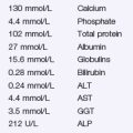
The full blood count is normal. U&Es show a Na+ of 131, K+ of 3.8, creatinine of 97 and urea of 5. Liver function tests reveal a total protein = 55, albumin = 23, ALP = 235, GGT = 187, ALT = 23, bilirubin = 12. INR = 1.3. BNP = 2187. Cardiac enzymes are normal.

A 12-lead ECG showed a sinus tachycardia, normal axis, normal intervals and no ST-T wave changes.

The chest X-ray showed an increased cardiothoracic ratio, a small left pleural effusion and interstitial and alveolar oedema.

A transthoracic echocardiogram was performed at the bedside in the emergency department, which showed a dilated and severely globally impaired left ventricle with a left ventricular ejection fraction (LVEF) of 20%. The right ventricle was also dilated and severely impaired. There was moderate mitral regurgitation, moderate tricuspid regurgitation and no pulmonary hypertension.

This patient had acute cardiogenic pulmonary oedema due to a recent onset of a viral dilated cardiomyopathy. As the patient was hypoxic, she should be given oxygen and be sitting upright. A bolus dose of 40 mg of intravenous furosemide was given, followed by an infusion of glyceryl trinitrate (GTN).

The patient’s breathlessness improved and she was transferred to the coronary care unit on telemetry. She was commenced on an ACE inhibitor (tritace 2.5 mg bd), regular oral furosemide (40 mg daily) and a heart failure beta blocker (carvedilol, initially starting at 3.125 mg bd). Over the next 5 days, the patient’s condition stabilized. Contact was made with the hospital multidisciplinary heart failure team (consisting of a heart failure cardiologist, heart failure nurse, pharmacist and physiotherapist) and they coordinated the patient’s ongoing inpatient and outpatient care, in consultation with the patient’s usual general practitioner. The patient was discharged on day 7 on ramipril 5 mg bd, carvedilol 6.25 mg bd, and furosemide 40 mg daily.

The patient had close follow-up through the heart failure outpatient service, which included outpatient visits to the heart failure cardiologist and heart failure nurse, telephone contact and monitoring by her general practitioner. A follow-up CXR showed cardiomegaly, no pleural effusions and clear lung fields. A repeat transthoracic echocardiogram at 3 months showed a moderately dilated left ventricle, a LVEF of 25%, mildly impaired right ventricular function and mild tricuspid regurgitation and mild mitral regurgitation only.

The patient re-presented to the emergency department 6 months later with severe acute cardiogenic shock, with a heart rate of 130 and a blood pressure of 70/40 mmHg. This did not respond to conventional therapy and after extensive consultation with the cardiac transplant team, the intensive care unit and cardiothoracic surgery, a left ventricular assist device was inserted. This was used as a bridge to transplantation and the patient underwent a successful heart transplantation 4 months later. The patient was discharged 2 weeks after her transplant, with no episodes of rejection, and is now back at work.

Answers

A.1

A.2 When examining an acutely unwell patient, both the severity of the cardiac dysfunction and the end organ consequences of reduced perfusion must be assessed. Starting at the peripheries: is there evidence of reduced perfusion (cool or warm peripheries) or cyanosis (although cyanosis is usually a manifestation of severe cardiorespiratory failure)? Are the peripheral pulses palpable? What is the cardiac rhythm (is there a sinus tachycardia, atrial fibrillation or a relative bradycardic state)? What is the blood pressure (this may be so low that it is not detectable unless via invasive means)? Is the jugular venous pressure (JVP) elevated? Palpation of the praecordium may reveal an altered apex beat (is it displaced, indicating a dilated heart)? Listen carefully to each component of the heart sounds in turn; it may help to say to yourself ‘I am listening to S1, to systole, to S2, to diastole, to any added sounds’. Ask yourself about the intensity of the heart sounds (is there a soft S1, which occurs in severe mitral regurgitation?). Are there any added heart sounds (either an S3/S4 or gallop rhythm)? Are there any murmurs (such as mitral regurgitation, aortic regurgitation, aortic stenosis or a ventricular septal defect)? Listening to the chest, is there evidence of pulmonary oedema or a pleural effusion? In severe cases of pulmonary oedema, there may be just severely reduced breath sounds or a wheeze.

It may not be possible to lay the patient flat in severe pulmonary oedema to examine the abdomen adequately. In congestive heart failure there may be evidence of tender hepatomegaly (due to congestion) or a pulsatile liver (due to severe functional tricuspid regurgitation). There may be evidence of ascites if there is significant right heart involvement. There may also be peripheral oedema.

Reduced perfusion from cardiac dysfunction can have significant consequences on other organ systems. The patient may be agitated or delirious due to reduced cerebral perfusion. The respiratory distress associated with pulmonary oedema can result in respiratory fatigue and exhaustion which may require management with mechanical ventilation. Reduced perfusion of the kidneys can result in pre-renal failure and a reduced urine output. Liver dysfunction can also occur from prolonged hypotension (although this is difficult to detect clinically).

A.3 The investigations required for heart failure are important because they will help establish a diagnosis, determine the severity of the illness, point toward an aetiology, find precipitating or exacerbating factors and help in guiding therapy.

The initial investigations in the emergency department should be an ECG, CXR and blood tests (full blood count, urea, creatinine and electrolytes, liver function tests, thyroid function tests and BNP). Investigations performed later include those that aim to determine the aetiology of the heart failure, and those that help establish severity and prognosis. These tests include assessing for coronary artery disease (such as a nuclear perfusion scan, stress echocardiogram, cardiac MRI, coronary CT angiogram, or invasive coronary angiogram), haemodynamic assessment with a right heart catheter, 6-minute walk test, cardiopulmonary exercise test, myocardial viability assessment and a cardiomyopathy blood screen (such as iron studies for haemachromatosis, autoantibody screen for disorder such as lupus, serum and urine electrophoresis for amyloidosis, and a viral screen).

A.4 By integrating the history, examination findings and initial investigations, the likely diagnosis is heart failure due to a dilated cardiomyopathy. This may have been caused or precipitated by a recent viral infection. It is important to recognize that heart failure is a syndrome and not a diagnosis. There is a three-stage process to assessing this patient. Firstly, establish the presence of a heart failure syndrome has been determined. Secondly, determine the type of cardiac dysfunction that is causing the heart failure. The third step is to determine the underlying aetiology of the cardiac dysfunction.

Not all heart failure is due to left ventricular systolic dysfunction (although this is the most common cause). Other important causes include diastolic heart failure (or heart failure with normal ejection fraction: HFNEF), right heart failure (without left heart involvement) or non-myocardial heart failure (such as pericardial disease, valvular disorders or a cardiac mass/tumour).

Once the cardiac mechanism has been identified, the cause of this dysfunction has to be determined. In this case, the heart failure due to a dilated cardiomyopathy is most likely due to the preceding viral illness. A viral aetiology is a common cause of a dilated cardiomyopathy and common aetiological agents include Coxsackie virus, enterovirus and adenovirus.

The other common causes of heart failure in Western countries are coronary artery disease, hypertension, valvular heart disease (such as untreated aortic stenosis or mitral regurgitation), toxicity from alcohol or chemotherapy, and other disorders affecting the myocardium – such as viral infections, infiltration (sarcoid and amyloid), metabolic abnormalities (such as thyroid dysfunction or haemochromatosis) or genetic disorders such as hypertrophic cardiomyopathy.

A.5 The next investigation that should be ordered is a transthoracic echocardiogram. This is a fundamentally important investigation in heart failure and should be performed in all patients that are suspected of having heart failure. It provides an accurate, safe, non-invasive, time-efficient evaluation of cardiac structure and function, valvular function, and assessment of cardiac haemodynamics and presence of pulmonary hypertension or intra-cardiac thrombus.

A.6 Acute pulmonary oedema is a medical emergency. The initial management requires oxygen, diuretics and a nitrate vasodilator. The oxygen is to correct the hypoxia and may be given via a face mask, or in more severe cases, positive pressure ventilation. Mechanical ventilation may be required for those in significant respiratory distress. Intravenous loop diuretics (such as furosemide) are required to increase sodium and water excretion (and furosemide also has an immediate venodilator effect). This may be given as either a bolus (such as 40–80 mg) or an infusion (starting as 5 mg/hour). Nitrates are also important as they are predominantly a venodilator and have very rapid onset of action. They are particularly useful in heart failure due to myocardial ischaemia as they also dilate the coronary arteries. The nitrate is usually given as a continuous intravenous infusion. If IV access cannot be immediately obtained, the nitrate can be given as a topical patch as a temporary measure.

If these measures do not result in improvement of the patient’s condition, other options need to be considered. These include non-invasive assisted ventilation, intubation and ventilation, inotropic therapy and finally mechanical support (such as an intra-aortic balloon pump or ventricular assist device).

A.7 Once the patient is more stable on the ward, the treatment for heart failure is both increased and broadened. Treatment centres around ACE inhibition and beta blockade. All patients with LV systolic dysfunction (irrespective of functional class) should be commenced on an ACE inhibitor, with the dose up-titrated to maximal dose or that which is tolerated. ACE inhibitors have been shown to improve survival, improve symptom class, reduce hospitalizations and increase left ventricular ejection fraction.

Beta blockers are also fundamental in the management of chronic heart failure. They help to inhibit the chronic activation of sympathetic nervous system (which in the long term is toxic to the myocardium). Beta blockers have also been shown to improve survival (both by reducing sudden arrhythmic cardiac death, and by reducing progressive heart failure). Beta blockers also improve symptom class and left ventricular ejection fraction. Beta blockers should not be commenced until the patient has recovered from the acute pulmonary oedema and are generally better tolerated when the patient is euvolaemic. To reduce the likelihood of beta blocker side-effects (such as hypotension, fatigue, worsening heart failure), they should be started at a low dose and gradually increased.

Diuretics in the chronic situation may be required to maintain euvolaemia and control symptoms. However, they do not confer a survival benefit in isolation. Additional pharmacological therapy that may be required, depending upon response and severity of heart failure, includes aldosterone antagonists (spironolactone and eplerenone), digoxin, angiotensin 2 receptor blockers, nitrates and hydralazine. Some patients may also require long-term anticoagulation, usually with warfarin.

A.8 When the patient is discharged, it is important that they are linked into a heart failure programme and closely monitored in conjunction with their general practitioner. Heart failure management is a dynamic process with alterations in treatment and dosing a common occurrence. Further up-titration of heart failure medications is continued in an outpatient setting. Patient education is important. This includes fluid intake control, daily weighing, exercise programmes and good lines of communication with their GP and heart failure programme.

Patient compliance with prescribed therapy is important and poor compliance is a common cause for re-presentation with worsening heart failure. Other precipitants for heart failure include arrhythmias, infection, myocardial ischaemia, anaemia, alcohol excess, thyroid disorders, pregnancy, medications (such as steroids or NSAIDs) and pulmonary embolism.

A.9 If the patient does not respond to conventional medical therapy, there are other important options to consider. These include device therapy and surgical treatment. In some patients who do not respond to appropriate medical therapy, biventricular pacing (cardiac resynchronization therapy – CRT) may be of benefit. In this form of pacing, both ventricles are paced to try and induce mechanical synchrony between the two ventricles. The right heart is paced via a right ventricular lead and the left heart is paced via a lead passing through the coronary sinus and into a cardiac vein, usually on the lateral aspect of the left ventricle. In this way, the left ventricle can be paced via access from the venous system. Some patients, whose LVEF remains below 35%, may also be candidates for insertion of an implantable cardiac defibrillator (ICD).

Surgical options are also important to consider in the refractory heart failure patient. Ventricular assist devices (VADs) are small mechanical pumps that provide circulatory support and can be either implanted within the thoracic cavity or abdomen, or be extra-corporeal and sit outside the chest or abdominal wall. There are various types of pumps available which can provide either pulsatile or continuous rotary blood flow support.

In the more chronic setting, cardiac surgery may be needed to correct the cause of the heart failure. Common examples include surgical revascularization for heart failure due to coronary artery disease, or an aortic valve replacement for severe aortic stenosis or severe aortic regurgitation.

The final management option is cardiac transplantation. There are strict criteria as to who is eligible for heart transplantation and this form of therapy is only offered to a select group of patients who have undergone an extensive screening process and for whom no other form of therapy is available. This is due to a shortage of donor organs and the complexity of the procedure and resources required to maintain a cardiac transplant programme. Following cardiac transplantation, 1-year survival is approximately 85% and 5-year survival 65%. Early complications include acute cellular rejection and infection, while more long-term complications include coronary artery vasculopathy (CAV – a form of chronic rejection), malignancy, hypertension and renal dysfunction. However, this therapy is both a lifesaver and provides the opportunity to have an excellent quality of life, neither of which would occur in the non-transplanted state.