Breast disease

Published on 17/03/2015 by admin

Filed under Basic Science

Last modified 22/04/2025

Print this page

rate 1 star rate 2 star rate 3 star rate 4 star rate 5 star
Your rating: none, Average: 0 (0 votes)

This article have been viewed 1453 times

chapter 51 Breast disease

BREAST HEALTH

Women of all ages should know the normal look and feel of their breasts—this is known as ‘breast awareness’. There is no evidence to support any specific technique for breast self-examination. For many women there is also a psychological barrier to conducting a technical systematic examination, often suggesting they are not confident in their technique. For these reasons women should be encouraged to get to know what is normal for them through normal activities such as showering, dressing, putting on body lotion, looking in the mirror. Most importantly, women should be encouraged to present early to their GP if they find a change in their breasts—they are not wasting their doctor’s time. This is important even if they have had a recent ‘normal’ mammogram.

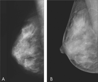
In Australia, two-yearly mammographic screening is available free to all women aged 40 years and over through BreastScreen Australia. For women aged 50–69 years, there is strong evidence from international randomised trials for the effectiveness of mammographic screening in reducing mortality from breast cancer by about 30%. For women aged 40–49 years, the benefit may be smaller, as the effectiveness of mammography in detecting cancer is affected by the density of the individual woman’s breast tissue. BreastScreen does not actively target women aged over 70 years. However, given that risk for breast cancer increases with age, it is important that older women do not presume they are no longer at risk. For women aged 70 years or over, the benefits of mammographic screening will depend on whether there are any significant medical comorbidities.

Women who have a significant family history should be referred to a familial cancer or genetics clinic, where an individual surveillance program and management advice can be provided and, if appropriate, genetic testing can be conducted.1

BENIGN BREAST DISEASES

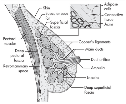
During a woman’s life there are three main phases of breast change. Breast development and early reproductive life is followed by mature reproductive life and finally by involution.

There are regular changes in relation to the menstrual cycle. Pregnancy causes a doubling of the breast weight at term, and the breast involutes after pregnancy.

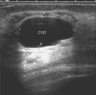
In nulliparous women, breast involution commences at around age 30 years. During involution the breast stroma is replaced by fat and the breast becomes softer and less radio-dense. Changes in the glandular tissue during involution include the development of areas of fibrosis, the formation of small cysts and an increase in the number of glandular elements (adenosis).

Most presentations to healthcare professionals are benign and are the result of aberrations of these normal physiological processes. The most common presenting symptoms are:

Despite the above statement, the most important consideration for the GP is to exclude cancer as the cause of the presenting symptom.

BREAST PAIN AND FOCAL BREAST NODULARITY

Premenstrual breast pain and nodularity, improving with the onset of menstruation, are so common in women as to be considered physiological, rather than being a disease. Severe pain and nodularity are aberrations of these normal cyclical changes, which occur in the breasts of all women during their reproductive years. Bilateral focal breast nodularity, sometimes referred to as fibroadenosis or fibrocystic disease, is the most common cause of a breast lump. If excised, these areas show either no pathological abnormality, or aberrations of the normal involutional process, such as focal areas of fibrosis or sclerosis.

Breast pain

The causes of breast pain can be cyclical or non-cyclical. The best way to assess whether pain is cyclical is to ask the patient to complete a breast pain record chart.

Cyclical mastalgia is the more common. It shows a definite relationship to the menstrual cycle and is often associated with nodularity of varying degree, maximal in the upper outer quadrant and showing similar cyclicity.

Non-cyclical mastalgia affects older women. The origin of the pain can be from the chest wall, as in costochondritis, the breast itself or outside the breast. The pain may be continuous or random in its time pattern. A careful history and examination is required, to exclude non-breast causes.

The aetiology of mastalgia is unclear. Abnormalities in the control mechanisms of the pulsatile secretion of gonadotrophins and/or prolactin are likely. Women with mastalgia have also been found to have abnormal fatty acid profiles, but the role of dietary factors such as caffeine and fats in the aetiology of breast pain is unclear.

Management

For the management of cyclical breast pain, diuretics, progestogens and vitamin B6 have not been shown to be any more efficacious than placebo. After excluding cancer, reassurance that the pain is not related to cancer, and an explanation of the hormonal basis of breast pain, may be the only treatment required. A soft support bra worn at night may also assist. Some women find stopping the Pill or changing formulations may assist.

Evening primrose oil (gamma-linolenic acid) has been shown to reduce pain, nodularity and tenderness, at a dose of 3 g daily.2 It has only minor side effects, including headache, nausea, gastrointestinal upset and possible drug interactions with anticoagulant and antiplatelet agents and phenothiazines. A trial of treatment should last 4 months and be monitored with a pain chart. It does not interact with oral contraceptives.

Several clinical studies in women have suggested that chasteberry (Vitex agnus castus) is efficacious in reducing symptoms associated with premenstrual symptoms (PMS) including mastalgia.2,3 This herb contains steroidal precursors and active moieties including progesterone, testosterone and androstenedione. Chasteberry may interact with oral contraceptives, other hormonal therapy and dopazmine antagonists such as haloperidol and prochlorperazine. Adverse effects reported include nausea, rash, headache and agitation.

If symptoms remain unresponsive to these therapies, consider referral. A range of medications are available to specialist practitioners, including bromocryptine, danazol, tamoxifen and goserelin.

Benign breast lumps

Fibroadenomata

Fibroadenomata result from a focal proliferation of benign breast elements, both epithelial and stromal, and are influenced by hormonal factors. They may fluctuate during the menstrual cycle and pregnancy. They are most common in the 20–30 year age group and are uncommon post menopause.

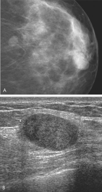
On clinical examination they are typically smooth, mobile, rubbery masses, which may be tender, especially premenstrually. They may be single or multiple. On breast ultrasound they appear as a well-defined ovoid homogenously hypoechoic mass with smooth margins and increased through transmission.

Diagnosis is confirmed by non-excisional biopsy. Ultrasound-guided core biopsy is used more commonly because of the high proportion of fibrous tissue to epithelial tissue, which increases the risk of not sampling the epithelial cells on fine needle aspiration.

Once the diagnosis is confirmed, fibroadenomata may be managed by either surgical excision or regular clinical and imaging review over 12–18 months until the lesion is proved to be stable. Should the lesion significantly increase in size or develop atypical features on imaging, it should undergo excision biopsy. New palpable fibroadenomas in women aged over 40 years should be referred to a breast surgeon for consideration of excision biopsy, because the likelihood of a new lump being cancer increases with age.4

Phyllodes tumour is a rare fibroepithelial tumour that produces a spectrum of diseases ranging from benign (with a significant risk of local recurrence) to malignant (sometimes with rapidly growing metastases). Clinically they may be indistinguishable from fibroadenoma, presenting as a smooth, rounded, painless breast lump that has continued to increase in size. Their appearance on mammogram and ultrasound may also resemble fibroadenomata. Diagnosis is confirmed by histology following excision biopsy.

BREAST CANCER

AETIOLOGY

Despite much research into causes and risk factors for breast cancer, we have no means of preventing this disease.6 There are a number of factors that bear on the probability of a particular symptom being due to a breast cancer.

In general, breast cancer is a disease of ageing, with 75% of breast cancers diagnosed in women aged 50 years or older, and about 6% in women aged under 40 years. However, breast cancer can occur at any age and it is important that younger women who present with symptoms have these adequately investigated. Other factors that may increase risk for a particular woman include whether she has a significant family history of breast or ovarian cancer1 or a relevant inherited gene mutation, whether she has had a previous invasive or in situ breast cancer or a previous biopsy that shows atypical proliferative disease or other marker for increased risk.

A number of other factors associated with risk for breast cancer are not modifiable, such as the age at menarche or menopause. Additionally some potentially modifiable factors are associated with reduced risk but are complex decisions for the individual woman. These include age at first pregnancy, number of children and breastfeeding. However, a number of lifestyle factors affecting risk for breast cancer are readily modifiable.

DIAGNOSTIC APPROACH

For the GP charged with assessing the presenting symptom, it is important to pursue a systematic approach to investigation that minimises the risk of missing a breast cancer. The ‘triple test’ is universally accepted as the most effective way to maximise the detection of cancer and to provide an accurate diagnosis for an abnormality. The correct sequencing of tests is important to the overall interpretation of the results.

The triple test refers to:

The sensitivity of the triple test approaches 100%. Triple test negative, where no test results are positive (that is, no suspicious or malignant results), provides very good reassurance that the symptom is not due to cancer. Less than 1% of women will be falsely reassured that they do not have cancer, based on correlation of all three test results that are negative.

If any of the tests is suspicious for cancer or malignant (a triple test positive result), or if the results do not correlate with one another, irrespective of whether there are normal or benign test results, further evaluation and referral to a breast surgeon or specialist breast clinic is required.

However, not all women will require investigation with all three tests. Indeed, as the majority of symptoms are due to normal physiological changes, most women will be able to be reassured after providing a history and having a clinical breast examination—for example, where a woman presents with bilateral breast pain that occurs cyclically before her menstrual period and there is no clinical abnormality on examination. Imaging may additionally support this diagnosis.

The sensitivity of each component of the triple test for any individual woman will depend on a number of factors, including the age and history of the patient, the characteristics of the woman’s breast tissue and the characteristics of the breast lesion. For example, the evaluation by clinical breast examination of a small lesion within lumpy breast tissue will be limited; and mammography in a young woman is less sensitive because of the increased density of the breast tissue.

TREATMENT

The vast majority of breast cancers are diagnosed ‘early’—that is, they are confined to the breast with or without local lymph node involvement, and have not metastasised to other organs. The primary goal of treatment of early breast cancer is to control the disease with the aim of achieving cure. The initial management of breast cancer offers the best hope of cure. There are significant differences in women’s views about, and need for, information, choice and support. The patient should be provided with adequate and appropriate information about treatment options in order to make an informed decision about her care.

The way in which a clinician relates to and communicates with a cancer patient can affect her wellbeing.9 Communication skills training is available to assist health professionals to manage difficult conversations such as breaking bad news and discussing prognosis.10

When organising referral for patients with breast cancer, GPs should consider both the preferences of the patient and the fact that patient outcomes are better if treated by clinicians who are part of a multidisciplinary team.11

The usual primary treatment of early breast cancer is surgery. Where appropriate, women should be offered the choice of either breast-conserving surgery followed by radiotherapy, or mastectomy, as there is no difference between them in the rate of survival or distant metastasis. Women who undergo mastectomy should be provided with information about, and the option of, breast reconstruction, either immediate or delayed.

Determining the extent of spread of disease is vital to informing treatment choices. Sentinel node biopsy is increasingly becoming the standard of care for the assessment of the axilla, as it has been shown to be as effective as axillary dissection in predicting lymph node involvement and has less morbidity.

The pathological features of the cancer and the involvement of lymph nodes will provide valuable information to guide decisions about the use of systemic adjuvant therapy, which includes all forms of hormonal and cytotoxic chemotherapy. The aim of this treatment is to treat undetectable remaining cancer and reduce the risk of recurrence.

New targeted systemic therapies, such as tamoxifen for oestrogen receptor positive breast cancer, are constantly evolving and have had a significant impact on breast cancer survival rates over the past two decades. The decisions about adjuvant systemic therapy are most appropriately made in a multidisciplinary setting, and will take into consideration both tumour factors and patient factors such as age and overall health, as well as patient preferences, if known. The suggested treatment plan should then be discussed with the patient such that she has an understanding of the side effects and potential benefits in her individual case. Issues that may affect younger patients’ decision-making, such as infertility and premature menopause, should be discussed and relevant referrals provided if necessary, prior to treatment commencing.

Note that where a woman has had surgery or radiotherapy involving the axillary lymph nodes, thereafter procedures including measurement of blood pressure or venepuncture should be avoided on that side.

Integrative management

Many patients who have been diagnosed with cancer use complementary and alternative therapies as adjuncts to their medical treatment. Most of these patients find this a way of maintaining a sense of control, particularly in relieving side effects of treatment and improving health-related quality of life, as well as facilitating physical and psychological rehabilitation.

Beyond the stage of active medical and surgical treatment and rehabilitation comes the question of enhancing wellbeing and, ultimately, longevity.

It is important to encourage patients to discuss any adjunctive therapies they may be considering or taking during treatment, as some interfere with conventional therapies and may cause harm. Other complementary therapies can work effectively alongside conventional treatment.

Many patients raise the question of antioxidant use during chemotherapy. Despite the concerns of some oncologists, no trials have reported evidence of significant decreases in efficacy from antioxidant supplementation during chemotherapy.12

Many of the studies indicated that antioxidant supplementation resulted in either increased survival times, increased tumour responses, or both, as well as less toxicity than controls; however, lack of adequate statistical power was a consistent limitation.

Patients enquiring about using supplements to control radiation side effects should be informed that they must abstain from smoking tobacco if they take antioxidants during radiotherapy.

It is important that patients discuss their motivation for enquiring about antioxidant and other supplement use, and discuss with their oncologist or radiotherapist. It is equally important for oncologists and radiotherapists to provide well-informed, evidence-based advice for patients, rather than a blanket ‘take nothing’.

Hormone therapies for breast cancer can result in a significantly increased risk of cardiovascular disease, obesity, type 2 diabetes, osteoporosis and sarcopenia, so active risk factor management is essential in this group.

General advice includes:

Supplements

Supplements may be recommended as adjunctive therapy for nutritional and immune system support, and for reducing the side effects and toxicity of cancer treatment (see ch 24, Cancer).

Beyond the medical and surgical treatment for cancer, patients usually become very focused on measures to improve their wellbeing and longevity.

The individual requirement for nutritional supplements will be determined by the woman’s general state of health, her age, stage of cancer treatment, diet, specific deficiencies and personal preferences.

A multivitamin19—daily, containing the antioxidant vitamins A, C, E, the B-complex vitamins, and trace minerals such as magnesium, calcium, zinc and selenium.

Psychosocial care

Psychosocial care should be considered alongside medical care for all patients with cancer, whose information and supportive care needs may change over time and should be monitored. It has been estimated that, at 3 months after diagnosis, 10–17% of women meet the criteria for major depression. Patients should be monitored for risk or symptoms of anxiety or depression, and referred to a clinical psychologist or psychiatrist for assessment. Psychological therapies have been shown to improve emotional adjustment and social functioning, reduce stress, and improve quality of life for patients with cancer.11

A range of therapies is available to improve quality of life for women undergoing treatment for breast cancer. The effectiveness of many of these therapies is influenced by the attitudes, beliefs and psychological make-up of the patient, and will need to be tailored accordingly. It is also important to enquire about the psychosocial needs of the woman’s partner and her family.

Psychoeducational programs, which provide both support and information, delivered on an individual or group basis, may reduce anxiety and depression. Patients have also reported feeling less anxious and more optimistic about the future after involvement in peer support programs, but they may not be universally helpful.

Cognitive behavioural techniques such as relaxation therapy, guided imagery, systematic desensitisation and problem solving have been demonstrated to be effective in reducing anxiety. Prayer and laughter are also effective for some individuals.11 Chemotherapy-induced nausea and vomiting may benefit from psychological interventions, including cognitive behavioural techniques, relaxation and meditation.11

Traditional Chinese medicine

Chinese medicinal herbs, when used together with chemotherapy, may improve bone marrow function and quality of life.23 Other complementary botanicals are commonly used by cancer patients, although most have not been tested in rigorous clinical trials.

REFERENCES

1 National Breast Cancer Centre. Advice about familial aspects of breast cancer and epithelial ovarian cancer: a guide for health professionals. Sydney: NBCC, 2006.

2 Newall C. Herbal medicines: a guide for health care professionals. London: Pharmaceutical Press, 1997.

3 Schellenberg R. Treatment for premenstrual syndrome with agnus fruit extract: prospective, randomised, placebo-controlled study. BMJ. 2001;322:134-137.

4 Houssami N, Cheung MN, Dixon JM. Fibroadenoma of the breast. Med J Aust. 2001;174:185-188.

5 Australian Institute of Health and Welfare & National Breast Cancer Centre. Breast cancer in Australia: an overview 2006. Cancer Series No. 34. Cat. no. CAN 29. Canberra: AIHW, 2006.

6 National Breast Cancer Centre. Risk factors report. Sydney: NBCC, 2007.

7 National Health and Medical Research Council. Making decisions: should I use hormone replacement therapy? Canberra: NHMRC, 2005.

8 National Breast and Ovarian Cancer Centre. Hormone replacement therapy (HRT) and risk of breast cancer. NBOCC Position Statement.

9 National Breast Cancer Centre. A guide for women with early breast cancer. Sydney: NBCC, 2003.

10 National Breast Cancer Centre. Communication Skills Training Initiative;. Online. Available: http://www.nbcc.org.au/bestpractice/commskills/, 2007. 10 October 2007.

11 National Breast Cancer Centre and National Cancer Control Initiative. Clinical practice guidelines for the psychosocial care of adults with cancer. Sydney: NBCC/NCCI, 2003.

12 Block KI, Koch AC, Mead MN, et al. Impact of antioxidant supplementation on chemotherapeutic efficacy: a systematic review of the evidence from randomized controlled trials. Cancer Treat Rev. 2007;33(5):407-418.

13 Berstad P, Ma H, Bernstein L, et al. Alcohol intake and breast cancer risk among young women. Breast Cancer Res Treat. 2008;108(1):113-120.

14 Ibrahim EM, Al-Homaidh A. Physical activity and survival after breast cancer diagnosis: meta-analysis of published studies. Med Oncol. 2010; Apr 22. [Epub ahead of print].

15 Chajès V, Thiébaut AC, Rotival M. Association between serum trans-monounsaturated fatty acids and breast cancer risk in the E3N-EPIC Study. Am J Epidemiol. 2008;167(11):1312-1320.

16 Blask DE. Melatonin, sleep disturbance and cancer risk. Sleep Med Rev. 2009;13(4):257-264.

17 Lambe M, Wigertz A, Holmqvist M. Reductions in use of hormone replacement therapy: effects on Swedish breast cancer incidence trends only seen after several years. Breast Cancer Res Treat. 2010;121(3):679-683.

18 Katalinic A, Rawal R. Decline in breast cancer incidence after decrease in utilisation of hormone replacement therapy. Breast Cancer Res Treat. 2008;107(3):427-430.

19 Harvard School of Public Health. The nutrition source. Vitamins: the bottom line. HSPH; 2010. Online. Available: http://www.hsph.harvard.edu/nutritionsource/what-should-you-eat/vitamins/index.html.

20 Nogues X, Servitja S, Peña MJ. Vitamin D deficiency and bone mineral density in postmenopausal women receiving aromatase inhibitors for early breast cancer. Maturitas. 2010; 14 Apr. [Epub ahead of print].

21 Hines SL, Jorn HK, Thompson KM, et al. Breast cancer survivors and vitamin D: a review. Nutrition. 2010;26(3):255-262.

22 Stevens RG. Circadian disruption and breast cancer: from melatonin to clock genes. Epidemiology. 2005;16(2):254-258.

23 Zhang M, Liu X, Li J, et al. Chinese medicinal herbs to treat the side effects of chemotherapy in breast cancer patients. Cochrane Database Syst Rev. 2007;2:CD004921.

24 Ezzo J, Richardson M, Vickers A, et al. Acupuncture-point stimulation for chemotherapy-induced nausea or vomiting. Cochrane Database Syst Rev. 2006;2:CD002285.

25 Carati CJ, Anderson SN, Gannon BJ, et al. Treatment of postmastectomy lymphoedema with low-level laser therapy: a double blind, placebo-controlled trial. Cancer. 2003;98(6):1114-1122.