Breast Biopsy: Open

Published on 31/05/2015 by admin

Filed under Obstetrics & Gynecology

Last modified 22/04/2025

Print this page

rate 1 star rate 2 star rate 3 star rate 4 star rate 5 star
Your rating: none, Average: 0 (0 votes)

This article have been viewed 1836 times

Chapter 232 Breast Biopsy: Open

TECHNIQUE

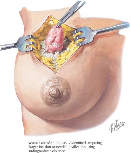
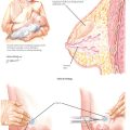
After appropriate informed consent has been obtained, the skin is disinfected and local anesthetic is injected at the chosen site. (The majority of biopsies can be performed with curvilinear incisions following the contours of the breast, often in the circumareolar area.) An open biopsy should be performed using a scalpel rather than electrosurgical energy because thermal effects on the biopsy material may blur the margin of normal tissue around the tumor and cause abnormally low receptor levels.

The dissection is carried to the area of concern through a combination of sharp and blunt techniques. A change in tissue character or a “gritty” sensation may be noticed as the tissue is dissected near some mass lesions. The mass or area of interest is excised and hemostasis is obtained through electrosurgical energy or the placement of hemostatic sutures to close dead space. The skin may be closed using a running subcuticular suture or self-adhesive skin tapes.

At the close of the procedure, samples are sent to the pathology laboratory for diagnosis and light dressing is applied. (A self-adhesive bandage often suffices.) Ice and gentle pressure may be applied for 15 to 30 minutes to minimize bruising.

It is important to send the pathology laboratory a small sample (1 g of suspect tissue) to determine the presence or absence of estrogen and progesterone receptors. These receptors are heat labile; therefore, the tissue must be frozen within 30 minutes.

Nonpalpable masses may be localized through the placement of a small needle or sterile J-wire under fluoroscopic or ultrasonographic guidance. These are then used as guides for the open dissection. The specimen is removed with the wire or needle in place, and it is radiographed to confirm the removal of the suspect area. (These techniques have been largely supplanted by computer-guided core biopsy techniques.)