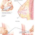
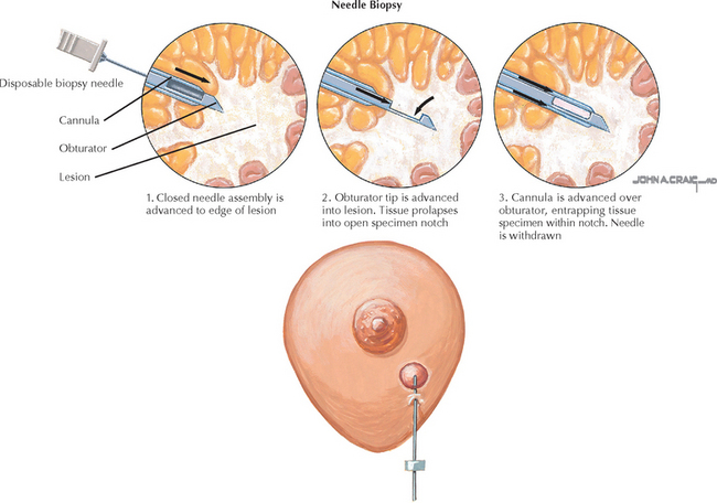
Chapter 231 Breast Biopsy: Core
REQUIRED EQUIPMENT
CPT CODE(S)
Brenner RJ, Fajardo L, Fisher PR, et al. Percutaneous core biopsy of the breast: effect of operator experience and number of samples on diagnostic accuracy. AJR Am J Roentgenol. 1996;166:341.
Frayne J, Sterrett GF, Harvey J, et al. Stereotactic 14 gauge core-biopsy of the breast: results from 101 patients. Aust N Z J Surg. 1996;66:585.
Jackman RJ, Nowels KW, Rodriguez-Soto J, et al. Stereotactic, automated, large-core needle biopsy of nonpalpable breast lesions: false-negative and histologic underestimation rates after long-term follow-up. Radiology. 1999;210:799.
Lee CH, Philpotts LE, Horvath LJ, Tocino I. Follow-up of breast lesions diagnosed as benign with stereotactic core-needle biopsy: frequency of mammographic change and false-negative rate. Radiology. 1999;212:189.
Liberman L, Dershaw DD, Rosen PP, Abramson AF, et al. Stereotaxic 14-gauge breast biopsy: how many core biopsy specimens are needed? Radiology. 1994;192:793.
Rich PM, Michell MJ, Humphreys S, et al. Stereotactic 14G core biopsy of non-palpable breast cancer: what is the relationship between the number of core samples taken and the sensitivity for detection of malignancy? Clin Radiol. 1999;54:384.
American College of Obstetricians and Gynecologists. Breast cancer screening. ACOG Practice Bulletin 42. Obstet Gynecol. 2003;101:821.
Britton PD. Fine needle aspiration or core biopsy. Breast. 1999;8:1.
Staren ED, O’Neill TP. Ultrasound-guided needle biopsy of the breast. Surgery. 1999;126:629.
Strong JW, Worsham GF, Austin RM, et al. Stereotactic core biopsy of nonpalpable breast lesions. J S C Med Assoc. 1995;91:489.