Chapter 3 Brachioplasty with bicipital groove scar
• The patient must be willing to accept the possibility of visible and even poor scarring.
• Improvement in contour must be significant to justify the visible scarring.
• The arm must be adequately deflated before brachioplasty.
• If there is laxity on the lateral chest wall consideration should be given to extending the excision to the inframammary fold.
• In massive weight loss patients, a Z plasty is not usually necessary in the axilla.
Introduction
The increase in obesity in the United States of America and the concurrent meteoric rise in bariatric surgery has led to an increasing interest and popularity of the brachioplasty procedure. Statistics from the American Society of Plastic Surgeons have borne this out, with an increase in the number of brachioplasties from 2516 in 1997 to 16 102 in 2009.1 Massive weight loss patients make up the bulk of brachioplasty patients. As is true in all parts of the body after massive weight loss, the deformities can be highly varied. Some patients do not have much loss of fat from their arms and require debulking before a formal brachioplasty. Other patients have truly deflated arms and describe “pinching” of the loose skin as it is folded into their clothing.
Esthetic brachioplasty was first described by Correa-Inturraspe and Frenandez2 in 1954. Lockwood (1995)3 described the superficial fascial repair of the arm, but as was the case with his work on lower body lifts, most of his patients were complaining of aging and not massive weight loss. However, until the avalanche of massive weight loss patients entered the plastic surgeon’s office there was little interest in the procedure. That said, many plastic surgeons were still reluctant to perform the procedure due to concerns about scars and axillary contractures. The massive weight loss patients have helped change the plastic surgeons’ minds, focusing the improvement of contour with fewer concerns about scars. Plastic surgeons of course, however, continue to try and improve the scars and argue about scar placement. Some plastic surgeons advocate a scar along the most inferior point of the upper arm4 and others have recommended a sinusoidal type of scar placement.5 At polls taken during plastic surgery meetings the most common scar placement has been reported to be within the bicipital groove (Downey S, personal communication). A survey taken of the general public, plastic surgeons and patients confirmed that the most acceptable position of a scar is along the bicipital groove.6 Many plastic surgeons are now continuing the excision proximally into the axillary area7 and in some massive weight loss patients this excision may be extended down the trunk to the inframammary fold.8,9
Preoperative Preparation
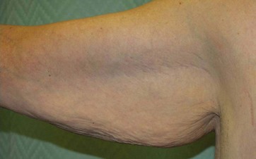
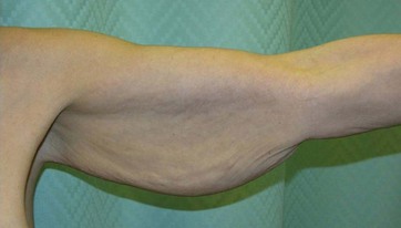
The evaluation of patients presenting for brachioplasty involves consideration of how much residual fat is present and the looseness of the skin. One of the first considerations will be whether there is enough laxity to justify a scar from elbow to axilla or longer (Figs 3.1 and 3.2). Patients with minimal looseness may benefit from looking at pictures of patients who have undergone brachioplasty in order to understand the length of the scars and the typical “rope-like” appearance of the scar near the elbow. Some clinicians have advocated drawing the proposed scar on the patient so they can live with the proposed scar for a while and see if it is something that they can accept.
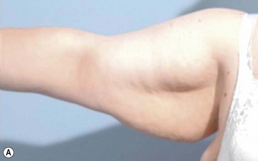
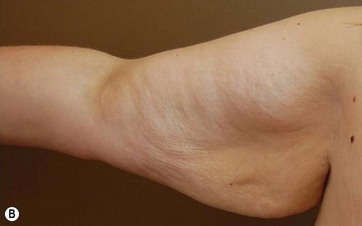
The second type of patient is the opposite – the patient who has had minimal deflation of their arms. These patients will not have a satisfactory result from a brachioplasty without deflation. Attempts to perform a brachioplasty will lead to an arm still very full, but now with the addition of a long scar. There are two schools of thought: liposuction at the time of brachioplasty, and liposuction or deflation followed at a later interval (usually several months) by the formal excision. In many cases the arms are not the patient’s first priority and the deflation can be combined with the first excisional procedure the patient is undergoing (for example, an abdominoplasty) and then the formal brachioplasty could occur at a later time. Often this is a very acceptable plan for the patient as the deflation will allow the patient to wear more normal clothes or clothes that better fit their torso even though the arms are still loose (Fig. 3.3).
The third type of patient has adequate deflation and enough looseness that they will have enough improvement in contour to justify the scar. Evaluation of these patients needs to include the skin of the forearm as well as the excess and laxity of the lateral chest wall. Rotation of the skin/fat flap in an upwards and inwards manner will somewhat improve the loose skin of the forearm, but not significantly. This upward/inward rotation will leave an excess or dog-ear in the axilla. To excise this dog-ear and to give patients an even better outcome the excess can be excised down to the inframammary fold. Patients often consider this a real benefit as they object to the “bra roll” fat.9 This fat or roll may not be visible to the plastic surgeon unless the patient has on or puts on their bra during the consultation. Even male weight loss patients may benefit from this extension down the chest wall as many male shirts now are more body revealing and a smooth contour in this area is desirable. Even in these patients who will have a significant improvement in contour it is advisable to show them pictures of brachioplasty patients so that they are aware of the length of the scars, the scar position, and the length of time that it will take the scars to mature.
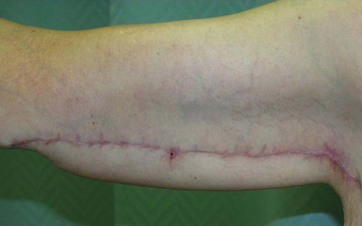
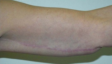
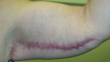
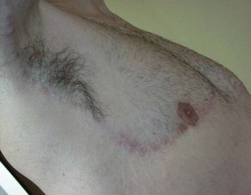
Preoperative consultation not only should include discussions as to the improvement to be seen in the arms but also the limits. Many patients are concerned about the lower arm. Often the lower arm exhibits laxity but not enough looseness to justify extending the incision below the elbow. Many patients who have experienced extreme weight loss will always have more laxity to their skin and no surgical procedure to date will improve this. The scar of a brachioplasty needs to be discussed in detail with patients. In general, the scar closest to the axilla will do the best while the scar closest to the elbow will do the worst (Figs 3.4 and 3.5). This is, of course, opposite to what the patient (and surgeon) wishes. Although the scar close to the elbow will eventually settle down and become flatter, it is often a long process and may require not only a lot time but topical therapy such as taping or even steroid injections. In some cases the scars are thick and wide (Fig. 3.6).
Surgical Technique
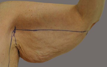
The markings for a brachioplasty should be done with the patient in the standing position (Fig. 3.7). Patients should hold their arms at 90° to their body with the elbow flexed. A horizontal line should be drawn from the axilla to the elbow marking the desired position of the final incision line along the bicipital groove. The high point of the axilla should be marked. This will represent the high point position of the flap to be excised from the upper arm as it is rotated up and in. A horizontal line should be marked, which corresponds to the inframammary fold, to mark the end of the lateral chest excision. The excess tissue of the back should be pulled forward and then a vertical line drawn, which now represents the desired position of the final incision line along the lateral chest. The excess skin and fat of the chest wall should then be pinched, utilizing the marked posterior line as the final extent of the excision. Care should be taken to observe the breast to make sure that the breast tissue is not pulled posterior and the lateral aspect of the breast is not violated. The excess tissue to be excised is then marked. This excision of the lateral chest wall tissue allows the skin and fat of the upper arm to be rotated up and in and the resultant dog-ear to be excised along the lateral chest wall.
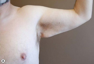
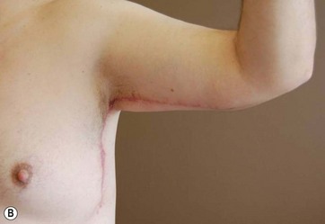
For female patients, the excision within the axilla will include all or most of the hair-bearing skin. For male patients this needs to be modified as the normal male has hair in his axilla (Fig. 3.8). The incision can be moved posterior to leave behind hair-bearing skin, which also helps camouflage the scar.
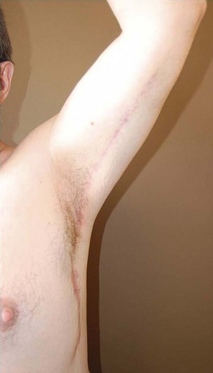
For massive weight loss patients a Z plasty has not been found to be necessary in the axilla. The incision and eventual scar in the axilla will recruit some of the residual laxity from the surrounding upper thorax area, which adds to the eventual cosmetic outcome of the procedure (Figs 3.9 and 3.10).
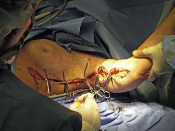
The procedure is done with the patient in the supine position with both arms extended. The arms should be prepped circumferentially to allow for movement and the lateral chest prepped and draped as well. The incision is made along the markings and dissection is carried down to the fascia of the arm musculature. At this juncture the loose inferior skin and fat is elevated off the arm musculature dissecting towards the inferior aspect of the arm. The skin and fat is then rotated up and in, in order to improve looseness around the elbow as well as remove the excess of the lower upper arm. This maneuver creates a dog-ear in the axilla, which is then taken out with the upper lateral chest excision. Once the excess skin and fat is elevated, the assistant should hold the upper skin and fat in place so that the incision lies in the desired position along the biciptial groove. The excess skin and fat can then be marked for excision without any fear of inability to close the wound. The excess of the upper lateral chest is excised directly using the preoperative marking. Once the excess skin and fat is excised then the wound is closed over a closed suction drain which is exited through the inferior portion of the lateral chest excision. Closure can be done in a single layer. Use of barbed sutures can make the closure quicker (Fig. 3.11). If cyanoacrylate is used to seal the wound then no dressings are needed. Many surgeons have found that patients do well without compression garments. Compression garments may pinch the arm skin, especially around the axilla and can in some cases cause areas of uneven swelling. A drain may be placed and, if it is used, the drain should exit along the chest wall for the patient’s comfort.
1 American Society for Aesthetic Plastic Surgery. Online. Available from http://www.surgery.org/sites/default/files/Stats2010_1.pdf (accessed 8 March 2012)
2 Correa-Inturraspe M, Fernandez JC. Dermatolipectomia braquial. Prensa Med Argent. 1954;34:24–32.
3 Lockwood T. Brachioplasty with superficial fascial system suspension. Plast Reconstr Surg. 1995;96:912–920.
4 Lockwood T. Brachioplasty with superficial fascial system suspension. Plast Reconstr Surg. 1995;96:912–920.
5 Aly A, Cram A. Brachioplasty. In: Aly A, ed. Body Contouring after Massive Weight Loss. St. Louis: QMP, 2006.
6 Strauch B, Greenspun D. Approach to the arm after weight loss. In: Rubin JP, Matarasso A. Aesthetic Surgery after Massive Weight Loss. Oxford: Elsevier, 2007.
7 Samra S, Sawh-Martinez RM, Kiu Y, et al. Plast Reconstr Surg. 2010;126:77.
8 Rubin JP, Michaels J. Correction of arm ptosis with a medial bicipital scar. In: Strauch B, Herman C. Encyclopedia of Body Sculpting after Massive Weight Loss. New York: Thieme, 2010.
9 Downey S, Gross J. Lateral thoracic excisions in the post massive weight loss patient. Clin Plast Surg. 2008;35(1):115–120.


