Brachial plexus anatomy

Published on 27/02/2015 by admin

Filed under Anesthesiology

Last modified 22/04/2025

Print this page

rate 1 star rate 2 star rate 3 star rate 4 star rate 5 star
Your rating: none, Average: 0 (0 votes)

This article have been viewed 1925 times

CHAPTER 14 Brachial plexus anatomy

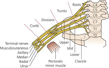
Before selection of technique, it is important to have a thorough understanding of brachial plexus anatomy. The plexus is composed of roots, trunks, divisions, cords, and branches (Fig. 14.1). The brachial plexus stems from the ventral rami of the C5 to T1 nerve roots in the majority of individuals. Approximately 15% of patients may have contributions to the brachial plexus from the C4 or T2 nerve roots, creating a ‘prefixed’ or ‘postfixed’ plexus (see Fig. 15.9). The cervical roots emerge from the intervertebral foramina and lie on a sulcus on the vertebral transverse processes between the anterior and posterior tubercles to which the scalene muscles are attached. Immediately lateral to the transverse processes of the cervical vertebrae, the nerve roots are sheathed in the prevertebral fascia. The nerve roots then travel between the scalene muscles and form three trunks (upper, middle, and lower). The interscalene groove is a palpable surface anatomy depression between the anterior and middle scalene muscles; it allows clinicians easy and reliable access to the roots and trunks of the plexus. The roots are posterior to the vertebral artery – an important anatomic relation for the interscalene block.

The nerve roots of the plexus, as they exit the intervertebral foramina, typically comprise a single large fascicle, surrounded by a scant amount of epineurium. These then divide and recombine along the length of the plexus until, eventually, most fascicles have been segregated into largely motor or sensory axons, bound for a particular area or structure. The number of fascicles increases and their cross-sectional area decreases, from proximal to distal in the plexus. At the same time, the amount of connective tissue of the epineurium progressively increases from proximal to distal.

The dorsal scapular nerve (to rhomboids) and the long thoracic nerve (to serratus anterior) arise from the C5 and C5, 6, and 7 roots, respectively. The upper (C5, 6) and lower (C8, T1) pairs of roots merge to form the upper and lower trunks of the brachial plexus, while the middle root (C7) continues as the middle trunk. The lower trunk is smaller than the others, and is frequently derived entirely from the eighth cervical nerve. The nerve to subclavius arises from the roots of C5 and C6, where they join to form the upper trunk, which also gives off the suprascapular nerve (to supra- and infraspinatus muscles and the shoulder joint). The trunks are formed in the lower part of the posterior triangle of the neck, between the sternocleidomastoid and trapezius muscles and above the middle third of the clavicle. The trunks travel to the margin of the first rib and divide into anterior and posterior divisions.

Relations

In the neck, the brachial plexus lies in the posterior triangle, being covered by the skin, platysma, and deep fascia; it is crossed by the supraclavicular nerves, the inferior belly of the omohyoid, the external jugular vein, and the transverse cervical artery. It emerges between the anterior and medial scalene muscles; its upper part lies above the third part of the subclavian artery, while the trunk formed by the union of the eighth cervical and first thoracic is placed behind the artery. The plexus next passes behind the clavicle.

Each trunk divides into an anterior and a posterior division behind the clavicle. Divisions then form three cords as they pass under the clavicle and around the humeral head, where they are joined by the axillary artery. The cords are labeled as the lateral, medial, and posterior cords; they are named as such based on the relative anatomic position to the axillary artery. At the outer border of the first rib, the upper two anterior divisions unite to form the lateral cord, the anterior division of the lower trunk runs on as the medial cord, while all three posterior divisions unite to form the posterior cord. The lateral and medial cords provide ventral innervation to the upper limb, with the posterior cord providing dorsal innervation. The cords enter the axilla from above the axillary artery and lie lateral, medial, and posterior to the middle part of the artery, behind the pectoralis minor muscle. The cords divide at the border of the pectoralis minor muscle into five major peripheral nerves, which provide innervation to the upper extremity. The five nerves of the brachial plexus are the axillary, musculocutaneous, radial, median, and ulnar nerves.

The cords and the artery are surrounded by a thin fascial sheath called the axillary sheath. The sheath is a collection of connective tissue surrounding the neurovascular structures of the brachial plexus. It is a continuum of the prevertebral fascia, which invests the scalene muscles in the neck. The sheath is a multicompartmental structure formed by thin layers of fibrous tissue surrounding the plexus in thin membranes and extending inward to create discrete fascial septae. Nerves are thus enmeshed in this tissue rather than lying separate and distinct. These compartments may limit the circumferential spread of injected solutions, thereby requiring separate injections into each compartment for optimal nerve block. However, proximal connections between compartments have been identified, which may account for the success of single-injection techniques.

Lateral and medial pectoral nerves from their respective cords supply the pectoralis major and minor. The musculocutaneous nerve from the lateral cord enters the coracobrachialis, supplying it and going on to supply the biceps and brachialis; it becomes the lateral cutaneous nerve of the forearm at the lateral side of the biceps tendon at the elbow. Lateral and medial roots from their respective cords unite to form the median nerve, which crosses in front of the brachial artery at the middle of the arm and lies medial to the artery at the elbow. The ulnar nerve arises from the medial cord between the axillary artery and vein, and passes down the medial aspect of the arm to lie behind the base of the medial epicondyle at the elbow. The radial nerve from the posterior cord passes posteriorly through an intermuscular space to spiral round the back of the humerus and enter the cubital fossa, where it lies in a deep plane between the brachioradialis and brachialis. The radial nerve gives off the posterior cutaneous nerve of the arm in the axilla, and the lower lateral cutaneous nerve of the arm and posterior cutaneous nerve of the forearm at the back of the humerus. The posterior cord also gives off two subscapular nerves (to subscapularis and teres major), the thoracodorsal nerve (to latissimus dorsi), and the axillary (circumflex humeral) nerve; the latter winds round the back of the humerus to supply the deltoid and a small area of skin at the upper lateral part of the arm.

Branches of the brachial plexus

The branches of the brachial plexus may be arranged into two groups: those given off above and those below the clavicle.

The radial nerve

The radial nerve (C5 to T1), the largest branch of the brachial plexus, is the continuation of the posterior cord of the plexus. It descends behind the first part of the axillary artery and the upper part of the brachial artery, and in front of the tendons of the latissimus dorsi and teres major. It then winds around from the medial to the lateral side of the humerus in a groove between the medial and lateral heads of the triceps. It pierces the lateral intermuscular septum and passes between the brachialis and brachioradialis to the front of the lateral epicondyle, where it divides into a superficial and a deep branch.

The deep branch of the radial nerve

The deep branch of the radial nerve winds to the back of the forearm around the lateral side of the radius between the two planes of fibers of the supinator, and continues downward between the superficial and deep layers of muscles to the middle of the forearm. Diminished in size, it descends as the dorsal interosseous nerve on the interosseous membrane, in front of the extensor pollicis longus, to the back of the wrist, where it presents a gangliform enlargement from which filaments are distributed to the ligaments and articulations of the wrist.

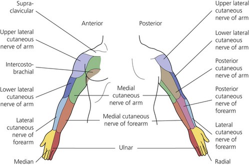
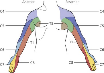
The sensory (Figs 14.2 and 14.3) and motor innervation of the upper limb is clinically important. Knowledge of sensory innervation helps determine which cutaneous nerve distributions within a surgical field require blockade. Motor innervation is clinically relevant as a means of matching a peripheral nerve stimulation response to a particular nerve being stimulated. As the arm has multiple innervation, assessment of block efficacy is best achieved by assessing function unique to each nerve. It is important to remember that significant variation in the structure of the brachial plexus occurs, with seven major configurations described.

The approaches to the brachial plexus include the interscalene, supraclavicular, infraclavicular, axillary, and midhumeral approaches. In relation to the brachial plexus anatomy, the interscalene block is performed at the level of the trunks, whereas the supraclavicular block is performed where the divisions are transitioning into cords. The infraclavicular block is performed at the proximal cord level and the axillary block is performed where the terminal nerves emerge. The midhumeral approach occurs well after the peripheral nerves have been formed.

Knowledge of anatomy remains the cornerstone of regional anesthesia. Variations and anomalies of the upper extremity that may impact on the performance of brachial plexus anesthesia include abnormalities of muscles, vessels, and nerves. Newer imaging modalities have allowed improved characterization of anatomic relationships for successful nerve blockade in various positions along the brachial plexus and its terminal nerves. Real-time ultrasonography is particularly useful in characterizing local anatomy, demonstrating nerve positions, and detecting anatomic variation that may affect block success or compromise patient safety.