Bone marrow aspiration and trephine biopsy

Published on 03/04/2015 by admin

Filed under Hematology, Oncology and Palliative Medicine

Last modified 22/04/2025

Print this page

rate 1 star rate 2 star rate 3 star rate 4 star rate 5 star
Your rating: none, Average: 0 (0 votes)

This article have been viewed 1867 times

53

Bone marrow aspiration and trephine biopsy

The indications for performing bone marrow aspiration and trephine biopsy procedures have previously been discussed (p. 19). In this section the practical aspects of obtaining these samples are outlined. More detailed accounts can be found in books of practical procedures, but ultimately the only way to perfect techniques is to practise under expert supervision.

Although the anterior iliac crest is occasionally preferred, most operators get the best specimens from the posterior iliac crest. The sternum is now rarely used. This is, in part, due to the small risk of causing catastrophic damage to the mediastinum, but mainly because it is not possible to obtain a trephine biopsy. Only the posterior iliac crest approach is described here.

Bone marrow aspiration

As for all procedures the sequence of events should be explained to the patient, reassurance given and consent obtained. A degree of discomfort should be acknowledged but it should be emphasised that this is transitory. In most adults, local analgesia is adequate but sedation is considered where patients are unusually anxious. A general anaesthetic is the norm in children. A clean, no touch technique is mandatory and operators should wear gloves. Stringent asepsis is needed in immunosuppressed cases.

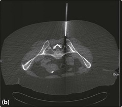
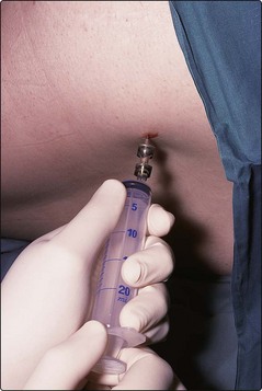
The patient lies in the left or right lateral position and the skin over the posterior iliac crest is cleaned with antiseptic prior to screening with sterile drapes. The crucial next stage is to properly identify the bony landmarks (Fig 53.1). This is straightforward in most patients but can be problematic in obese subjects. If there are real difficulties in locating the posterior iliac crest then the anterior crest or the sternum may be considered or the procedure may be performed under CT guidance. A local anaesthetic is infiltrated into the skin and then down to the periosteum. Before use it should be checked that the marrow aspirate needle stylet is easily withdrawn and the guard is removed (this is only required for sternal aspirates). The needle (Fig 53.2) is inserted through the skin and subcutaneous tissues at the site of local anaesthetic infiltration until the periosteum is encountered. It is pushed through the periosteum with a deliberate screwing motion (alternating clockwise and anti-clockwise) – a ‘give’ is felt as the marrow cavity is entered. The stylet is withdrawn and a syringe attached to the needle (Fig 53.3). Approximately 0.5 mL of marrow is aspirated into the syringe. The patient should be warned that this stage often causes pain but that it is momentary.
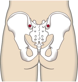
Marrow aspirate smears must be made promptly at the bedside before the marrow clots. If a larger volume is needed for tests such as cytogenetics and immunophenotyping, it is best to use a second syringe as large samples dilute the marrow with peripheral blood and reduce the quality of the morphological preparations. If it proves difficult or impossible to aspirate marrow it is worth replacing the stylet and carefully advancing or retracting the needle a short distance before repeating aspiration. It is important to remember that a ‘dry tap’ can result from marrow pathology (particularly fibrosis or solid malignancy) and is not always caused by poor technique.

Once the aspirate needle is withdrawn, firm pressure is applied to the site for a few minutes and then a sterile dressing or plaster used as protection. The patient lies on his back for 15 minutes to ensure a period of recuperation and that further light pressure is applied to the puncture site. Outpatients should probably be observed for at least an hour before being allowed home (more if sedated). Troublesome haemorrhage from the site is rare but it is sensible to correct a severe coagulation defect before undertaking the procedure. Thrombocytopenia alone is generally not a problem.

Patients often ask how quickly the ‘results’ will be available. Aspirate slides can be processed for microscopy (see p. 19) within a few hours but most ancillary tests (Table 53.1) take longer.

Bone marrow trephine biopsy

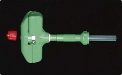
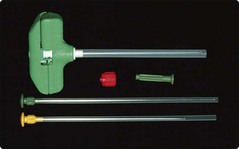
In practice the trephine procedure is usually performed immediately following the aspirate at the same site. It is helpful to enlarge the aspiration puncture site slightly with a scalpel blade. There is sometimes more prolonged discomfort than in the aspirate procedure and sedation is indicated in anxious adults, and a general anaesthetic is necessary in children. A number of different disposable needles are available – the Jamshidi type is illustrated in Figure 53.4. Smaller needles are available for paediatric use.

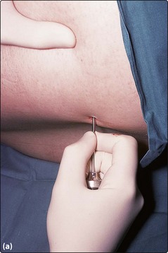
It is important to ensure that the device is complete and that the stylet can be easily withdrawn. The trephine needle is inserted in a similar fashion to the aspirate needle through the periosteum and approximately 0.5 cm into the cortex (Fig 53.5a) – when properly inserted the needle should easily support its own weight. The stylet is removed prior to advancing the needle 2–3 cm using the same oscillatory movement. The needle is aimed towards the anterior iliac crest. The method for breaking off the biopsy varies with the needle used. Some have devices designed to grip the biopsy and ensure its retention. The needle is then withdrawn taking care not to catch the skin and lose the biopsy in subcutaneous tissue. A special blunt probe is provided to push the biopsy out of the needle. The probe is inserted (with great care to avoid injury to the operator) at the sharp end of the needle so as not to traumatise the sample.

If the aspirate is a ‘dry tap’ it is worthwhile gently dabbing the trephine biopsy onto a glass slide before putting it into histological fixative. This ‘touch preparation’ is not useful for subtle morphological diagnosis but can permit rapid identification of malignant infiltration. It usually takes several days to process the trephine biopsy. Aftercare is the same as for the aspirate, although as it is a slightly more invasive procedure the patient also having a trephine may require a longer period of recuperation. Nevertheless, trephine biopsies are routinely performed in the outpatient clinic.

Bone marrow harvesting

Bone marrow can be harvested from a patient (for autologous stem cell transplantation) or from a donor (for allogeneic stem cell transplantation). The procedure is performed under a general anaesthetic, the marrow being collected from the iliac crests using multiple punctures with specialised harvest needles. Normally, approximately 1 litre is harvested from an adult in under an hour. Donors are hospitalised for around 48 hours. Serious side-effects are rare but some short-lived discomfort over the aspiration sites is common. The procedure is now undertaken less often as peripheral blood stem cells are more commonly used than bone marrow in both autologous and allogeneic transplants (see page 56).