Blood transfusion

Published on 11/04/2015 by admin

Filed under Surgery

Last modified 22/04/2025

Print this page

rate 1 star rate 2 star rate 3 star rate 4 star rate 5 star
Your rating: none, Average: 0 (0 votes)

This article have been viewed 1474 times

9

Blood transfusion

Principles of blood transfusion

The ability to safely transfuse blood and blood products revolutionised outcomes from major trauma. It also enabled extraordinary advances in areas of surgery involving heavy blood loss such as arterial reconstruction, open-heart surgery and organ transplantation.

Nevertheless, blood transfusion carries a range of potential hazards, including transfusion reactions, transmission of infection, clerical errors leading to incompatible transfusion, and potential immunosuppression in cancer patients. For replacing blood loss, stored blood has the advantage over gelatin and electrolyte solutions that it remains within the vascular compartment, although most ‘blood’ for transfusion consists of concentrated red cells with platelets and clotting factors removed. Potentially lethal side-effects mean that a decision to transfuse blood or blood products must be based on clear indications and after considering alternatives (see Reducing the need for bank blood transfusion, p. 120). The types of transfusion components available and the general indications for their use are summarised in Box 9.1.

Box 9.1   Types of transfusion and indications for their use

Whole blood

Very rarely used because of the demand for separate blood components. Whole blood carries greater risks of adverse reactions owing to the presence of leucocytes

Packed or concentrated red cells (the most common transfusion product)

For use in substantial haemorrhage and severe anaemia. Whole blood is collected then centrifuged to separate the red cells (and platelets and plasma). Packed/concentrated red cells are then suspended in a preservative solution ready for reinfusion. A bag of red cells is approximately 300 ml and raises the haemoglobin concentration by approximately 1 g/dl

Human albumin solution, available as 4.5% and 20% solutions

Albumin is expensive and is derived from human donor blood. In terms of efficacy, mortality and cardiorespiratory function, there is little evidence that albumin solutions are better than plasma substitutes or normal saline for resuscitating patients with traumatic or septic shock or requiring large perioperative volume replacement

Transfusion of albumin solutions should probably be restricted to patients with hypoproteinaemic oedema with nephrotic syndrome or with ascites in chronic liver disease. Other uses are difficult to justify in the absence of scientifically proven benefit

Fresh-frozen plasma (FFP)

Plasma is separated from fresh whole blood and then frozen. FFP contains near normal amounts of all clotting factors and other plasma proteins. It should be blood group compatible when possible and should not be used simply for volume replacement. FFP is often used to replace clotting factors exhausted during major haemorrhage (due to a combination of consumption of clotting factors by attempted haemostasis and the lack of clotting factors in transfused blood). This is likely when blood loss exceeds 1.5 times the blood volume, and the loss has been replaced rapidly with red cells and crystalloids or colloids. Clotting studies usually demonstrate a coagulopathy

FFP is also used to replace deficiencies of coagulation factors when there is continued bleeding and the necessary specific factor concentrates are unavailable. This may occur in liver disease, thrombotic thrombocytopenic purpura (TTP) and acute disseminated intravascular coagulation (DIC)

Platelet concentrates

Used for platelet exhaustion during major haemorrhage (e.g. ruptured abdominal aortic aneurysm) and in thrombocytopenia. Indicated if the platelet count is < 50 × 109/L, or massive blood loss is occurring. Platelets should be avoided in autoimmune platelet disorders except in the presence of life-threatening haemorrhage

Cryoprecipitate, fibrinogen and other specific clotting factor concentrates

Used for various specific coagulation deficiencies, e.g. severe hypofibrinogenaemia, haemophilia. These should only be used in consultation with a haematologist

Plasma substitutes

These are solutions of macromolecules with colloid osmotic pressure and viscosity characteristics similar to plasma. Gelatin solutions (e.g. Haemaccel, Gelofusine) and etherified starch solutions (e.g. hetastarch) are used for initial restoration of circulating volume in haemorrhage or burns and to maintain volume, blood pressure and renal perfusion intraoperatively where blood transfusion is not indicated

Laboratory aspects of blood transfusion

Blood grouping and compatibility testing

Transfusion of ABO incompatible blood may be fatal. Transfusion practice aims to minimise this risk and involves two steps: first, determining the patient’s ABO and Rhesus groups; second, screening the patient’s blood (and each unit of donor blood) for antibodies. Traditionally, each unit of group-compatible donor blood has been directly cross-matched against the patient’s serum to ensure compatibility. Fortunately, in donor blood shown to be antibody-free, 97% is completely compatible with a group-matched recipient and can safely be transfused without cross-matching. Thus grouped and antibody screened blood can be supplied quickly from a prepared pool, rather than individually cross-matching it. This saves time and money and most hospitals are adopting this practice.

After transfusion of about 10 units of blood, the patient’s antibodies are so depleted that group-compatible blood can safely be given safely. In an emergency (e.g. obstetric haemorrhage) where group-compatible blood is unavailable, group O, Rh-negative blood can be given with comparative safety.

Storage and useful life of blood

Blood and blood products have exacting requirements for preserving their quality. Their usefulness is easily impaired if handled inappropriately.

Packed red cells are stored between 2°C and 6°C, and have a shelf life of 35 days. Improved preservative solutions mean red cell quality deteriorates little during storage but pH changes occur and potassium leaches from the cells. The latter is only significant in neonates undergoing exchange transfusion or in patients with renal failure.

To ensure vitality, blood should be retained in the designated blood refrigerator until just before use. Blood removed for more than 30 minutes and not transfused should be returned to the laboratory for disposal because of the risk of bacterial proliferation. As soon as it becomes clear that blood products will not be required, the laboratory should be informed so they can be made available for other patients. Empty blood packs should be retained for 48 hours for examining and testing in the event of a transfusion reaction.

Blood transfusion in clinical practice

Blood transfusion and elective surgery

Attitudes to transfusion in elective surgery are becoming more conservative. Blood is expensive and its use carries risks. In elective surgery, patients fall into one of three categories: transfusion not anticipated (e.g. hernia repair), transfusion possible but unlikely (e.g. cholecystectomy), and transfusion probable (e.g. major arterial reconstruction). For patients in the second category, blood should be sent for ABO and Rhesus grouping and antibody screening, and serum retained for compatibility testing if required later (‘group and save’). For patients in the third category, an appropriate number of units is requested for the operation; many hospitals operate a maximum blood ordering schedule (MSBOS) to ensure appropriate ordering. The blood is prepared a day or so before operation. If the antibody screen is negative and criteria for electronic cross-match are met, a serological cross-match is unnecessary and group-specific blood can be immediately issued. If a further transfusion is required more than 72 hours later, a new blood sample must be tested for new antibodies.

To prevent the disaster of giving blood to the wrong patient, transfusion blood samples must be scrupulously labelled in the presence of the patient. Immediately before transfusion, the label of each unit of blood must be checked against the identity and blood group of the patient (along with any compatibility slip).

Volume and rate of transfusion

The volume of blood required and the rate of transfusion depend on age, the indications for transfusion and the patient’s general and cardiovascular condition.

Volume and rate in haemorrhage

Haemorrhage can be classified according to rate and volume of loss. Loss of less than 1500 ml (30% of blood volume) normally requires only crystalloids/colloids except in pre-existing anaemia. Loss of 30–40% of blood volume requires red cell transfusion, and more than 40% loss, equivalent to 2 L in an adult, requires rapid volume replacement with crystalloids/colloids followed by urgent provision of blood and blood products according to the local Massive Blood Loss protocol. Massive blood loss is defined as haemorrhage of 2 L blood in 3 hours, 4 L in 24 hours or > 150 ml blood per minute. Urgent provision of blood products for life-threatening haemorrhage needs focus and good communication between transfusion laboratory and clinicians.

A sample massive loss protocol is to follow immediate resuscitation with 4 units of red cells via a blood warmer. O-negative blood can be given if the blood group is unknown, followed by group-specific or cross-matched red cells. If bleeding continues, further red cells should be accompanied by fresh-frozen plasma (FFP) to prevent coagulopathy. The ideal ratio of red cells to FFP is unclear but is about 4 : 4 or 4 : 3. Platelet concentrates are given to maintain levels at > 100 × 109/L. Repeat coagulation screens are needed after every 4 units to determine the need for other blood products, e.g. cryoprecipitate for fibrinogen levels. Hypothermia and hypocalcaemia should be corrected. If bleeding persists despite adequate blood product support, recombinant activated factor VII is occasionally recommended.

Reducing the need for bank blood transfusion

Deaths and serious reactions still occur from incompatible transfusions (particularly when blood products are administered under general anaesthesia). In addition, serious infections such as hepatitis, malaria, HIV and variant Creutzfeldt–Jakob disease (vCJD) can be transmitted despite careful screening of donors and donations. The risk increases proportionate to the number of units transfused and can be reduced by critical scrutiny of the indications for transfusion.

Autologous transfusion

Transfusion methods involve ‘recycling’ the patient’s own blood (autologous transfusion) by one of the following methods:

Acute normovolaemic haemodilution (ANH)

This involves collecting whole blood from the patient immediately before surgery and replacing it afterwards if necessary. At collection, normovolaemia is restored with colloid or crystalloid. This allows autologous transfusion without the disadvantages of pre-donation or need for expensive intraoperative collecting and cell washing equipment. In adults without cardiac disease, up to 2 litres of blood can safely be removed.

The procedure reduces the venous haematocrit and increases cardiac output. When about 300 ml of low-haematocrit blood has been lost at operation, reinfusion is begun. The procedure is inexpensive, convenient and flexible as regards operation scheduling. The blood is fresh and contains functioning platelets and clotting factors. The collected blood is retained in the operating theatre, minimising the risk of clerical error. The technique has been used extensively in cardiac and vascular surgery but other types of surgery with a potential blood loss of more than 20% of blood volume could benefit.

Intraoperative cell salvage (IOCS)

UK government directives and national guidelines have endorsed intraoperative cell salvage as effective and appropriate in cardiac, vascular and orthopaedic surgery.

Intraoperative cell salvage involves collecting blood spilled at operation by suction, processing and reinfusion. There are two main techniques:

Hazards and complications of blood transfusion

Haemolytic reactions

If there is major ABO incompatibility, massive haemolysis may be fatal. Incompatibility of minor determinants causes a lesser degree of haemolysis. Almost all haemolytic reactions are caused by human error and incompatible transfusion. Clinical features are summarised in Box 9.2. The diagnosis is confirmed by blood tests—finding hyperbilirubinaemia and a positive Coombs’ test, and demonstrating a new antibody. With massive haemolysis, haemoglobinaemia and haemoglobinuria may occur. The transfusion must be halted immediately and the patient resuscitated. Oliguria is treated by osmotic diuresis (e.g. mannitol), sometimes aided by a loop diuretic.

Infection

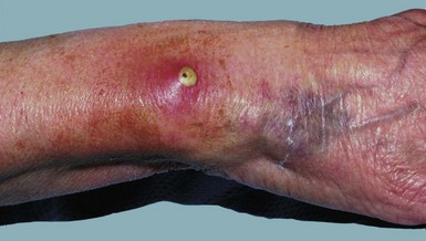
Infection may arise from three sources: the donor, contamination during blood preparation and storage, or from the giving set or cannula site (Fig. 9.1).

Infections transmitted by donor blood or blood product transfusion

Donated blood can now be screened for most significant transmissible infectious agents. Many tests rely on detecting antibodies to viruses and cannot identify disease acquired too recently for the antibody response to develop.

Donor infections are most likely when a chronic latent carrier state exists in the donor. Diseases include:

The more frequent of these infections are discussed below.

Hepatitis viruses: In carriers or infected individuals, hepatitis B virus is transmitted by any blood product. Donors have been screened for hepatitis B virus (HBV) by all transfusion services in the developed world since the late 1960s and this has dramatically reduced transfusion-related hepatitis B. Despite screening, infections occasionally occur because the surface antigen HBsAg, the first marker to appear, may be absent early after infection. For this reason, patients needing multiple transfusions or blood products should be vaccinated against hepatitis B.

Hepatitis C used to be transmitted via blood transfusion twice as often as hepatitis B. The illness caused by hepatitis C is often mild but up to half of all cases progress to chronic hepatitis (10% for hepatitis B), and 10% develop cirrhosis or hepatoma or both. Since 1991 all UK donations have been screened and the incidence of post-transfusion hepatitis C has fallen from 1.3 per 1000 units transfused to 0.1. However, the ‘window’ for HCV antigen to appear after infection can be up to 3 months.

Human immunodeficiency virus (HIV): In 1981, the HIV epidemic and its link with blood transfusion was recognised, with reports of opportunistic pneumonias in haemophiliacs. By the mid-1980s, the risk of HIV transmission via transfused blood in the USA was as high as 1 in 2500. Luckily, transmission via surgical blood transfusion has been rare in the UK. Haemophiliacs were much less fortunate, being infected via clotting factor VIII preparations, often collected abroad.

In the UK, all blood donors and donations have been screened for HIV antibody since 1985. About 5 per million donors in the UK have proved HIV positive. Current HIV antibody detection is very reliable but there is a ‘window’ lasting 1–3 months after infection when antibody may be undetectable. The most effective method of reducing risk where donors are unpaid is self-exclusion by donors if they have transmissible infections or fall into high-risk categories. When combined with a low prevalence of HIV in the donor pool (as in the UK), the risk of transmitting HIV via a screened blood or blood component donation is small but not negligible.

Contamination of blood or giving sets with microorganisms

Low-grade environmental contamination of blood packs rarely has serious consequences because of the low temperature of blood storage and the self-sterilising properties of blood. However, microorganisms may proliferate if storage conditions are poor.

Transfusion of bacterially infected blood products is rare but can be catastrophic, leading rapidly to death. Platelet transfusions have been incriminated most often. Gram-negative aerobic bacilli, particularly Pseudomonas and Salmonella species, have been identified. Accumulation of exotoxins or endotoxins may also lead to life-threatening complications.

Giving sets may become contaminated unless strict aseptic technique is maintained during setting-up or changing of any component of transfusion equipment. Finally, peripheral intravenous cannulae, and particularly central venous lines, may become infected by opportunistic skin commensals (e.g. coagulase-negative staphylococci) or contaminating pathogens (e.g. Staph. aureus, E. coli) causing local infection (see Fig. 9.1), bacteraemia or even systemic sepsis. Cannula sites should be inspected daily and changed at least every 72 hours. In a patient with a central venous line and pyrexia of unknown origin, the line should be removed and tested bacteriologically.

The clinical result of transfusing a bacterially contaminated donation resembles a haemolytic reaction, with fever, chills, hypotension, nausea and vomiting, oliguria and disseminated intravascular coagulation. If a patient develops these symptoms, investigations should include culture of the patient’s blood and the blood product as well as rechecking compatibility.

Delayed transfusion reactions