6 Blood Supply of the Brain
The Internal Carotid Arteries and Vertebral Arteries Supply the Brain
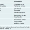
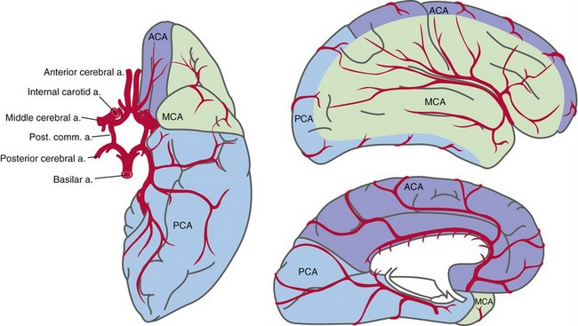
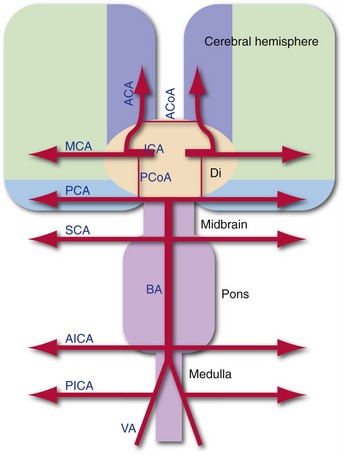
Two interconnected arterial systems provide the blood supply to the brain (Fig. 6-1). The internal carotid system of each side supplies the ipsilateral cerebral hemisphere, except for the medial surface of the occipital lobe and the medial and inferior surfaces of the temporal lobe. The vertebral-basilar system supplies those parts of the occipital and temporal lobes, as well as the brainstem and cerebellum. The supply of the diencephalon is shared by the two systems, with the vertebral/basilar system supplying most of the thalamus and the internal carotid system supplying most of the hypothalamus.

The Internal Carotid Arteries Supply Most of the Cerebrum
The internal carotid has two major terminal branches, the anterior and middle cerebral arteries. These are two of the three arteries that supply the cerebral cortex. On their way to their cortical areas of supply, both give rise to lots of small perforating or ganglionic arteries that supply most of the hypothalamus, basal ganglia, and internal capsule. (A comparable set from the vertebral-basilar system supplies the thalamus and brainstem.) The anterior and middle cerebral arteries have large terminal areas of distribution that are suggested to some extent by the name of each artery (Fig. 6-2).
The Vertebral-Basilar System Supplies the Brainstem and Parts of the Cerebrum and Spinal Cord
The two vertebral arteries fuse near the pontomedullary junction to form the single basilar artery, which ascends to the midbrain and terminates by bifurcating into the two posterior cerebral arteries. As they move along the brainstem, the vertebral and basilar arteries give off a series of branches; each has a name suggesting its terminal area of distribution (e.g., the superior cerebellar artery supplies the superior surface of the cerebellum). Along the way, each of these arteries provides perforating branches that supply the part of the brainstem it passes over. So knowing the levels at which these vertebral-basilar branches emerge (Fig. 6-3) provides a first approximation of the supply of the brainstem.
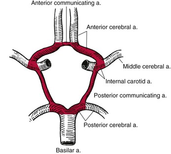
The Circle of Willis Interconnects the Internal Carotid and Vertebral-Basilar Systems
The internal carotid and vertebral/basilar systems are interconnected by a posterior communicating artery on each side. In addition, the two anterior cerebral arteries are interconnected by the anterior communicating artery. These communicating arteries complete the arterial circle of Willis (Fig. 6-4). The communicating arteries are ordinarily fairly small and not capable of carrying much blood. However, in cases of slowly developing occlusions within the circle or in arteries leading into the circle, they can enlarge and provide a major alternative pathway for blood flow.
Imaging Techniques Allow Arteries and Veins To Be Visualized
Increasing the contrast between blood and brain by any of several methods makes it possible to produce images of cerebral arteries and veins. Injecting radiopaque dyes makes blood vessels stand out when the head is examined with x-rays (THB6 Figures 6-15 and 6-16, pp. 133 and 134). Alternatively, MRI can also pick up flowing blood, even without the injection of contrast agents (THB6 Figure 6-17, p. 134).
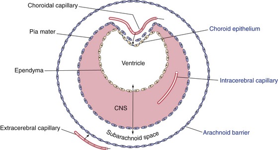
A System of Barriers Partially Separates the Nervous System from the Rest of the Body
The arachnoid barrier layer (see Chapter 4) prevents things from diffusing into subarachnoid space from outside the CNS. The choroid epithelium (see Chapter 5) regulates what gets into newly formed cerebrospinal fluid. The last part of the barrier system between the CNS and the rest of the body is an array of tight junctions between the endothelial cells of CNS capillaries. This blood-brain barrier prevents substances from diffusing out of CNS capillaries and allows the endothelial cells to regulate what gets into CNS extracellular space. Collectively these three barriers (Fig. 6-5) make up a system that separates the extracellular spaces of the CNS from the general extracellular spaces of the body. A few small sites in the walls of the third and fourth ventricles don’t have a blood-brain barrier (THB6 Figures 6-29 and 6-30, p. 143), allowing these circumventricular organs to either monitor the composition of the systemic circulation or add things to it.
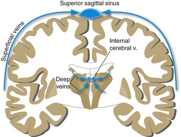
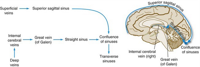
Superficial and Deep Veins Drain the Brain
Two sets of veins cooperate in the drainage of the brain (Figs. 6-6 and 6-7). Superficial veins, as their name implies, lie on the surface of each cerebral hemisphere and mostly drain upward or backward into the superior sagittal sinus (THB6 Figure 6-33, p. 145). Deep veins, in contrast, mostly drain structures in the walls of the ventricles and converge on the internal cerebral veins above the roof of the third ventricle (THB6 Figures 6-34 and 6-35, p. 146).