[level-membership-for-internal-medicine-category]
Rectal Bleeding
Rectal bleeding is a common symptom. The majority of patients with rectal bleeding have a simple condition such as haemorrhoids, but the symptoms should always be taken seriously and investigated. Rectal bleeding with a change in bowel habit and colicky abdominal pain should be regarded as due to colorectal cancer until proved otherwise.
Causes
History
Anus
Haemorrhoids
Piles occur at any age. Bleeding from piles is noted either on the toilet paper or as splashes in the toilet after defecation. Uncomplicated piles are not painful.
Fissure-in-ano
Fissure-in-ano is most common under the age of 40 years. It is quite common in children. The patient experiences pain on defecation, which may persist for minutes or hours afterwards. Constipation is usually a precipitating cause, the constipation being made worse by the fissure as the patient avoids defecation because of pain. Blood is noticed on the toilet paper or streaked on the stool.
Carcinoma of the anal canal
Carcinoma of the anal canal usually occurs in the elderly. It presents with pain on defecation and streaking of blood on the stools and blood on the toilet paper. Initially, in the early stages, it may be mistaken for a fissure-in-ano.
Trauma
There may be a history of a penetrating injury to the anal canal. Sexual abuse or homosexual practices may be relevant.
Colorectal
Carcinoma
With colonic carcinoma, the blood may be mixed with the stool. There is usually a history of accompanying change in bowel habit and colicky abdominal pain. With rectal cancer, the blood is usually streaked on the stool and there may be a history of tenesmus, i.e. a sense of incomplete evacuation of the rectum.
Polyps
The history may be similar to that of carcinoma.
Diverticular disease
Bleeding associated with diverticular disease is typically acute, massive and fresh. There may be a past history of diverticular disease.
Inflammatory bowel disease
With ulcerative colitis and Crohn’s disease, there is often sudden onset of diarrhoea with watery, brown motions containing mucus and fresh blood. There is usually colicky abdominal pain. With ulcerative proctitis, the patient may complain of tenesmus.
Ischaemic colitis
This usually occurs in the elderly. There is colicky abdominal pain associated with the passage of dark-red venous blood PR.
Angiodysplasia
The patient is usually elderly. There is bleeding PR, which may be torrential but is more often repeated small bleeds.
Irradiation proctitis or colitis
There will be a history of irradiation, often for carcinoma of the cervix. The patient passes blood and mucus PR and complains of tenesmus.
Rectal prolapse
The patient will be aware of something hanging out of the back passage that comes down on defecation. Bleeding is due to trauma.
Solitary rectal ulcer
Bleeding occurs after defecation, usually in small volumes. This may be associated with mucus discharge and a feeling of a lump in the anus.
Small bowel
Meckel’s diverticulum
This usually results in painless bleeding in young adults. The blood tends to be dark red and may on occasions have the characteristics of melaena.
Intussusception
This usually occurs in infants, but may rarely occur at any age. The child has colicky abdominal pain and draws the legs up, screams and passes a stool consisting of mixed blood and mucus (‘redcurrant jelly’ stool).
Mesenteric infarction
The patient is usually elderly, or a younger patient who has a history of heart disease (embolism). The patient develops severe central abdominal, colicky pain associated with diffuse tenderness and later collapse and shock.
Aortoenteric fistula
This usually occurs following repair of an infrarenal aortic aneurysm with a Dacron graft. The Dacron graft becomes infected, the fistula forms between the aorta and duodenum. Depending on the speed of the bleed, there may be either melaena or profuse red rectal haemorrhage with shock.
Upper gastrointestinal tract
Massive haemorrhage from the upper gastrointestinal tract, e.g. bleeding oesophageal varices, duodenal ulcer, may present with bright-red bleeding PR. This is due to extremely fast intestinal transit and the patient will always be shocked.
Others
Check for a history of anticoagulants. The patient may have a bleeding disorder. Check for bleeding from other sites or spontaneous bruising.
Examination
Anus
Haemorrhoids
There may be obvious prolapsed haemorrhoids; however, sigmoidoscopy and proctoscopy are usually required to make the diagnosis.
Fissure-in-ano
Separate the buttocks. A chronic fissure-in-ano with a sentinel pile may be seen in the midline posteriorly, or more rarely, in the midline anteriorly. Digital rectal examination, if attempted, will be extremely painful.
Carcinoma
This may show a hard ulcer in the anal canal with everted edges; however, in the early stages, carcinoma of the anal canal may be difficult to distinguish from a chronic fissure-in-ano. Biopsy should be undertaken.
Trauma
Bruising may be apparent around the anal canal. There may be a split in the skin or mucosa. Digital rectal examination will be painful.
Colon and rectum
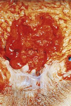
Carcinoma
An abdominal mass may be palpable. There may be signs of intestinal obstruction. Digital rectal examination may reveal a hard, irregular, ulcerating mass.
Polyps
A polyp may be palpable in the rectum. There may be no findings on physical examination.
Diverticular disease
The patient may be tender in the left iliac fossa. Often, there are no abdominal findings.
Inflammatory bowel disease
There may be a palpable abdominal mass with Crohn’s disease. There may be localised abdominal tenderness. If toxic dilatation has occurred, the abdomen will be distended and tender, and there may be signs of peritonitis if perforation has occurred.
Ischaemic colitis
Physical examination may show left-sided abdominal tenderness and the patient may be shocked.
Angiodysplasia
There may be little to find other than rectal bleeding. There are usually no abdominal signs.
Irradiation colitis or proctitis
There may be some abdominal tenderness. Digital rectal examination will reveal a granular mucosa and blood on the glove.
Rectal prolapse
There will be obvious prolapse of the rectum with ulcerated, bleeding rectal mucosa.
Solitary rectal ulcer
There are no abdominal findings. Digital rectal examination may give the impression of a polypoid swelling just inside the rectum, which may be mistaken for carcinoma. Proctoscopy will reveal redness and oedema of the mucosa and, in about 50% of patients, frank ulceration will be noted.
Small bowel
Meckel’s diverticulum
There will usually be nothing to find on abdominal examination.
Intussusception
A mass may be palpable in the right iliac fossa. Eventually, as the intussusception proceeds, the right iliac fossa becomes ‘empty’.
Mesenteric infarction
The patient may be in atrial fibrillation and this suggests embolism. There will be diffuse abdominal tenderness, later accompanied by collapse and shock.
Aortoenteric fistula
There will usually be the long midline scar of a recent aortic aneurysm repair. Otherwise, there will be little to find on abdominal examination.
Upper gastrointestinal tract
Massive haemorrhage
There may be signs of liver failure associated with a massive bleed from varices. There may be epigastric tenderness associated with duodenal ulceration.
Others
With anticoagulants there may be bleeding from other orifices, as may also occur with bleeding diatheses. Look for signs of bruising. Uraemic bleeding is often seen in patients with established uraemia and they may already be on dialysis and have an arteriovenous fistula or a CAPD tube in situ. Rectal bleeding from collagen diseases is rare. Polyarteritis nodosa is probably the most common. Bleeding is due to necrotising vasculitis and there may be signs of vasculitis elsewhere, e.g. skin.
General Investigations
■ FBC, ESR
Hb ↓ may occur in almost any type of rectal bleeding. WCC ↑ inflammatory bowel disease, ischaemic colitis, aortoenteric fistula. Platelets ↓ in bleeding diatheses. ESR ↑ carcinoma, collagen disease.
■ U&Es
Urea and creatinine raised with uraemic bleeding. The urea may be raised due to absorption of blood from the bowel. Usually in this case, the creatinine is normal.
■ LFTs
Liver failure associated with oesophageal varices.
■ Sigmoidoscopy/proctoscopy
Anorectal tumours. Haemorrhoids. Distal colitis. Solitary rectal ulcer. Biopsy.
■ AXR
Obstruction associated with carcinoma. Inflammatory bowel disease (toxic dilatation of the colon).
Specific Investigations
■ Barium enema
Carcinoma. Diverticular disease. Polyps. Inflammatory bowel disease. Ischaemic colitis.
■ Colonoscopy
Diverticular disease. Colonic tumours. Angiodysplasia. Colitis.
■ Angiography (in the acute bleeding phase)
Angiodysplasia. Bleeding Meckel’s diverticulum.
■ Labelled red cell scan
Angiodysplasia. Meckel’s diverticulum.
[/level-membership-for-internal-medicine-category][not-level-membership-for-internal-medicine-category]
Rectal Bleeding
Rectal bleeding is a common symptom. The majority of patients with rectal bleeding have a simple condition such as haemorrhoids, but the symptoms should always be taken seriously and investigated. Rectal bleeding with a change in bowel habit and colicky abdominal pain should be regarded as due to colorectal cancer until proved otherwise.
Causes
History
Anus
Haemorrhoids
Piles occur at any age. Bleeding from piles is noted either on the toilet paper or as splashes in the toilet after defecation. Uncomplicated piles are not painful.
Fissure-in-ano
Fissure-in-ano is most common under the age of 40 years. It is quite common in children. The patient experiences pain on defecation, which may persist for minutes or hours afterwards. Constipation is usually a precipitating cause, the constipation being made worse by the fissure as the patient avoids defecation because of pain. Blood is noticed on the toilet paper or streaked on the stool.
Carcinoma of the anal canal
Carcinoma of the anal canal usually occurs in the elderly. It presents with pain on defecation and streaking of blood on the stools and blood on the toilet paper. Initially, in the early stages, it may be mistaken for a fissure-in-ano.
Trauma
There may be a history of a penetrating injury to the anal canal. Sexual abuse or homosexual practices may be relevant.
Colorectal
Carcinoma
With colonic carcinoma, the blood may be mixed with the stool. There is usually a history of accompanying change in bowel habit and colicky abdominal pain. With rectal cancer, the blood is usually streaked on the stool and there may be a history of tenesmus, i.e. a sense of incomplete evacuation of the rectum.
Polyps
The history may be similar to that of carcinoma.
Diverticular disease
Bleeding associated with diverticular disease is typically acute, massive and fresh. There may be a past history of diverticular disease.
Inflammatory bowel disease
With ulcerative colitis and Crohn’s disease, there is often sudden onset of diarrhoea with watery, brown motions containing mucus and fresh blood. There is usually colicky abdominal pain. With ulcerative proctitis, the patient may complain of tenesmus.
[/not-level-membership-for-internal-medicine-category]
