Biliary Hamartoma

Published on 09/08/2015 by admin

Filed under Radiology

Last modified 22/04/2025

Print this page

rate 1 star rate 2 star rate 3 star rate 4 star rate 5 star
Your rating: none, Average: 3 (2 votes)

This article have been viewed 5080 times

 a.k.a. von Meyenburg complex

image Asymptomatic and of no clinical concern

IMAGING

• Multiple, near water density/intensity liver lesions < 15 mm in diameter

image Varied enhancement based on cystic and solid components
image No communication with biliary tree
• US: Small and well-circumscribed lesions

image Often have echogenic walls with small fluid content
• US shows much more echogenicity and fewer cystic lesions than anticipated based on prior CT or MR

TOP DIFFERENTIAL DIAGNOSES

• Autosomal dominant polycystic liver disease

image Larger, more numerous cysts in liver and other organs
• Multiple simple hepatic cysts

image Fewer cysts of varying size; no mural nodules
• Caroli disease

image Central dot sign on CECT and MR
image ERCP and MRCP: Communicating bile duct abnormality
• Multiple/solitary, small metastatic lesions

image More complex and varied in size
• Opportunistic infection (microabscesses)

image Must be considered in immunosuppressed patient with fever

DIAGNOSTIC CHECKLIST

• No further evaluation needed when seen as isolated finding in healthy, nononcologic patient
• Should be considered as likely diagnosis in setting of innumerable small, slightly complex “cysts” in healthy patient
• Lesions appear more echogenic than expected on sonography
image
(Left) Axial T2WI MR shows innumerable tiny bright foci throughout the liver image, representing biliary hamartomas. This patient also had evidence of congenital hepatic fibrosis on imaging and liver biopsy, both part of the congenital hepatic and renal fibropolycystic disease spectrum.

image
(Right) MRCP shows small spherical cyst-like lesions image 
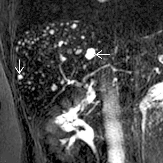
that do not communicate with the (normal) biliary tree. This feature helps to distinguish biliary hamartomas from Caroli disease.
image
(Left) Sonographic image shows innumerable tiny echogenic foci image 
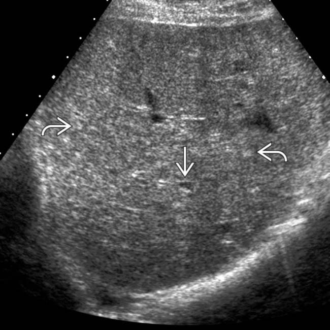
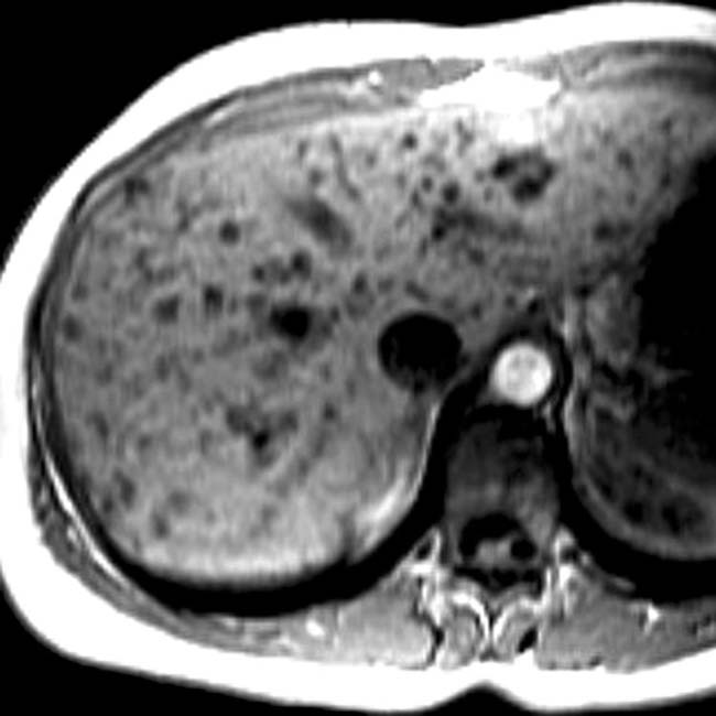
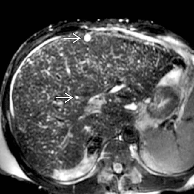
throughout the liver and 1 of ∼ 10 cyst-like lesions image, though even these have small foci of echogenicity within the wall. MR on this patient showed many more cystic-appearing biliary hamartomas.

image
(Right) The branching, angulated glands in biliary hamartomas are lined by a single layer of flattened cuboidal epithelium. These glands may expand or rupture to produce small “cysts.” There is no nuclear atypia. (Courtesy S. Kakar, MD.)

TERMINOLOGY

Synonyms

• Bile duct microhamartoma
• von Meyenburg complex

Definitions

• Uncommon benign malformations of biliary tract

IMAGING

General Features

• Best diagnostic clue

image Multiple near water density/intensity liver lesions < 15 mm in diameter
• Location

image Subcapsular or intraparenchymal in location
image Scattered throughout both lobes of liver
• Size
• Irregular spherical
• Usually multiple to innumerable

CT Findings

• NECT

image Density of lesions depends on predominance of cystic or solid component

– Predominantly cystic: Water density
– Predominantly solid (fibrous stroma): Soft tissue attenuation
• CECT

image Cystic components remain near water density

– No enhancement of contents
image Solid (fibrous stroma) components enhance

– Usually become nearly isodense to liver
image Punctate calcifications may be seen

MR Findings

• T1WI

image Hypointense (both cystic and solid lesions)
• T2WI

image Very hyperintense (cystic components)
image Intermediate intensity (solid lesions)
image Heavily T2WI: Remain hyperintense (equal to fluid)

– 
• T1WI C+

image No enhancement of cystic components

– ± nodular enhancement of fibrous nodules
• MR cholangiography (MRC)

image Markedly hyperintense lesions
image No communication with biliary tree

Ultrasonographic Findings

• Grayscale ultrasound

image Multiple small and well-circumscribed lesions
image Fibrotic parts of lesions are very echogenic

– Reflect solid and cystic components
– Fluid or cystic component is sonolucent

image ± posterior acoustic enhancement
– US shows much more echogenicity and fewer cystic lesions than anticipated based on prior CT or MR

Angiographic Findings

• Conventional

image Lesions show abnormal vascularity: Grape-like clusters of small rings

Imaging Recommendations

• Best imaging tool

image Thin section, multiplanar CECT or MR
image Multiplanar reformations are helpful
• Protocol advice

image Helical CT

– 0.65-1.25 mm collimated scans reconstructed every 2.5 mm
– View multiplanar reformations as well
image MR: Heavily T2WI/MRCP

DIFFERENTIAL DIAGNOSIS

Autosomal Dominant Polycystic Liver Disease

• Larger and more numerous hepatic cysts
• Do not communicate with each other or biliary tract
• Usually have cysts in kidneys and other organs
• ± family history

Multiple Simple Hepatic Cysts

• Rarely as numerous or limited to small size as biliary hamartomas
• US: Anechoic lesion with increased through transmission; no mural nodularity

Caroli Disease

• Multiple, small, rounded, hypodense/hypointense (T1WI), saccular dilatations of intrahepatic ducts
• Central dot sign on CECT and MR

image Enhancing tiny dots (portal radicles) within dilated cystic intrahepatic ducts
• ERCP and MRCP: Communicating bile duct abnormality

Multiple/Solitary Small Metastatic Lesions

• Metastases are more varied in size and distribution, more mural nodularity and complexity

Opportunistic Infection (Microabscesses)

• Must consider in immunosuppressed patient with fever
• Difficult to distinguish by imaging alone; need aspiration/biopsy

PATHOLOGY

General Features

• Etiology

image Congenital malformation of bile ducts

– Due to failure of involution of embryonic bile ducts
image Considered part of spectrum of fibropolycystic disease of liver and kidneys

– May coexist in multiple forms in same patient

image Autosomal dominant (or recessive) polycystic disease
image Caroli disease
image Congenital hepatic fibrosis
image Choledochal cyst
• Uncommon benign malformations of bile ducts

image Can be found in 3% of unselected autopsy population
• Usually diagnosed at imaging as incidental finding
• Biliary hamartomas are 1 variant of congenital hepatic fibropolycystic disease

image Other variants include polycystic liver disease and Caroli disease
• Increased prevalence of bile duct hamartomas in polycystic liver disease cases

Staging, Grading, & Classification

• Classification based on degree of hamartoma consistency/biliary dilatation (not widely used)

image Class 1: Predominantly solid lesions and narrow bile channels
image Class 2: Intermediate lesions (both solid and cystic foci) with mild dilatation
image Class 3: Predominantly cystic lesions with prominent dilated bile channels

Gross Pathologic & Surgical Features

• Multiple grayish-white nodules of varying sizes ranging from solid to cystic
• Subcapsular or intraparenchymal location

Microscopic Features

• Noncommunicating ducts and glands embedded within dense fibrous stroma

image May contain proteinaceous debris or bile
image Trapped bile ducts are irregularly shaped, angulated, and may be dilated to form cysts
• Rarely develop cellular atypia or cholangiocarcinoma
• Cystic hamartomas: Lined by cuboidal/flattened epithelium

CLINICAL ISSUES

Presentation

• Most common signs/symptoms

image Asymptomatic with normal liver function
• Clinical profile

image Lab data

– Normal liver function tests; normal tumor markers
image Diagnosis: Wedge or core needle biopsy and histologic exam
image No symptoms related to biliary hamartomas

– May have problems related to coexistence of Caroli disease, congenital hepatic fibrosis, polycystic disease
• Asymptomatic with normal liver function

Demographics

• Age

image Any age group (usually incidental finding)
• Gender

image M = F
• Epidemiology

image More common than realized; up to 3% of general population; M = F; any age

– Encountered more frequently due to common use of high-resolution CT, US, and MR

Natural History & Prognosis

• Usually occurs as multiple lesions; may slowly increase in size
• Complications

image Malignant transformation to cholangiocarcinoma (very rare)
• Prognosis: Excellent

Treatment

• No treatment needed for biliary hamartomas

DIAGNOSTIC CHECKLIST

Consider

• MRC to differentiate biliary hamartomas from other biliary or cystic abnormalities

Image Interpretation Pearls

• Should be considered as likely diagnosis in setting of multiple small, slightly complex “cysts” in healthy patient
• Lesions appear more echogenic than expected on US
image
(Left) Axial CECT of a 76-year-old woman with a history of renal cancer shows innumerable, tiny hypodense lesions image 
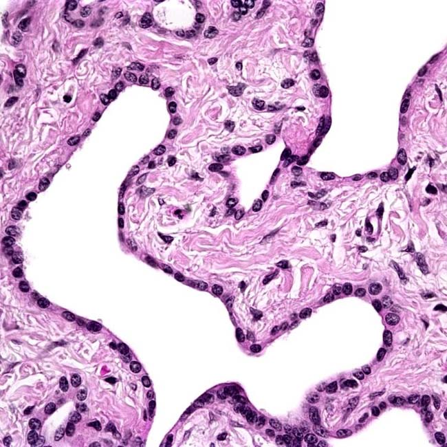
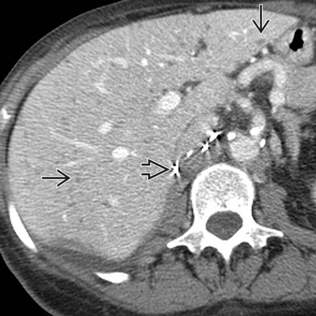
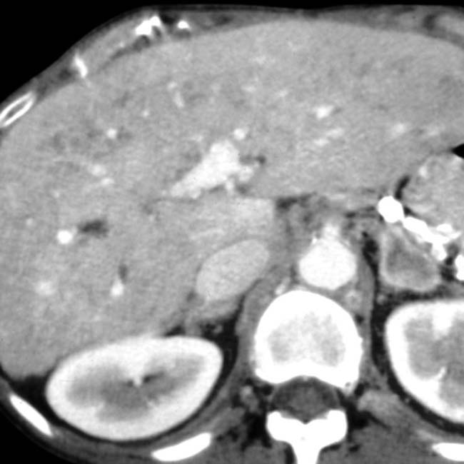
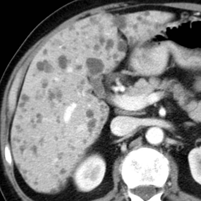
throughout the liver. Incidental note is made of surgical clips image from the prior nephrectomy. These biliary hamartomas should not be mistaken for metastases.

image
(Right) Axial CT in the same patient shows more of the lesions image 
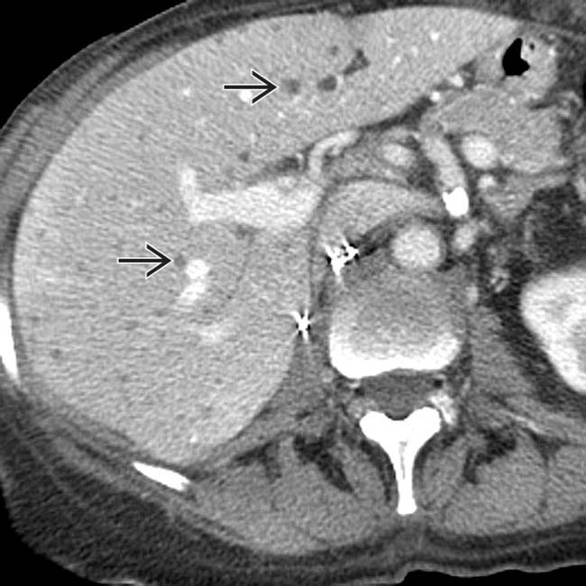
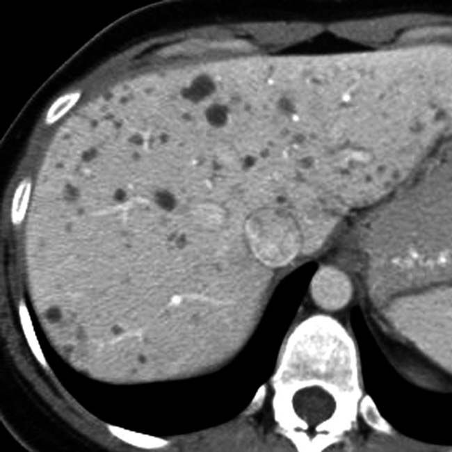
that almost certainly represent biliary hamartomas. These have remained stable for years, and the patient is not immunosuppressed.
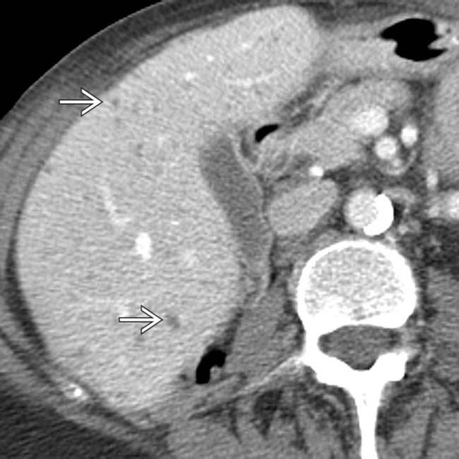
image
(Left) CT section in the same case shows the relatively uniform size of these small lesions. Careful attention to thin sections through the larger lesions shows mural nodularity image.

image
(Right) Transverse sonogram in the same patient, performed the same day as the CT scan, shows only a few “cystic” lesions image
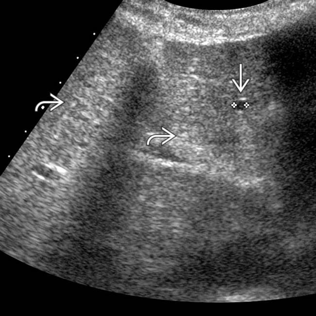
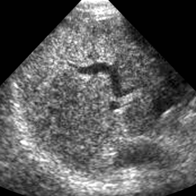
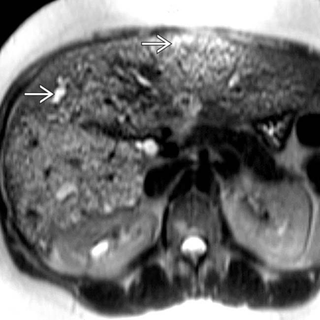
, while there are innumerable tiny echogenic foci image throughout the liver that correspond to the smaller biliary hamartomas, which presumably contain relatively more fibrous tissue and less fluid.
image
(Left) CT in a patient with chronic viral hepatitis shows innumerable small cystic lesions, some with nodularity and irregularity of the “cyst” walls image
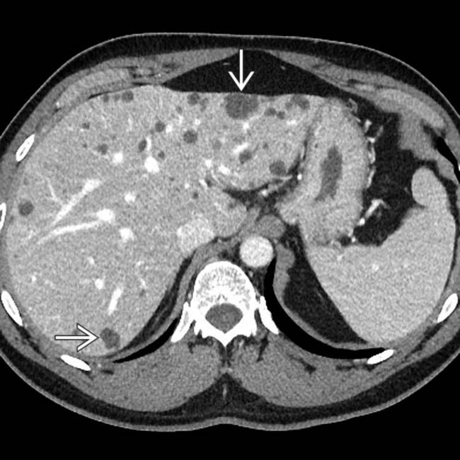
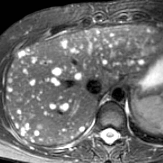
. These are typical findings for biliary hamartomas and would be a very rare (unreported) feature of hepatocellular carcinoma (HCC).

image
(Right) Transverse US of the same patient shows 1 of several echogenic foci image 
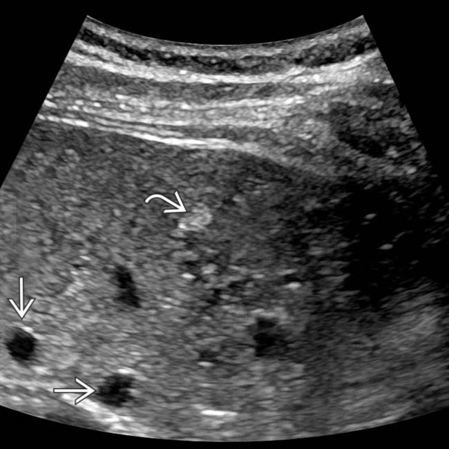
and many more cystic lesions image. None of these lesions showed interval change on subsequent imaging studies over several years and likely represent biliary hamartomas.
image
(Left) Axial CECT shows innumerable small (≤ 15 mm in diameter) hepatic lesions of near water density throughout the liver. They have remained stable and asymptomatic and are presumed to represent biliary hamartomas. The patient has no evidence of polycystic disease.

image
(Right) Axial CECT in the same patient shows more biliary hamartomas. In the absence of cancer history, biopsy proof is not necessary in most cases.
image
(Left) Axial CECT shows multiple tiny, distinct, hypodense lesions image 
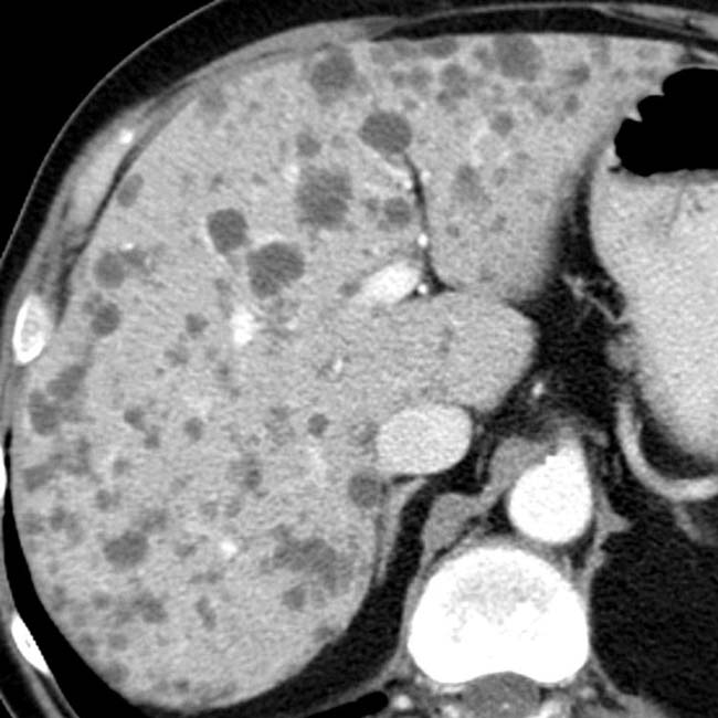
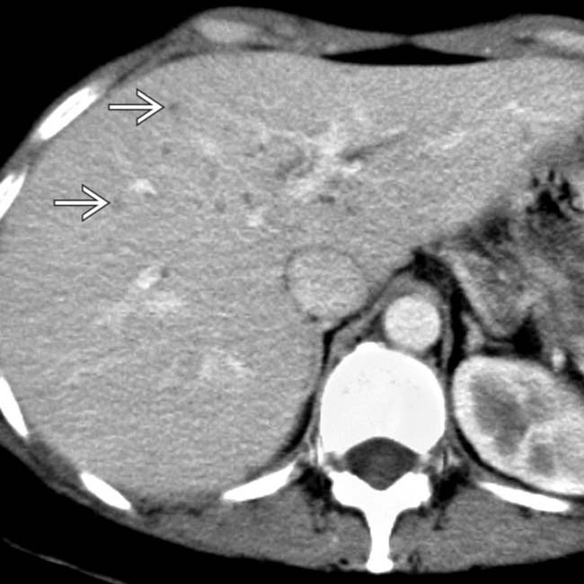
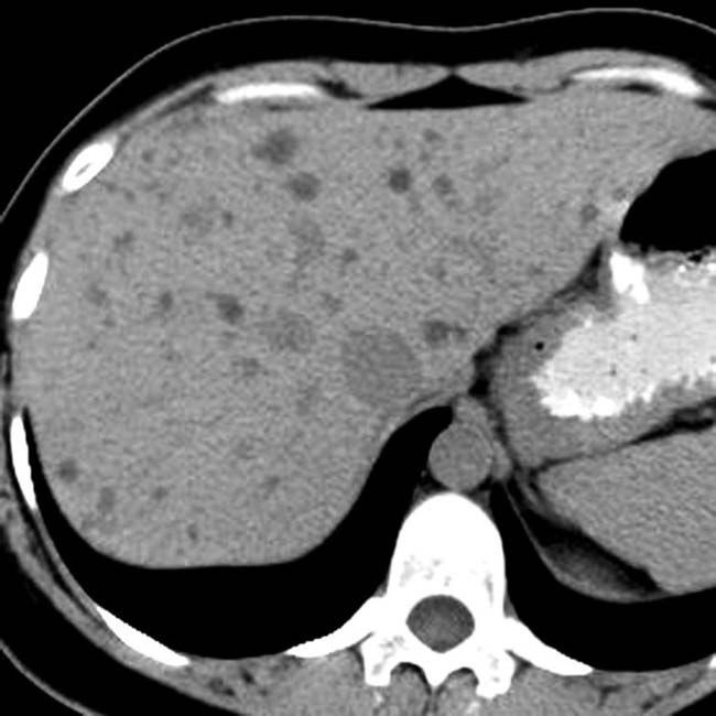
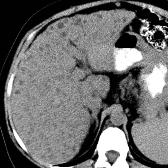
throughout the liver, too small to permit accurate density measurements. In a healthy, nononcologic patient, they likely represent biliary hamartomas.

image
(Right) Axial NECT in an asymptomatic patient shows innumerable subcentimeter, hypodense foci in both lobes of the liver. A liver biopsy confirmed the diagnosis of biliary hamartomas.
image
(Left) Axial T2WI MR shows high-intensity, small biliary hamartomas image
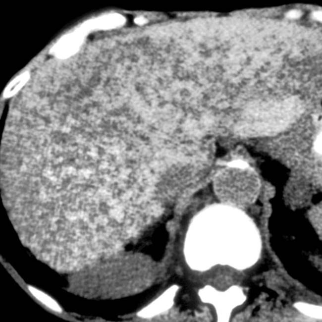
. This patient also had an enlarged, dysmorphic liver and supernumerary and enlarged hepatic arteries as signs of congenital hepatic fibrosis.

image
(Right) T2WI MR in the same patient shows larger cystic lesions image 
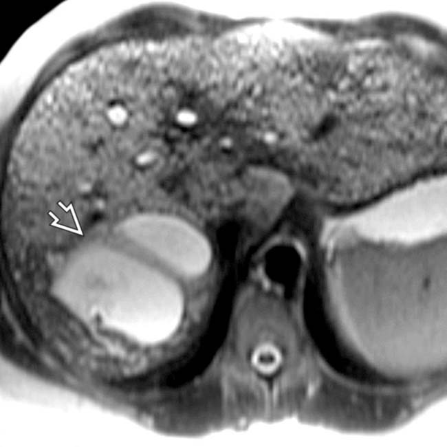
that communicate with the biliary tree and represent aberrant bile duct cysts of Caroli disease. Congenital hepatic fibrosis, Caroli disease, and biliary hamartomas are all manifestations of fibropolycystic disease.
image
Axial CECT shows many hamartomas isodense with the liver.

image
Axial NECT shows multiple small hypodense lesions in the liver.
image
Axial CECT in the same patient shows multiple, small, nonenhancing lesions of varied sizes distributed predominantly in the right lobe of the liver, findings consistent with biliary hamartomas.
image
Axial T1WI C+ MR shows numerous small, nonenhancing, hypointense lesions diffusely involving the liver.
image
Axial T2WI MR in the same patient demonstrates multiple small hyperintense foci of varied sizes throughout both lobes of the liver (biliary hamartomas).
image
Axial NECT in a cirrhotic liver shows multiple subcentimeter hypodense lesions. Biopsy specimen proved them to be biliary hamartomas.
image
Transverse ultrasound of the liver shows diffuse replacement of normal parenchyma with small hyperechoic foci, findings consistent with biliary hamartomas.

SELECTED REFERENCES

1. Lin, S, et al. A study of multiple biliary hamartomas based on 1697 liver biopsies. Eur J Gastroenterol Hepatol. 2013; 25(8):948–952.

2. Tohmé-Noun, C, et al. Multiple biliary hamartomas: magnetic resonance features with histopathologic correlation. Eur Radiol. 2008; 18(3):493–499.

Gil-Bello, D, et al. Calcification in biliary hamartomatosis. Br J Radiol. 2012; 85(1012):e99–101.

Desmet, VJ. Ductal plates in hepatic ductular reactions. Hypothesis and implications. III. Implications for liver pathology. Virchows Arch. 2011; 458(3):271–279.

Venkatanarasimha, N, et al. Imaging features of ductal plate malformations in adults. Clin Radiol. 2011; 66(11):1086–1093.

Drenth, JP, et al. Congenital fibrocystic liver diseases. Best Pract Res Clin Gastroenterol. 2010; 24(5):573–584.

Hughes, NR, et al. An immunohistochemical profile of the so-called bile duct adenoma: clues to pathogenesis. Am J Surg Pathol. 2010; 34(9):1312–1318.

Orii, T, et al. Cholangiocarcinoma arising from preexisting biliary hamartoma of liver—report of a case. Hepatogastroenterology. 2003; 50(50):333–336.

Mortele, B, et al. Hepatic bile duct hamartomas (von Meyenburg Complexes): MR and MR cholangiography findings. J Comput Assist Tomogr. 2002; 26(3):438–443.

Allgaier, HP, et al. Ampullary hamartoma: A rare cause of biliary obstruction. Digestion. 1999; 60(5):497–500.

Semelka, RC, et al. Biliary hamartomas: solitary and multiple lesions shown on current MR techniques including gadolinium enhancement. J Magn Reson Imaging. 1999; 10(2):196–201.

von Schweinitz, D, et al. Mesenchymal hamartoma of the liver—new insight into histogenesis. J Pediatr Surg. 1999; 34(8):1269–1271.

Cheung, YC, et al. MRI of multiple biliary hamartomas. Br J Radiol. 1997; 70(833):527–529.

Principe, A, et al. Bile duct hamartomas: diagnostic problems and treatment. Hepatogastroenterology. 1997; 44(16):994–997.

Iha, H, et al. Biliary hamartomas simulating multiple hepatic metastasis on imaging findings. Kurume Med J. 1996; 43(3):231–235.

Lev-Toaff, AS, et al. The radiologic and pathologic spectrum of biliary hamartomas. AJR Am J Roentgenol. 1995; 165(2):309–313.