8 Biliary Diseases
Anatomy of the Extrahepatic Biliary System
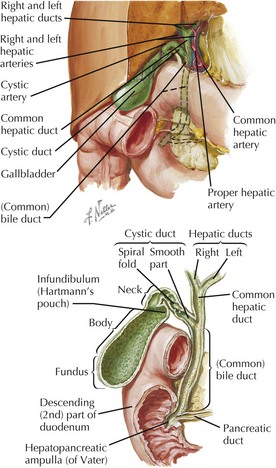
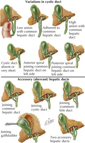
• Anatomy of the biliary system is highly variable, and this includes ducts, arteries, veins, and lymphatics.
Gallbladder
• Normally lies between hepatic segments IV and V, in a ventral fossa between the anatomical right and left lobes


• Parasympathetic preganglionic innervation from left (anterior) vagus fibers contracts gallbladder and relaxes bile duct sphincter.
Cystic Duct
• Typical cystic duct joins the common hepatic duct well below the right and left hepatic duct junction.


(Common) Bile Duct
Hepatopancreatic Ampulla (Vater)
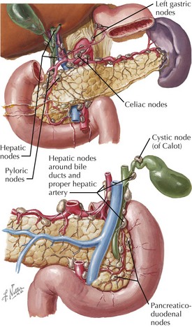
Vessels and Lymphatics
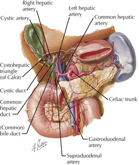
Arteries
• Gallbladder is supplied by cystic artery, typically a branch of the right hepatic artery (from the hepatic artery proper, off common hepatic, celiac axis).
• Source and course of the cystic artery vary widely: this must be carefully determined in cholecystectomy.
Clinical Correlates
Normal Bile Production
• Major salts: cholic, deoxycholic, and chenodeoxycholic acids; anionic and conjugated with taurine or glycine


Control of Bile Secretion
Cholelithiasis
• Diabetics not at increased risk, though inflammatory responses can complicate late-detected cases, with higher incidence of open surgery


Diagnostic Procedures
• Liver and biliary function tests


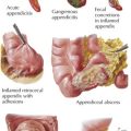
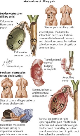
Cholecystitis
• Pain mediated by segmental visceral afferent fibers traveling with the splanchnic nerves (to thoracic spinal segments)
Cholecystectomy
• Laparoscopic cholecystectomy (lap chole) has been the treatment of choice for many years, preferred to formerly traditional open cholecystectomy.
• Open cholecystectomy uses conventional surgical instruments with a right upper quadrant or midline abdominal incision.