[level-membership-for-opthalmology-category]16.3
Best Disease
Clinical Features:
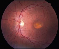
There are multiple clinical phenotypes, which represent different stages of disease and include vitelliform, pseudohypopyon, scrambled egg, and atrophic appearances. The vitelliform stage has an egg yolk appearance of subretinal material, whereas the pseudohypopyon stage exhibits a gravitational layering of the yellow subretinal material with a fluid layer above (Fig. 16.3.1). The disease can be multifocal and asymmetric leading to diagnostic uncertainty. CNV can rarely complicate the course in the atrophic state.
OCT Features:
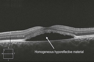
In the vitelliform stage, the subretinal material has a homogeneous, hyper-reflective appearance. In the pseudohypopyon stage, there is a homogeneous, hyporeflective layer above the hyper-reflective layer (Figs 16.3.2 and 16.3.3) that can be confused with subretinal fluid secondary to CNV. The scrambled egg stage exhibits a mix of retinal pigment epithelium atrophy, pigment clumping, and subretinal fibrosis. The atrophic stage exhibits central atrophy.
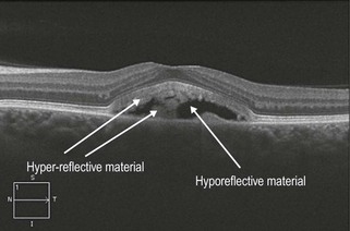
Figure 16.3.2 In the pseudohypopyon stage, a vertical OCT cut in the middle of the lesion would show a hyporeflective top layer (likely to be fluid) and a hyper-reflective bottom layer (likely to be more proteinaceous material) that are sharply demarcated. This example is a horizontal slice, which goes through both layers and shows a mix of both hypo- and hyper-reflective material in the subretinal space.
Ancillary Testing:
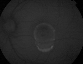
Fundus autofluorecence exhibits dramatic hyperautofluorescence (Fig. 16.3.4) and can be very helpful in assisting with the diagnosis. An electro-oculogram typically shows a reduced Arden ratio.
[/level-membership-for-opthalmology-category][not-level-membership-for-opthalmology-category]16.3
Best Disease
Clinical Features:
There are multiple clinical phenotypes, which represent different stages of disease and include vitelliform, pseudohypopyon, scrambled egg, and atrophic appearances. The vitelliform stage has an egg yolk appearance of subretinal material, whereas the pseudohypopyon stage exhibits a gravitational layering of the yellow subretinal material with a fluid layer above (Fig. 16.3.1). The disease can be multifocal and asymmetric leading to diagnostic uncertainty. CNV can rarely complicate the course in the atrophic state.
[/not-level-membership-for-opthalmology-category]
