Benign tumours
Skin tumours are common, and their incidence is rising in western countries (p. 24). The treatment of skin tumours makes up a large part of current dermatological practice (p. 24). Many skin tumours are benign, and these are described in this section. Viral warts, actinic keratoses and naevi are mentioned elsewhere.
Benign epidermal tumours
Seborrhoeic wart (basal cell papilloma)
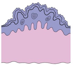
A seborrhoeic wart (seborrhoeic keratosis) is a common, usually pigmented, benign tumour consisting of a proliferation of basal keratinocytes (Fig. 1). The cause is unknown, although they may be ‘naevoid’. Seborrhoea is not a feature.
Clinical presentation
Seborrhoeic warts have the following features:
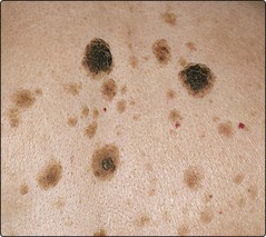
 often multiple (Fig. 2), sometimes solitary
 affect the elderly or middle-aged
 mostly found on the trunk and face
 generally round or oval in shape
 start as small papules, often lightly pigmented or yellow
 become darkly pigmented warty nodules, 1–6 cm in diameter
 have a ‘stuck-on’ appearance, with keratin plugs and well-defined edges.
Skin tags
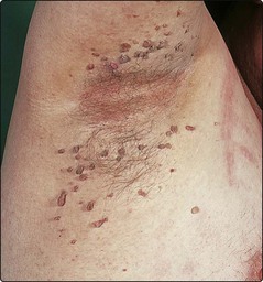
Skin tags are pedunculated benign fibroepithelial polyps, a few millimetres in length. They are common, mainly seen in the elderly or middle-aged, and show a predilection for the neck, axillae, groin and eyelids (Fig. 3). The cause is unknown, but they are often found in obese individuals. Occasionally, skin tags are confused with small melanocytic naevi or seborrhoeic warts. The treatment, usually for cosmetic reasons, is by snipping the stalk with scissors or cutting through it with a hyfrecator (under local anaesthetic if necessary), or using cryotherapy.
Benign dermal tumours
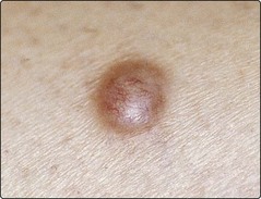
Dermatofibroma (fibrous histiocytoma)
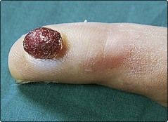
Pyogenic granuloma


Keloid
 Present as protuberant and firm smooth nodules or plaques (Fig. 6).
 Occur mainly over the upper back, neck, chest and ear lobes.
 Develop more commonly in black Africans.
 Have their highest incidence in the second to fourth decades.
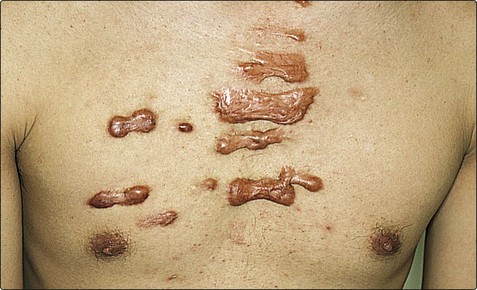
Treatment is with a topical silicone sheet or gel, or steroid injection (p. 21).
Campbell-de-Morgan spot (cherry angioma)
Campbell-de-Morgan spots are benign capillary proliferations, commonly seen as small bright-red papules on the trunk in elderly or middle-aged patients (see Fig. 2). If necessary, they can be removed by hyfrecation or cautery.
Chondrodermatitis nodularis
Chondrodermatitis nodularis is not a neoplasm, but presents as a painful small nodule on the upper rim of the helix of the pinna, usually in elderly men (p. 118). It is due to inflammation in the cartilage that may be a response to degenerate dermal collagen induced by pressure or chronic sun exposure. They are often confused with basal cell carcinomas. Excision is curative.
Benign tumours
| Lesion | Age at onset | Main features |
|---|---|---|
| Epidermal | ||
| Viral wart | Childhood mainly | Usually on hands or feet (p. 52) |
| Actinic keratosis | Old age | Sun-exposed areas (p. 106) |
| Seborrhoeic wart | Old/middle age | Keratosis, often on trunk or face |
| Milia | Childhood | White cysts, often on face |
| Epidermal cyst | After childhood | Mostly on face or scalp |
| Skin tags | Middle/old age | Seen on neck, axillae and groin |
| Dermal | ||
| Dermatofibroma | Young adult, F > M | Nodule, often on leg |
| Melanocytic naevus | Teens/young adult | Brown macule or papule (p. 96) |
| Cherry angioma | Old/middle age | Small red papule on trunk |
| Pyogenic granuloma | Child/young adult | Red nodule, often on finger |
| Keloid | Second–fourth decades | Chest/neck, affects black Africans |
| Lipoma | Any age | Soft tumour on trunk or limbs |
| Chondrodermatitis nodularis | Old/middle age | Nodule on upper pinna, M > F |
