Belt lipectomy/circumferential abdominoplasty

Published on 23/05/2015 by admin

Filed under Plastic Reconstructive Surgery

Last modified 22/04/2025

Print this page

rate 1 star rate 2 star rate 3 star rate 4 star rate 5 star
Your rating: none, Average: 0 (0 votes)

This article have been viewed 3053 times

Chapter 32 Belt lipectomy/circumferential abdominoplasty

Introduction

Body contouring’s deeply rooted history involves the evolution of specialized surgical techniques to sculpt and reshape the human physique. The concepts of panniculectomy and dermolipectomy were defined at the tail end of the 19th century as pioneers of this craft began to remove pendulous adipocutaneous tissue, resulting in improved functionality.1 In the early 20th century, contour surgery began its transformation from a purely functional modality to an esthetic procedure. As such, Thorek described the first umbilicus preserving abdominoplasty in 1924.2 The 1960s and 1970s saw major advances in this field as techniques involving aponeurotic plication,3 refinement in scar placement36 and tissue anchoring utilizing the anatomy of the superficial fascial system7 were made popular. At the turn of the 21st century, the explosive growth and widespread acceptance of bariatric surgery created a new group of patients, collectively referred to as “massive weight loss patients.” New procedures were necessary to excise the redundant tissue in all areas of the body while achieving an esthetic result. The range of contour deformities seen in these patients experienced a dramatic expansion from “anterior only” to “circumferential,” establishing the circumferential dermatolipectomy as a mainstream operation in plastic surgery today.

Body contour deformities of the lower trunk exist on a spectrum from “anterior only” to “circumferential” deformities. If the lower truncal deformity is limited to moderate lipodystrophy deposits, then liposuction may be the only treatment modality required. Abdominoplasty techniques are useful if anterior skin laxity and/or abdominal wall weakness are encountered. If lateral and posterior lipodystrophy accompany the anterior deformities, then liposuction may be added to abdominoplasty in order to achieve the best possible contour. If circumferential skin and subcutaneous laxity is additionally present, then belt lipectomy procedures are usually required to adequately address the deformities.

Belt lipectomy procedures treat deformities of the lower trunk, from the ribcage to the pelvic rim, and improve the contour of the lower back and the anterior, medial and lateral thighs. It does not, however, address upper back rolls/lateral back rolls, breast, or arm contour. The belt lipectomy procedure, therefore, will not address the upper trunk, which anatomically originates at the inferior border of the neck and extends to include the inframammary fold. To correct deformities of this anatomic unit, the authors suggest using an “upper body lift” procedure.

Relevant Anatomy

It is critical to understand the anatomy, in particular the blood supply, of the abdomen and all its variations when considering a circumferential dermatolipectomy of the lower trunk. This chapter will stop short of a detailed description of the vascular supply as this is covered elsewhere in this textbook. Preservation of the vascular supply as well as the peripheral innervation to the abdomen is a critical consideration in thwarting major complications of these procedures. The superficial fascial system, called Scarpa’s fascia in the abdomen, is a connective tissue network that exists between the subdermal plane and the deep fascia and it separates subcutaneous fat into superficial and deep layers. In thin patients, the two layers are of relatively similar thickness. Both layers demonstrate vertical or oblique fibrous septae; however, the vertically oriented compartments of the superficial layer thicken with increasing body weight and grow proportionally with the body mass index (BMI). Therefore, the superficial layer is often thicker with respect to the deep layer in patients with increased BMI. In well-defined areas of the trunk the superficial fascial system is tightly tethered to the underlying musculoskeletal surface, resulting in restriction of either descent or elevation, which typically occurs with age, fluctuations in weight, or surgical manipulation. These areas are referred to as “zones of adherence,” which behave like “hooks” that grasp the fat/skin envelope as it succumbs to gravitational forces, most noticeably following skin deflation from massive weight loss. It is important to know the anatomy of these zones and how they influence tissue draping. The zones of adherence are located in the inframammary, inguinal, suprapubic, and gluteal regions. The zones of adherence overlying the spine and sternum as well as the area located between the hip and lateral thigh fat deposits are strong and consistently present. On the other hand, the zone of adherence overlying the midline linea alba of the abdomen is relatively weak and occasionally nonexistent. Another strong zone of adherence is in the inguinal region, which plays an integral role in the final scar position following a belt lipectomy. A weaker zone of adherence exists in the horizontal suprapubic area, creating a suprapubic crease, which varies between individuals. Along with the inguinal zones of adherence, the suprapubic zone is responsible for the panniculus overhanging the mons pubis. The zone of adherence between the hip and the lateral thigh fat deposits mentioned above is an important anatomic structure because it prevents superior and inferior movement of lateral thigh tissue, especially during surgical manipulation. Destruction of this zone is often necessary if substantial elevation and redraping of the lateral thigh soft tissue envelope is desired, such as with the lower body lift type II as described by Lockwood.

Disease Process

In general, there are three groups of patients for which a belt lipectomy is beneficial.8 The majority are massive weight loss patients,8 who are not a homogeneous group. Although the pathophysiologic processes are similar and the indications and principles in operative design are comparable, their individual BMI at presentation, fat deposition pattern, and quality of their skin–fat envelope can result in a wide divergence in the nature of their physical deformity. Massive weight loss patients must therefore be well informed regarding the expected body contour outcome to avoid disappointment. Bariatric surgeons tend to emphasize overall weight reduction and often neglect to inform patients that many of the changes in their body habitus are not reversible with weight loss and that normalcy from an esthetic standpoint is often unattainable. The redundant skin with irreversible laxity is almost always circumferential in these patients, resulting in a truncal pattern of an inverted cone.

The second group of patients that may benefit from body lift/belt lipectomy are the 9–13 kg (20–30 pounds) overweight patients (BMI of 26–28). These patients have never lost significant weight despite reasonable exercise and nutritional support, which very often results in circumferential lipodystrophy and an overall lack of definition of the lower trunk. A third group is comprised of normal weight patients who desire more dramatic improvements in lower truncal contour than a conventional abdominoplasty can achieve. These normal BMI patients often seek drastic improvement in the contour of their anterior and lateral thighs, buttocks and lower back. Some of these patients can be treated with an abdominoplasty combined with liposuction of specific areas; however, if significant lifting and contour manipulation is required, a circumferential excisional procedure becomes necessary. A subset of this group is made up of older patients whose skin laxity requires the pull created by the circumferential excision. Liposuction alone will not sufficiently address the deformity as irreversible skin laxity precludes the contraction necessary to achieve the desired contour.

Diagnosis and Presentation

The discussions to follow will focus on the massive weight loss patient, with occasional reference to the other groups when appropriate. As discussed previously, massive weight loss patients vary in their BMI level, fat deposition pattern and quality of skin–fat envelope, which influences their presentation. Even though massive weight loss patients, through lifestyle changes or bariatric surgery, will tend to eventually attain a stable BMI, they will still present to the plastic surgeon at different levels. It is important for the plastic surgeon to elucidate a timeline of weight stabilization, as some patients will have a false plateau after which they will resume weight loss. Therefore, it is wise to consult with the bariatric surgeon before considering body-contouring procedures. If the patient loses weight through bariatric surgery, the type of procedure chosen for the patient will likely affect their final BMI. Lap-Band® procedures tend to induce the least amount of weight loss, followed by gastric sleeve resection and gastric bypass, while duodenal switch patients tend to have the most dramatic fall in BMI.

The unique fat deposition pattern also plays an important role in determining the specific contour deformity in massive weight loss patients. A patient’s fat deposition as well as fat loss pattern is genetically regulated and is therefore quite individualistic. For example, men tend to deposit fat intraperitoneally and in the flank region (“love handles”), presenting with mostly central, or “apple shaped,” obesity. Women, on the other hand, tend to store fat in a “pear shaped pattern” in the extraperitoneal space, the lower abdomen, hips, and thighs. These patterns are just two of many potential fat deposition presentations. Weight loss varies similarly, with some patients losing fat in equal proportions throughout their body while others experience more of an asymmetric pattern with relatively more or less lost in different areas. While fat deposition undoubtedly plays a major role in determining overall body contour, it is important to recognize that the underlying musculoskeletal foundation also contributes to the overall appearance of the individual and therefore must be considered when establishing expectations of the final cosmetic result.

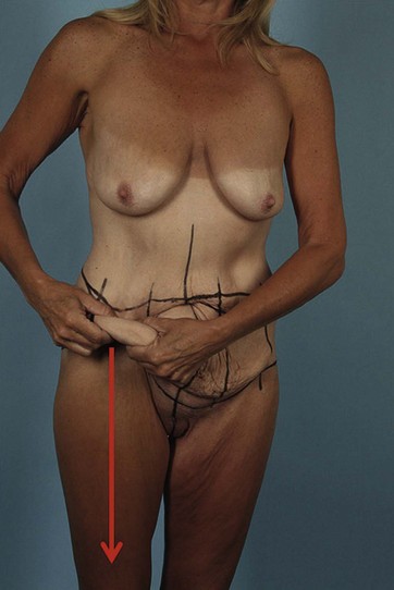
The characteristic of the skin–fat envelope is a third factor that affects the presentation of massive weight loss patients. The envelope is of major interest to the plastic surgeon and its intrinsic qualities are critical as it influences overall cosmetic outcome. The thickness of the post-weight loss subcutaneous fat tissue is important, as thin layers generally allow for greater pliability and movement of overlying skin, which is important in predicting the final cosmetic result. The authors introduced the concept of “translation of pull” as an excellent predictor of the final results. Prior to surgery, the lateral abdominal tissues are pinched in order to simulate the effect of the lateral abdominal resection on the distal thigh contour (see Fig. 32.1). Thicker nonpliable skin–fat envelopes, which are typical of patients with higher presenting BMIs, have little translation of pull in these areas, indicating that the potential overall improvement in body contour is limited. As a general guideline, the greater the drop in BMI for an individual patient, the greater the “translation of pull.”

Nearly all massive weight loss patients present with a “hanging panniculus,” the size and shape of which varies with the patient’s intrinsic fat deposition pattern. Additionally, almost every massive weight loss patient will present with “ptotic mons pubis, most commonly exhibiting vertical excess and varying degrees of horizontal excess. The hanging skin/fat envelope often blunts the waistline as it drapes from the ribs to below the pelvic brim. Despite weight loss, there are often residual fat deposits in the hip and lower back region that add to this blunting. The anterior and lateral thighs are greatly affected by the weight gain/weight loss process and are therefore typically ptotic. The effect on the thighs is less dramatic in the “20–30 pound overweight group” and the “normal weight group,” but nevertheless it remains an area of concern for these patients.

Similar to the thighs, the buttock in the massive weight loss patient is usually ptotic as a result of the weight loss process. The projection of the buttocks is generally correlated with the patient’s BMI, with patients above a BMI of 35 presenting with overly projected buttocks and patients with BMI under 26 with flat appearing and underprojected shape. Many massive weight loss patients also display poor demarcation between the lower back and buttocks.

Back rolls can be located in the lower, mid or upper back and are particularly displeasing to many patients. High back rolls, which are usually contiguous with breast rolls, are not affected by the belt lipectomy and require an upper body lift. Low back rolls are generally improved with the belt lipectomy. Few patients present with mid back, or “no man’s land,” rolls, which are difficult to treat with either an upper body lift or a belt lipectomy.

The psychiatric presentation of the patient requires special consideration. Although the plastic surgeon may be successful in reshaping the patient’s abnormal contour, psychological issues may persist. An obesity-associated depression may persist as well as estrangement from family and friends who might feel as if they have lost a fellow “sufferer.”

Preoperative Preparation

Patient Selection

Patients should be selected for their suitability to undergo circumferential excisional body contouring. Medical, as well as psychiatric, stability is of high importance. Conditions with a documented impact on wound healing such as diabetes, smoking, or connective tissue disorders must be approached with caution as they greatly increase the complication rate. Similarly, the BMI at presentation must be a factor in the plastic surgeon’s decision to operate, as complications are positively correlated with BMI.9 Reasonably, few plastic surgeons agree to operate on patients with a BMI higher than 32. Higher BMIs require surgeon acceptance of increased complication rates, with nearly 100% of patients experiencing some complication with a BMI above 35.

Low intraabdominal fat content and weight stability at the time of the surgery are critical to achieving the best overall outcome. Therefore, it may be necessary to delay body-contouring procedures until the patient can achieve weight stabilization without “heroic efforts,” such as unconventional diets and overly excessive exercise. A stable weight plateau is often difficult to identify with certainty and many patients will have “false plateaus” during which weight stabilization will be briefly apparent before the patient experiences further weight loss. If the patient had bariatric surgery, it may be beneficial to consult the bariatric surgeon to assist with delineation of true and false plateaus. Generally speaking, the length of weight stabilization is positively correlated with the patient’s chances for long-term success and most surgeons recognize 3 months as an acceptable minimum period of weight maintenance. The length of time necessary to reach maximum weight loss differs between the various bariatric surgical options. Empiric data suggest that an average of 2 years is necessary for Lap-Band® patients, 12–14 months for duodenal switch patients, and roughly 18 months for gastric sleeve resection and gastric bypass patients. These ranges are not absolute and can vary considerably between patients. Duodenal switch procedures are no longer routinely performed at most centers. Additionally, gastric sleeve resection is gaining considerable popularity over the past few years and is currently considered the treatment of choice by many bariatric surgeons.

Intraabdominal content is an important determinant of overall abdominal contour. Adequate flattening of the abdominal contour via muscle wall plication is heavily dependent on the amount of intraabdominal content. If excess exists, then the result of a circumferential procedure is similar to that attainable by panniculectomy.10 Therefore, in these situations it would be sensible to limit the procedure to a panniculectomy and avoid the increased risk that accompanies the circumferential excision.

Workup

Although many massive weight loss patients experience significant improvement in their medical problems following weight loss, they still require a thorough medical workup by the plastic surgeon during preoperative planning and evaluation. Patients should be carefully examined for cardiovascular disease, high blood pressure, diabetes, collagen vascular disease, bleeding disorders, smoking history, alcohol history, history of deep venous thrombosis/pulmonary embolus, previous abdominal scars, bariatric surgery, psychological problems, and nutritional habits. Bariatric surgery patients are typically treated with oral vitamins, so a history devoid of oral supplementation probably reflects poor follow up with resultant nutritional deficits. This should prompt the plastic surgeon to thoroughly evaluate their laboratory workup. Massive weight loss patients should additionally be questioned about signs and symptoms that may point to a history of anemia, hypocalcemia, iron deficiency, vitamin B deficiency, albumin/protein deficiency, hypomagnesemia, elevated liver function tests, and thiamine deficiency. It is also important to elucidate abnormalities in the patient’s bowel habits such as frequent diarrhea, which can put the patient at risk for nutritional deficiencies and wound contamination.

Many weight loss patients have life-long psychiatric problems, even after losing weight. The evaluation of the patient’s psychiatric stability is critically important as belt lipectomy surgery is a major life event and is associated with an arduous recovery. The authors have found it necessary to obtain psychiatric clearance for each belt lipectomy patient prior to consideration for surgical intervention. Additionally, mental health care providers must be alerted that maintenance may be required in the postoperative period.

An extensive history of the patient’s weight should be elicited from patients presenting for lower truncal contouring. First, it is important to ascertain the specific etiology of the patient’s lower trunk abnormalities. These can include aging, childbirth, sun-induced skin laxity, and massive weight loss. If the major cause is weight loss, then it is prudent to obtain a more specific history regarding the patient’s weight progression. This includes determining the patient’s weight at presentation, length of time at present weight, method of achieving weight loss and the patient’s historical maximum and minimum weights.

A careful physical exam should be followed by an evaluation specific to the body contouring procedure. It is important to note the degree of skin laxity, amount of subcutaneous fat tissue and resultant translation of pull in the various areas of interest. Waist definition, the presence of abdominal and/or back rolls, degree of rectus diastasis, presence of hernias, degree of buttock projection and ptosis, degree of anterior and lateral thigh lipodystrophy and ptosis, and the presence of any scars must be additionally noted. Any abdominal scar will alter the anatomy of the subcutaneous fat and the superficial fascial system, which ultimately influences the final contour result. Subcostal scars from gallbladder, gastric, liver, or pancreatic procedures can have a detrimental effect on the vascularity and the viability of the flaps. Additionally, vertical midline scars from previous surgeries may limit the inferior mobility of the abdominal flap. The amount of intraabdominal content must be ascertained as it considerably influences the final cosmetic result. Rather than performing the traditional “diver’s test,” as the surgeon would do in preparation for an abdominoplasty, the authors find it more effective to evaluate the patient’s abdominal contours while in a supine position. The diver’s test is less effective in a massive weight loss patient due to the thickness and laxity of the abdominal wall and subcutaneous fat. If the exam reveals a scaphoid abdominal contour and the abdominal wall falls below the ribcage, then rectus fascia plication during body contouring surgery will likely be effective in flattening the abdomen.11 However, if the abdominal tissues do not fall below the ribcage, then it can be inferred that the intra-abdominal contents are considerable and will blunt the effect of plication in this area.

The altered absorption and dietary manipulation in the massive weight loss patient can create significant abnormalities that must be addressed prior to surgery.12 Exhaustive lab work should be obtained early on in the care of the patient so that ample time exists for correction of any observed abnormalities. The labs should include: CBC, BUN, creatinine, electrolytes, glucose, urinalysis, LFT, iron, calcium, albumin, prealbumin, total protein, vitamin B, magnesium, and thiamine. Chest X-ray and EKG should be obtained as indicated on an individualized basis.

It is important to elicit and address the patient’s priorities. Massive weight loss patients often present with multiple concurrent deformities in areas such as the arms, breasts, upper back rolls, thighs, face, and lower trunk. The lower trunk and hanging panniculus is usually the chief complaint; however, many patients are fixated on improving just a few areas with less concern over others. In some situations, it may be necessary to plan and stage multiple procedures in order to achieve the maximal desired result and this should be communicated and discussed with the patient. Typically, the lower trunk deformity is addressed first, followed by arms, the thoracic region, and then the thighs.

Facial rejuvenation is also a frequent concern for patients. The authors often address this after the body contouring procedures are completed. In addition to a discussion regarding the risks and complications associated with each procedure, it should be made clear that the agreed plan might require modification as the body’s contour changes with each stage. For all major complications, a full disclosure of its occurrence, prevention, and treatment should be provided, as proper consultation should result in a well-informed patient who understands each stage of the contouring process.

Surgical Plan and Goals

The belt lipectomy is essentially a circumferential wedge excision of the lower trunk with variations in the operative technique.13 On one end of the spectrum exists the “lower body lift type II” as originally described by Lockwood and on the other the “belt lipectomy” as described by the authors, originally in Iowa.8,14,15 Neither procedure is superior to the other and they are instead used to accomplish different operative objectives. We will cover the belt lipectomy here in detail.

Markings

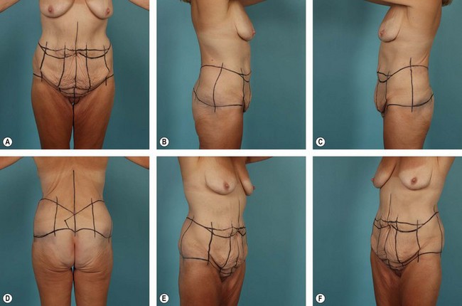
The importance of properly marking the patient prior to a belt lipectomy procedure cannot be overstated. Although there are often adjustments to be made intraoperatively, the majority of the planning and decision making is accomplished through the marking process. This is also a time for the patient and the surgeon to agree on details such as final scar position and for the surgeon to reaffirm previously discussed expectations. Preoperative photographs should assist the surgeon in identifying areas of special concern and potential pitfall. Once the patient has been marked, the surgeon should again photograph the patient. This set of photos in particular can be used for preoperative evaluation and last minute adjustments and also for postoperative comparison at 12 months, allowing the surgeon to critique and improve his or her technique (see Fig. 32.2).

Markings are only guidelines and do not represent a standardized pattern to be applied uniformly. It is most important to learn and understand the principles behind the technical details so that they may be individually tailored to each unique presentation. One exceedingly important skill used in the marking process is the simulation of tissue dynamics at closure in order to estimate and approximate the final scar position. Although some basic principles apply universally, it is important that surgeons strive to master the art of scar positioning by integrating simulated tissue dynamics with their individualized surgical technique.

The abdomen is often the patient’s foremost concern and the surgeon should be reluctant to compromise abdominal contour for improved lateral or posterior contour. There is often inferomedial rotation of tissues around the sacrum, which requires generous lateral resections in order to attain a harmonious truncal contour.

The Superior Back Marking

The superior back marking extends from the posterior midline to the lateral extent of the superior anterior mark. Initially, the inferior mark is pinched, simulating the amount of ascent required of the buttocks and lateral thigh tissues to establish the best possible lateral buttocks/thigh contour. The superior aspect of the pinched tissue is marked at several points, which are subsequently connected by a line extending from the midline to the lateral aspect of the superior anterior mark, bilaterally. A final “V”-shaped midline scar that lies at the natural junction of the lower back, sacrum, and buttocks is preferred by the authors.

In contrast to the anterior markings, the superior marks define the final posterior scar position in a belt lipectomy. This is the direct result of greater buttock mobility as compared to the superior back tissues, which have relatively stronger zones of adherence. Therefore, if markings for a belt lipectomy/central body lift are made such that the intended scar is to be just superior to the widest aspect of the pelvic brim, the final scar is most often within 2–3 cm of the superior mark. This concept is unique to the belt lipectomy/central body lift and does not apply if a lower body lift is being performed, as the area of resection is further from the restrictive zones of adherence in the lower back. As a result, the final scar is often more than 3 cm inferior to the superior mark. Therefore, prior to surgery surgeons must evaluate and adjust the position of the posterior superior mark so that the final scar is adequately placed (Fig. 32.2).

Surgical Technique

Positioning Sequence

There are a wide variety of positioning sequences available to the plastic surgeon performing a belt lipectomy. It is generally recommended that the surgeon remain fairly consistent with the chosen technique so that greater control over the final contour will be achieved. Regardless of the chosen method, there must be adequate operating room personnel on hand to complete the turns efficiently and effectively.

Prone/supine positioning completes the circumferential procedure with only one turn and may afford the surgeon ease in judging the symmetry of the back excision. Position injuries are a serious risk of this positioning technique, especially if the operative time is prolonged. Leaving the patient in one position for a protracted period puts them at risk for developing respiratory difficulties, shoulder injuries, ulnar nerve injuries, and an increased potential for eye damage.

A second single-turn technique is the supine/prone positioning sequence. Using this sequence, the anterior excision is completed first, allowing for a more aggressive anterior resection and superior anterior contour. Compared to the prone/supine technique, the supine/prone technique has the additional disadvantage of placing the patient in the prone position after an anterior resection, potentially dehiscing the anterior closure during the turn.

The authors prefer the supine/lateral decubitus/lateral decubitus positioning because it allows for the best possible contour to be achieved anteriorly. The remainder of the resection follows and is tailored accordingly. Furthermore, the lateral decubitus position allows for hip abduction, which enables a more aggressive lateral resection of redundant tissue. Since a prone position is not assumed during this sequence, visibility of the entire back and buttocks during the resection is limited, creating the possibility of an asymmetric result. However, the authors have not found that they had more asymmetry using this sequence as compared to when they utilized prone/supine, or supine/prone techniques. In addition, an extra turn may increase the operative time but again the authors have not found that to be the case in their practice. No matter what sequence is utilized, a well-trained and coordinated team is necessary in order to effectively maneuver the patient through the turns with minimal exposure and heat loss.

Anesthesia and DVT Prophylaxis

In the authors’ experience, adequate anesthesia for a belt lipectomy is best attained using a general anesthetic with a thoracic epidural placed preoperatively to aid with postoperative discomfort.16 Early evidence suggests that epidural anesthesia has significant deep vein thrombosis (DVT)/pulmonary embolism (PE) risk reduction; however, its use in plastic surgery remains relatively unstudied. If an epidural is not utilized, then the surgeon must decide whether to add chemoprophylaxis to alternating compression stockings and early ambulation protocols. The ideal chemoprophylaxis regimen has yet to be elucidated.

Step-By-Step Technique

The belt lipectomy procedure as performed by the authors is described here.

Following successful induction of general anesthesia, the patient is placed in the supine position with the arms abducted to 90°. The preoperative markings are re-enforced and tattooed using methylene blue. Sequential compression boots are applied and turned on prior to the induction of the general anesthetic. An indwelling urinary catheter is inserted, after which the patient is prepped and draped in the usual sterile fashion.

The procedure begins with a circumumbilical incision. Using scissors, the umbilical stalk is freed from the surrounding tissue down to the anterior fascia. Care must be taken when dissecting the umbilical stalk as patients may present with an undetected periumbilical hernia.

Next an incision is made along the inferior abdominal mark and dissection is carried down to, or just deep to, Scarpa’s fascia, and a superior based abdominal flap is elevated. The authors believe that leaving a fatty layer of tissue on the rectus fascia reduces seroma formation along with quilting sutures placed between the abdominal flap and the abdominal wall at closure.17

The supraumbilical dissection is dependent upon the thickness of the abdominal flap. A more traditional elevation towards the costal margins and the xiphoid process typically accompanies thin flaps. For thick flaps that need to be thinned with liposuction a very limited dissection tunnel from the umbilicus to the xiphoid, exposing the medial edges of the rectus muscle is performed. Abdominal flap elevation should not exceed the amount necessary for proper flap advancement, proper scar placement and attainment of a superior contour result. Careful attention must be paid to the tradeoff between vascular perforator preservation and the creation of the most ideal abdominal contour.

Following abdominal flap elevation, vertical plication of the abdominal wall from the xiphoid to the pubis is performed. While some surgeons prefer a single layer plication, the authors favor a two-layer plication using a long-lasting, nonpermanent, barbed, running suture. However, the choice of suture is ultimately based on surgeon preference. If deemed appropriate at the time of surgery, horizontal plications are performed.

Maximum anterior flap advancement is best achieved with the waist flexed. The flap is advanced inferiorly, tailored and temporarily tacked, with the upper mark as a guide. If the flap is thick, it may be necessary to bevel the resection. A thick mons pubis can be reduced using liposuction or through direct excision of sub-Scarpal fat.

A 1.5–2 cm vertical midline incision is made in the abdominal flap overlying the umbilical stalk. Blunt dissection, without fat removal, is carried out through this incision until a path sufficient for the umbilical stalk has been created. The authors emphasize the preservation of fatty tissue in this area as it may stave off vascular compromise of the flap. Inversion of the umbilicus is achieved using a three-point fixation technique that requires placement of inverted 3-0 nonpermanent monofilament sutures through the abdominal wall fascia, the subcuticular layer of the abdominal flap and the subcuticular layer of the umbilicus at the 3, 6, and 9 o’clock positions. This maneuver pulls the scar inside the umbilical depression while simultaneously affording the surgeon control over the degree of umbilical inversion. Using interrupted, inverted subcuticular sutures, the remainder of the umbilicus is sutured to the surrounding abdominal flap. External sutures are avoided in order to minimize suture marks. It should be noted that there are several, effective umbilicoplasty techniques that have been reported and may be used successfully. Whatever method is selected, the resultant umbilicus should be relatively small and somewhat vertical, with an internally placed suture line.

One or more closed suction drains are inserted and brought out laterally at the level of the ASIS. Prior to a layered abdominal closure quilting sutures from the abdominal flap to the underlying muscle fascia are placed to close dead space and reduce the risk of seroma. During the abdominal closure scarpa’s fascia is approximated using a synthetic absorbable suture material. One to two superficial layers are subsequently re-approximated for skin closure using an interrupted 2-0 or 3-0 synthetic absorbable suture material. Skin-glue is applied to achieve a watertight closure and the lateral dog-ears created by the anterior resection are closed temporarily with skin staples prior to turning the patient.

Positioning the patient into the lateral decubitus position is a coordinated effort led by the anesthesiologist and should involve a minimum of four additional operating room personnel. At least one person should be assigned to the crucial task of ensuring waist flexion throughout the turning process. All pressure points need to be adequately padded, which includes an axillary roll. Pillows are placed between the patient’s knees in order to achieve hip abduction, which as previously discussed allows for maximal excision of the lateral tissues. The patient is then re-prepped and draped for the posterior excision from the lateral dog-ear to the midline of the back. Many patients will also require liposuction of the lateral thighs, which is often performed at this time and involves infiltration of the lateral thigh region with a tumescent fluid.

The authors prefer to begin the posterior excision by incising the superior back mark. The extent of the subsequent dissection depends on the amount of fat that must remain in order to achieve a desirable buttock projection. Reduction of an over-projected buttock is best achieved by dissecting down to the muscle fascia with subsequent elevation of an inferiorly based flap to the level of the proposed excision line. The ultimate goal is attenuation of the projection with the creation of a depression at the waist by cinching above the hip. Further narrowing of the waist is achieved by beveling the resection margin when tailoring the flap. Extension of the superior mark incision down to the superficial fascia level with subsequent dissection of the flap inferiorly in this plane to the point of the proposed excision will serve to preserve buttock projection in normal or deficient patients.

The inferior dissection into the lateral thigh region is typically limited to the proposed inferior excision line. Some surgeons will undermine the entire lateral thigh region, disrupting the zones of adherence, in order to achieve more dramatic elevation of the lateral thigh tissues. Deep permanent sutures in the superficial fascial system are used to advance the lateral thighs superiorly onto the muscle fascia.

Closure is performed in a similar fashion to the anterior incision and is preceded by drain placement. The patient is then maneuvered to the second lateral decubitus position for the contralateral posterior resection. At the conclusion of the procedure the patient is returned to the supine position, with care taken to ensure hip flexion. Under the supervision of the operating surgeon, the patient is subsequently transferred to a flexed hospital bed for transport out of the operating room.

Postoperative Care

During recovery from anesthesia and while the patient is drowsy, they are unable to protect the wounds from mechanical dehiscence. Once the patient is alert, they should be able to sense wound tension and make adjustments to prevent dehiscence. Patients should be educated prior to surgery regarding the ability to sense tension and its importance in preventing postoperative wound complications. This is especially important during ambulation, which should occur, with assistance, on the day of surgery. The anesthesia team manages the epidural catheter, which is removed on the morning of the second postoperative day. Four to six hours after the removal of the epidural, the indwelling urinary catheter is removed. Ambulation with only one person’s assistance, tolerance of oral pain medication, the capacity to take food and drink by mouth and the ability to spontaneously urinate are the criteria used to assess discharge status. Patients are typically sent home 2 to 3 days postoperatively. Prior to discharge, patients are educated on proper drain care. Once drain output falls below 40 ml/day or the patient reaches the 14th postoperative day, the drains are removed.

For the first week postoperatively, the patient remains flexed at the waist during ambulation. Gradually during the second week, the patient is allowed to carefully resume an erect posture. Throughout the first month, patients should slowly advance their activity as tolerated and can be expected to return to nonphysical work 4 weeks postoperatively. Beginning a few days after surgery, an abdominal binder is worn by the patient until it is no longer tolerable.

Complications and Their Management

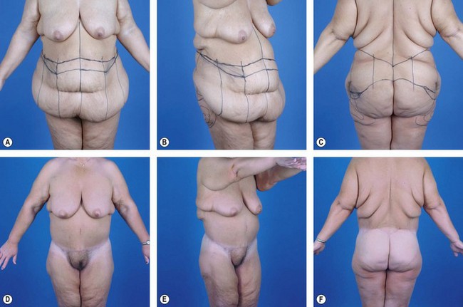
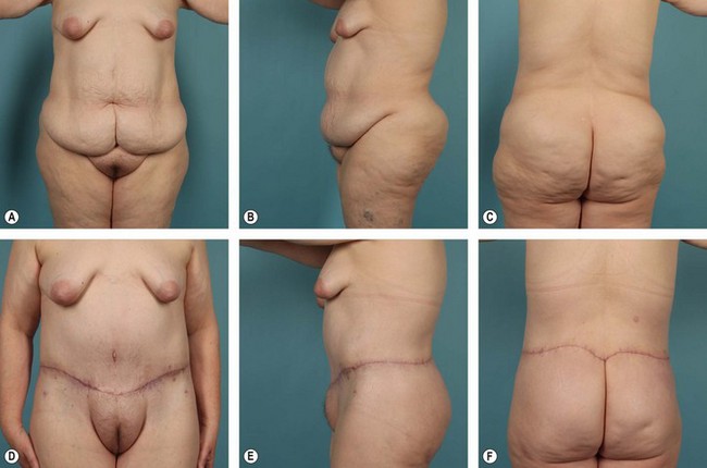
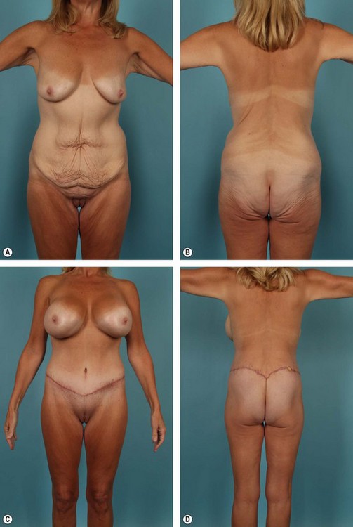
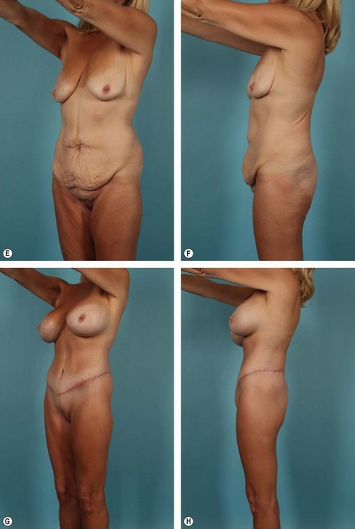
Complications, prognosis, and quality of result after belt lipectomy depend heavily on the patient’s presenting BMI, fat deposition pattern, and quality of the fat/skin envelope. Of these three factors, BMI is the most important. The authors have categorized massive weight loss patients according to presenting BMI as follows: Group I, BMI ≥36; Group II, BMI 30 to 35; Group III, BMI ≤29 (see Figs 32.332.5). Although relatively arbitrary, this schema is effective in generating discussion between the patient and the surgeon regarding the expected cosmetic result and potential complications. Group I, for instance, can expect less improvement in overall contour and a higher complication rate after a belt lipectomy than patients in Group II. The comparison is similar between Groups II and III. Within each group, however, BMI is less predictive and does not follow the same logic. Prior to surgery, it is important to explain to patients that although belt lipectomy will significantly improve their contour, their skin quality will remain unchanged. The inexperienced surgeon and the uninformed patient may seek additional tissue excision to address the unchanged skin laxity, which will ultimately lead to disappointment.

Seromas

Seroma formation is the second most common complication observed following a belt lipectomy and is a near certainty in patients exceeding a BMI of 35. Although the mechanism underlying seroma formation is poorly understood, the surface area of the operative field, concomitant liposuction and the shearing forces on the lower trunk during ordinary postoperative movement may all be contributing factors. Despite efforts to decrease and obliterate dead spaces using quilting sutures, the circumferential nature of this procedure is believed to be a significant contributor to seroma formation. The authors traditionally leave a thin layer of fat on the rectus fascia during the infraumbilical dissection of the abdominal flap, which in conjunction with quilting sutures, has reduced seroma frequency. Serial aspiration is the mainstay of treatment for seroma formation. If this is unsuccessful, however, a sclerosing agent, dicloxacillin, is injected in an attempt to close down the pocket. It is the author’s experience that small or nonexpanding fluid collections call for watchful waiting, as they will typically resolve spontaneously. Conventionally, the presence of a seroma capsule was an indication for operative intervention. However, the authors have noted the presence of capsules in almost all patients that have undergone belt lipectomy, whether they experienced a seroma or not. Therefore, the presence of a capsule is no longer an indication for surgery. In the rare situation that a seroma becomes infected, incision and drainage with antibiotic coverage and a “wick” type dressing is the most effective management. For real persistent seromas, the authors will open up part of the incision and place a Penrose drain into the pocket and allow it to heal from the “inside out”.

Wound Dehiscence

In contrast to a superficial wound separation, a dehiscence is defined as wound separation at the superficial fascial system or deeper. Competing tensions between the anterior and posterior closures increase the risk of wound dehiscence in circumferential procedures. Dehiscence of a wound may occur immediately postoperatively or a few weeks into recovery. Fortunately, the incidence of wound dehiscence is rare under the care of an experienced surgeon who can take several preemptive measures to reduce the risk of dehiscing in the early postoperative period. First, the importance of preoperatively marking the posterior midline resection with the patient in the flexed position after completion of the anterior markings cannot be overstated. Second, active measures must be taken to prevent patient manipulation immediately postoperatively until the patient is awake and can actively respond to tissue tension. Finally, the patient and the nursing staff should be educated preoperatively on the correct procedure for rolling out of bed in order to avoid positions that acutely strain the closures. A dehiscence that occurs a few weeks into the healing process is most often due to rapid bending at the waist, presumed to occur as the patient begins to experience significant milestones in recovery. It is therefore advisable to instruct patients to keep their movements slow and deliberate for the first 3 months following surgery. Unhurried movements will give the patient time to react to excessive wound tension before it reaches the level required to dehisce. If dehiscence occurs in the early postoperative period, the patient may be returned to the operating room for surgical closure or application of a wound vac. Later occurrences are best treated with wound care or the application of negative pressure wound therapy. The involved area may require scar revision at a later date.

A delicate balance exists between contour improvement and wound dehiscence. Generous resection of tissue will produce the tension necessary to create improved contour, while simultaneously increasing the likelihood of wound dehiscence, as tissue tension is common to both processes. The best, most consistent results are therefore attained if the surgeon comfortably operates between “not enough” and “too much” tissue resection.

Tissue Necrosis

Tissue necrosis following a belt lipectomy most commonly occurs along the inferior aspect of the abdominal flap at the anterior midline. Tissue necrosis ultimately results from vascular compromise, which is caused by a variety of factors including smoking, excessive wound tension, acute angulation of the superolateral excision line and old abdominal scars interfering with the natural blood supply to the abdominal flap. The belt lipectomy procedure requires more extensive undermining and lateral elevation compared to the traditional abdominoplasty procedures. As a result, the flap’s blood supply from the intercostal, subcostal, and lumbar vessels is increasingly compromised. The anteromedial aspect of the flap is farthest from these vessels and is therefore most commonly necrosed. Surgeons who choose to operate on smokers tend to modify their technique by using discontinuous undermining to mobilize the flap and limiting the midline supraumbilical elevation.

Belt lipectomy procedures are no different than the traditional abdominoplasty in the sense that horizontal subcostal scars can and will strain the remaining vascular supply to the flap after elevation. As mentioned previously, this remaining vascular supply includes the intercostal, subcostal, and lumbar vessels. Necrosis of the remaining tissue inferior to the scar may therefore ensue following flap elevation. Subcostal scars can sometimes be low enough to act as the superior border of the anterior resection, which eliminates all potentially compromised tissues. Some surgeons use a fleur-de-lis approach in which the subcostal scar is used as one of the vertical limbs brought together at the midline, but the authors have no experience with this technique. The authors have been most successful in handling subcostal scars through alterations in the sequence of surgical steps. The superior mark incision is made first with conservative elevation of the supraumbilical flap, just to the medial edges of the rectus fascia. The blood supply to the sub scar tissues is then evaluated and if felt to be compromised, the ischemic tissue is resected and the inferior flap is advanced superiorly. Patients should be counseled prior to surgery that this will result in a fairly high and undesirable final scar position. If, however, the sub scar tissue appears to be adequately perfused, then the supraumbilical flap is advanced inferiorly to the proposed inferior mark. The inferior flap is then tailored accordingly.

References

1 Kelly HA. Report of gynecological cases (excessive growth of fat). John Hopkins Med J. 1899;10:197.

2 Thorek M, ed. Plastic Reconstruction of the Female Breast and Abdomen Wall. Springfield, IL: Thomas, 1924.

3 Grazer FM. Abdominoplasty. Plast Reconstr Surg. 1973;51(6):617–623.

4 Pitanguy I. Trochanteric lipodystrophy. Plast Reconstr Surg. 1964;34:280–286.

5 Psillakis J. Abdominoplasty: some ideas to improve results. Aesth Plast Surg. 1978;2:205–215.

6 Regnault P. Abdominoplasty by the W technique. Plast Reconstr Surg. 1975;55(3):265–274.

7 Lockwood TE. Superficial fascial system (SFS) of the trunk and extremities: a new concept. Plast Reconstr Surg. 1991;87(6):1009–1018.

8 Aly A, Cram AE, Chao BS, et al. Belt lipectomy for circumferential truncal excess: The University of Iowa experience. Plast Reconstr Surg. 2003;111:398.

9 Aly AS, Capella JE. Staging, reoperation and treatment of complications after body contouring in the massive weight loss patient. In: Grotting JC, ed. Reoperative Aesthetic & Reconstructive Plastic Surgery. St. Louis: Quality Medical Publishing; 2007:1701.

10 Aly AS. Belt lipectomy. In: Aly AS, ed. Body Contouring after Massive Weight Loss. St. Louis: Quality Medical Publishing; 2006:83.

11 Aly AS. Belt lipectomy. In: Aly AS, ed. Body Contouring after Massive Weight Loss. St. Louis: Quality Medical Publishing; 2006:86.

12 Sebastian JL. Bariatric surgery and work-up of the massive weight loss patient. Clin Plast Surg. 35(1), 2008.

13 Aly AS. Option in lower truncal surgery. In: Aly AS, ed. Body Contouring after Massive Weight Loss. St. Louis: Quality Medical Publishing; 2006:59.

14 Lockwood T. Lower body lift with superficial fascial system suspension. Plast Reconstr Surg. 1993;92:1112.

15 Lockwood T. Lower body lift. Oper Tech Plast Reconstr Surg. 1996;3:132.

16 Michaud A-P, Rosenquist RW, Cram AE, Aly AS. An evaluation of epidural analgesia following circumferential belt lipectomy. Plast Reconstr Surg. 2007;120(2):538–544.

17 Pollock TA, Pollock H. No-drain abdominoplasty with progressive tension sutures. Clin Plast Surg. 2010;37(3):515–524.