13. Behçet’s Disease
Definition
Behçet’s disease is believed to be an autoimmune disorder characterized by uveitis and retinal vasculitis, optic atrophy, and aphthous lesions of the mouth and genitalia. Frequently there are other signs and symptoms that suggest diffuse vasculitis. Actual signs and symptoms vary according to the organ system involved.
Incidence
The incidence of Behçet’s disease varies according to ethnicity. The highest incidences are among Middle Eastern and Far Eastern populations.
Etiology
Behçet’s disease is believed t o be of autoimmune origin. As such, the true etiology remains unknown. Various aspects of the disease seem to indicate that it is an infectious process, but in the 80 years since it was first described, no specific causative agent has been identified. Genetic factors also seem to contribute to the development of this disease. Middle and Far Eastern populations have a higher rate of occurrence of this disease and have a higher prevalence of human leukocyte antigen B 5 (HLA-B 5); the Israeli population has a higher prevalence of HLA-B 51.
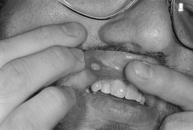 |
| Behçet’s Disease. Oral aphthous lesions in a patient with Behçet’s disease. |
Signs and Symptoms
Arthritis
• Enthesopathies
• Joint fluid turbidity
• Sacroilitis
Cardiac
• Coronary thrombosis
• Coronary vasculitis
• Diastolic dysfunction
• Endocarditis
• Myocarditis
• Pericarditis
• Valvular regurgitation
Gastrointestinal
• Abdominal pain
• Bloody diarrhea
• Intestinal perforation
• Ulcerative lesions
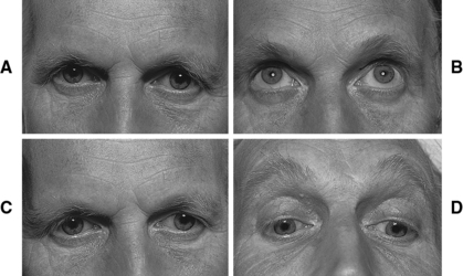 |
| Behçet’s Disease. Failure of horizontal gaze ( A and C) associated with preservation of vertical gaze ( B and D). |
Genitalia
• Penile lesions
• Perianal lesions
• Scrotal lesions
• Vulvar/vaginal lesions
Mouth
• Aphthous lesions/ulcers
Neurologic (10% to 30% of cases)
• Acute deafness
• Behavioral changes
• Clonus
• Cranial nerve lesions
• Dementia
• Emotional liability
• Meningoencephalitis
• Positive Babinski sign
• Seizures
• Spastic paralysis/pyramidal tract lesions
• Speech difficulty
• Swallowing difficulty
Ocular
• Anterior uveitis
• Cataracts
• Glaucoma
• Hypopyon
• Infarctions
• Posterior uveitis
• Retinal detachment
• Retinal edema
• Retinal vasculitis
• Synechiae
Pulmonary
• Hemoptysis
• Pleural effusions
• Pulmonary hypertension
• Pulmonary vasculitis
Renal
• Epididymitis
• Glomerulonephritis
• Immunoglobin A nephropathy
Skin
• Erythema nodosum
• Pseudofolliculitis
• Pustular, acnelike lesions
Vascular
• Aneurysms
• Claudication
• Ischemia
• Myocardial infarction
• Thrombotic disease
• Transient ischemic attack
• Vasculitis
Medical Management
Management of Behçet’s disease varies according to the organ system(s) involved:
Central nervous system: Typically treated with prednisone or prednisolone, chlorambucil, or cyclophosphamide.
Erythema nodosum: Treatment is generally with colchicine or dapsone.
Gastrointestinal system: Treating lesions in this system is usually accomplished using corticosteroids, sulfasalazine, or thalidomide.
Joints: Localized intra-articular injections of corticosteroids, prednisone, and/or nonsteroidal anti-inflammatory drugs (NSAIDs) may be used for treatment of joint pain. Colchicine, sulfasalazine, interferon-Q, levamisole, and azathioprine may also be utilized.
Ophthalmic manifestations: Topical drops, consisting of both mydriatic and corticosteroid medications, are utilized. Immunosuppression is attempted because of the autoimmune nature of the disease and is generally achieved using azathioprine, systemic corticosteroids, cyclosporine, chlorambucil, tacrolimus, or cyclophosphamide.
Systemic manifestations (skin disease or severe ulcers): Colchicine or azathioprine is used with some degree of success. Antileprosy drugs, such as dapsone and thalidomide along with the immune system–modifying drug levamisole, have demonstrated varying degrees of usefulness.
Thrombosis/thrombotic events: Anticoagulants are the treatment of choice.
Vasculitis: Immunosuppression is the treatment goal, utilizing suppressive medications such as cyclophosphamide, azathioprine, cyclosporine, and others. If anticoagulants are required in the treatment of pulmonary vasculitis, the mortality of patients may be increased.
Tumor necrosis factor antagonists, such as etanercept or in-fliximab, have demonstrated efficacy in treating Behçet’s disease.
Surgical intervention may be required to treat a variety of manifestations of Behçet’s disease, including gastrointestinal perforation(s) and the resulting peritonitis, aneurysms, endocardial fibrosis, coronary thrombosis, glaucoma, cataracts, and retinal detachment.
Complications
Three manifestations of Behçet’s disease can be especially devastating: aneurysms, thrombolic events, and vasculitis. Aneurysms may develop essentially at any location: thoracic, abdominal, cranial, ophthalmic, or the extremities. The effects, should an aneurysm rupture, can range from vision impairment and/or loss to death.
Anesthesia Implications
As with any patient, a thorough preoperative history is indispensable. Prior knowledge that the patient has Behçet’s disease should prompt heightened concentration on and investigation of all organ systems. Magnetic resonance imaging (MRI) or computed tomography (CT) may be warranted to assess for possible spinal cord compression, intracranial pressure increases, or intervertebral disk herniation. Initial cardiac manifestations may be indicated by electrocardiography and echocardiography. Potential pulmonary manifestations may necessitate arterial blood gas analysis, chest x-ray, spirometry, and pulmonary function testing. Renal involvement may be initially assessed via laboratory determination of blood urea nitrogen (BUN) and creatinine concentrations, particularly for the patient being treated with NSAIDs and/or antineoplastic medications. Prophylactic steroid coverage is necessary for the patient being treated with corticosteroids.
The patient with an autoimmune disorder may experience an exacerbation as a result of any skin puncture, such as insertion of an intravenous line or arterial pressure line or instillation of a regional anesthetic. Securing the patient’s airway during administration of general anesthesia can be a considerable challenge.
Direct laryngoscopy or placement of a laryngeal mask airway (LMA) can aggravate and rupture lesions that may be present in the mouth and/or oropharynx, producing significant bleeding. The patient treated with NSAIDs and/or anticoagulants may experience even greater blood loss. The risk versus benefit of continuing these classes of medications within 7 to 10 hours of a surgical procedure must be given consideration. Lesions in the oropharynx may extend to and into the glottis and trachea, resulting in glottic stenosis, which will make passage of an endotracheal tube more difficult. If the stenosis is severe enough, intubation may only be achieved by fiberoptic means; however, if intubation proves impossible, a surgical tracheostomy may be necessary to secure the airway to provide anesthesia for the surgical intervention(s) that are required. The patient may be more susceptible to pulmonary injury in the form of barotrauma, particularly if the patient has pulmonary vasculitis. The patient may benefit considerably from pressure-controlled ventilation rather than volume-controlled ventilation.