126
Basal cell carcinoma
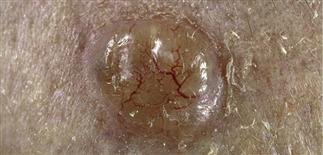
Common features of a nodular basal cell carcinoma with a smooth transleucent pink and pearly nodule with a sharp border and telangiectasias.
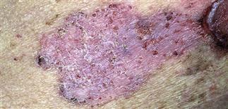
Superficial basal cell carcinoma spreads peripherally, sometimes several centimeters, and invades after considerable time. May resemble patch of eczema or psoriasis.
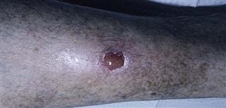
A basal cell carcinoma may present as a slow to heal ‘wound’ on the anterior shin with beefy-red, smooth erosive nodule.
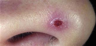
Nodular basal cell carcinomas are common on the nose. This lesion had been present for many months, ulcerated, then began to heal, which further delays diagnosis.
DESCRIPTION
Basal cell carcinoma (BCC) is the most common cutaneous malignancy. Locally invasive, slow-growing, rarely metastasizes (unless patient is immunocompromised). Neither life-threatening nor trivial.
HISTORY
• More common after age 40. • Highest incidence in the fair-skinned. • Cumulative sun exposure is primary risk factor. Occur mostly on sun-exposed skin of face, scalp, ears, neck. • Clinical variants include nodular, pigmented, superficial, sclerotic basal cell carcinoma.
PHYSICAL FINDINGS
• Nodular BCC. Most common variant. A pearly white, almost translucent, dome-shaped papule with overlying telangiectasias. Papule or nodule enlarges slowly, may become flattened in center or may develop a raised, rolled, translucent border. Frequently ulcerates, bleeds, becomes crusted in center. • Pigmented BCC. Contains melanin, may therefore resemble melanoma. • Superficial BCC. Least aggressive form. More commonly on trunk, extremities. Circumscribed, round to oval, red, scaling plaque resembles eczema, psoriasis, extramammary Paget disease, or Bowen disease. • Sclerosing BCC. Most subtle and least common variant. Smooth, pale white to yellow papules. Resembles scar tissue. Borders may be difficult to discern. Higher rate of recurrence.
TREATMENT
Without treatment, BCCs persist, enlarge, ulcerate, invade, destroy surrounding structures. Treatment determined by size and location of tumor, tumor variant, patient’s concerns. Clinical aggressiveness correlates with histologic pattern.• Electrosurgery involves electrodesiccation and curettage of obvious tumor. The 5-year cure rates approach 92%. • Primary excision is preferred for non-facial, well-defined nodular. The 5-year cure rates approach 90%. • Mohs micrographic surgery is a highly specialized, tissue-sparing method of excision used for difficult tumors with contiguous growth, especially BCCs. Mohs micrographic surgery is used for recurrent BCC, histologically aggressive forms of BCC, such as sclerotic BCCs, and tumors in anatomically important locations such as around eyes, nasal ala, mouth, and ears. Also used for tumors with high risk of recurrence. Treatment of choice for sclerotic and recurrent BCC and most BCCs of the central face, near functionally important structures. The 5-year cure rates approach 99%.
Non-surgical options are increasing. These include radiation therapy, photodynamic therapy, and topical immune modulators. • Radiation therapy may be useful for difficult-to-treat tumors, such as on eyelids, and for patients unwilling or unable to tolerate surgery. The 5-year cure rates are roughly 90%. Hedgehog inhibitors are newer oral medications that show great promise for slowing down or inhibiting BCC progression, especially for advanced and inoperable BCCs.
• Photodynamic therapy is an evolving chemotherapeutic modality for superficial BCC that is not widely available today but may be useful in the future.
• Topical imiquimod 5% cream is an immune response modifier shown to be about 85% effective or better for superficial BCC. It is less effective for nodular BCC.
All patients with BCC require follow-up to monitor for recurrence at the treated site, regardless of which treatment is used, and for the development of new tumors.


