Barotrauma

Published on 20/07/2015 by admin

Filed under Radiology

Last modified 22/04/2025

Print this page

rate 1 star rate 2 star rate 3 star rate 4 star rate 5 star
Your rating: none, Average: 0 (0 votes)

This article have been viewed 2248 times

 Free air in pleural and mediastinal spaces

image 
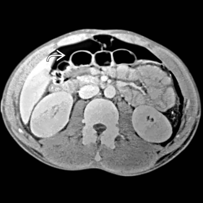
Extraluminal ectopic gas in subcutaneous soft tissues and muscles, lung interstitium, retroperitoneum, intraperitoneal spaces, and bowel wall
• Radiographic findings

image Pneumothorax

– Radiolucent gas between visceral and parietal pleura
– Inferiorly displaced costophrenic angle on supine films (deep sulcus sign)
image Pneumomediastinum

– Radiolucent streaks outlining heart and trachea
image Pneumoperitoneum

– Best seen on upright and left decubitus films
– Supine films: Air outlining bowel or falciform ligament
image Subcutaneous emphysema

– Radiolucent streaks outlining fat and muscles

TOP DIFFERENTIAL DIAGNOSES

• Perforated duodenal or gastric ulcer
• Iatrogenic introduction of ectopic gas
• Diverticulitis
• Other causes of pneumothorax, pneumomediastinum, pneumoperitoneum, or pneumatosis
• Ischemic enteritis

PATHOLOGY

• Positive pressure ventilation → alveolar rupture → air leakage into pulmonary interstitium
• Interstitial air can dissect along perivascular sheaths into mediastinum
• Mediastinal and pleural air can leak into peritoneal and retroperitoneal cavities
• Primary risk factors include interstitial lung disease, asthma, acute respiratory distress syndrome (ARDS), and mechanical ventilation with high tidal volumes
image
(Left) Axial CECT in a young man on a ventilator following a motor vehicle crash shows a tension pneumothorax image 
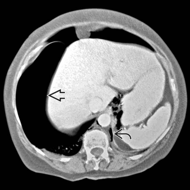
on the right side and a smaller pneumothorax on the left. Gas dissects under pressure along the peridiaphragmatic fat image.

image
(Right) Axial CECT in the same patient shows the extraluminal air from the thorax dissecting into the peritoneal cavity image to outline bowel loops. There was no intraabdominal injury.
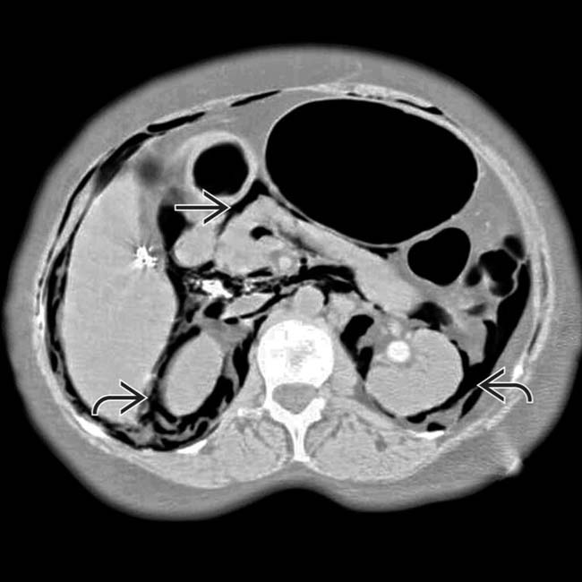
image
(Left) Axial CECT in an elderly man on positive pressure ventilation and with known large bilateral pneumothoraces and gas in the mediastinum shows the gas dissecting under pressure into the abdomen, including the retroperitoneum image and mesentery image.

image
(Right) Axial CECT in the same patient shows that in addition to the extensive retroperitoneal gas, intraperitoneal gas is also present image
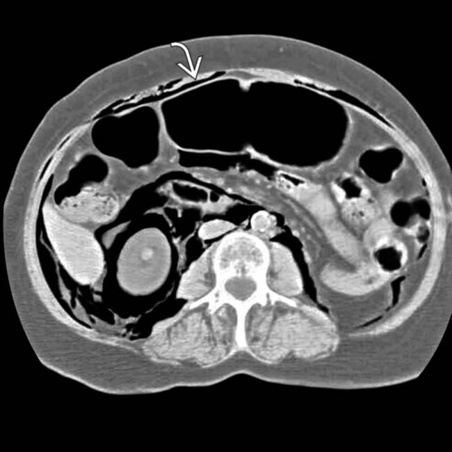
. In some cases, gas can dissect into the bowel wall, simulating pneumatosis from bowel ischemia.

TERMINOLOGY

Synonyms

• Pulmonary barotrauma

Definitions

• Alveolar rupture caused by elevated transalveolar pressure during mechanical ventilation

IMAGING

General Features

• Best diagnostic clue

image Extraluminal air in patient treated with positive pressure ventilation
• Location

image Ectopic gas in pleural space, mediastinum, subcutaneous soft tissues, intraperitoneal and retroperitoneal spaces, bowel wall

Radiographic Findings

• Radiography

image Diagnosis is difficult because patient is usually supine and being ventilated
image Pneumothorax

– Radiolucent collection of gas between visceral and parietal pleura
– Inferiorly displaced costophrenic angle on supine films (deep sulcus sign)
image Pneumomediastinum

– Radiolucent streaks outlining heart and trachea
image Pneumoperitoneum

– Best seen on upright and left decubitus films
– Supine films: Air outlining bowel, falciform ligament
image Subcutaneous emphysema

– Radiolucent streaks outlining subcutaneous fat and muscles

CT Findings

• Free air in pleural and mediastinal spaces

image Easily detected on “lung windows” (wide window width; low level)

– Lower density than lung, without visible vessels or parenchyma
• Free (extraluminal) gas in subcutaneous tissues and muscles, lung interstitium, retroperitoneum and mesentery, intraperitoneal spaces, and bowel wall (pneumatosis)

DIFFERENTIAL DIAGNOSIS

Perforated Duodenal or Gastric Ulcer

• Gas bubbles and infiltration of fat planes adjacent to duodenal bulb or in lesser sac (gastric ulcer)
• May extend into perirenal space (through renal hilum)

Iatrogenic Introduction of Ectopic Gas

• Introduction of gas or air via surgery, catheterization, peritoneal lavage, or endoscopy

Diverticulitis

• Free intraperitoneal gas is uncommon result of diverticulitis
• Usually has other signs of inflammation adjacent to involved portion of colon (infiltrated sigmoid mesocolon, bowel wall thickening, etc.)

Other

• Multiple other causes of pneumothorax, pneumomediastinum, pneumoperitoneum, or pneumatosis (e.g., pneumoperitoneum due to bowel perforation, pneumothorax due to trauma)

Ischemic Enteritis

• Pneumatosis can result from ischemia, but also other causes, including medications and barotrauma
• Barotrauma is usually accompanied by pneumothorax and other sites of extraluminal air

PATHOLOGY

General Features

• Etiology

image Positive pressure ventilation → alveolar rupture → air leakage into pulmonary interstitium
image Interstitial air can dissect along perivascular sheaths into mediastinum
image Mediastinal and pleural air can leak into peritoneal and retroperitoneal cavity
image Rare complications include tension pneumothorax, bronchopleural fistula, subpleural air cyst, and air embolus

Staging, Grading, & Classification

• Risk factors

image Interstitial lung disease; asthma
image Acute respiratory distress syndrome (ARDS)

CLINICAL ISSUES

Presentation

• Most common signs/symptoms

image Some patients may be asymptomatic
image Tachypnea, tachycardia, hypertension or hypotension, oxygen desaturation
image Abdominal distension, tenderness
image Subcutaneous emphysema (crepitus on palpation)

Demographics

• Epidemiology

image Older literature reported barotrauma seen in 3% of patients undergoing mechanical ventilation, but frequency has decreased due to low tidal volume ventilation becoming more common

Natural History & Prognosis

• Associated with ↑ morbidity and mortality

Treatment

• Conservative management

image Close monitoring of pneumothorax for progression
image Pneumomediastinum, pneumoperitoneum, and subcutaneous emphysema are self-limited
• Chest tube insertion for large pneumothorax
• Prevention: Maintaining plateau airway pressure < 30 cm H₂O

DIAGNOSTIC CHECKLIST

Consider

• Pneumoperitoneum and pneumatosis are rare complications of barotrauma, and abdominal source must be excluded

Image Interpretation Pearls

• Almost all patients have pneumothorax and other sites of air

SELECTED REFERENCES

1. Santa Cruz, R, et al. High versus low positive end-expiratory pressure (PEEP) levels for mechanically ventilated adult patients with acute lung injury and acute respiratory distress syndrome. Cochrane Database Syst Rev. 2013.

Barcia, SM, et al. Pulmonary interstitial emphysema in adults: a clinicopathologic study of 53 lung explants. Am J Surg Pathol. 2014; 38(3):339–345.

Frutos-Vivar, F, et al. [Prognosis for acute exacerbation of chronic obstructive pulmonary disease in mechanically ventilated patients.]. Med Intensiva. 2006; 30(2):52–61.

Anzueto, A, et al. Incidence, risk factors and outcome of barotrauma in mechanically ventilated patients. Intensive Care Med. 2004; 30(4):612–619.

Bejvan, SM, et al. Pneumomediastinum: old signs and new signs. AJR Am J Roentgenol. 1996; 166(5):1041–1048.

Satoh, K, et al. CT appearance of interstitial pulmonary emphysema. J Thorac Imaging. 1996; 11(2):153–154.

Tocino, I, et al. Barotrauma. Radiol Clin North Am. 1996; 34(1):59–81.

Gammon, RB, et al. Clinical risk factors for pulmonary barotrauma: a multivariate analysis. Am J Respir Crit Care Med. 1995; 152(4 Pt 1):1235–1240.

Cho, KC, et al. Extraluminal air. Diagnosis and significance. Radiol Clin North Am. 1994; 32(5):829–844.

Levine, MS, et al. Diagnosis of pneumoperitoneum on supine abdominal radiographs. AJR Am J Roentgenol. 1991; 156(4):731–735.