[level-membership-for-dermatology-category]
Chapter 27 Bacterial infections
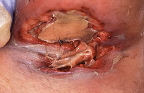
As always, the diagnosis should be confirmed with a culture and antibiotic sensitivities performed at the initial visit, since not all follicular-based abscesses are due to staphylococci. The initial antibiotic choice before the culture and antibiogram results is usually oral dicloxacillin, oral cephalexin, or oral amoxicillin/clavulanate. Oral erythromycin, azithromycin, or clarithromycin can be used if the patient is allergic to penicillin. However, if MRSA infection is clinically suspected then the initial antibiotic choice is different (see question 16).
El-Gilany AH, Fathy H: Risk factors of recurrrent furunculosis, Dermatol Online J 15:16, 2009.
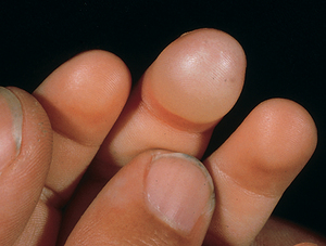
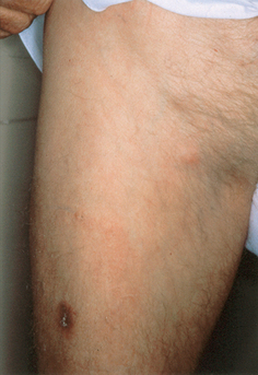
Figure 27-6. Blistering distal dactylitis demonstrating a characteristic tender superficial blister on the volar fat pad.
Wormser GP, Dattwyler RJ, Shapiro ED, et al: The clinical assessment, treatment, and prevention of Lyme disease, human granulocytic anaplasmosis, and babesiosis: clinical practice guidelines by the Infectious Diseases Society of America, Clin Infect Dis 43:1089–1134, 2006.
[/level-membership-for-dermatology-category][not-level-membership-for-dermatology-category]
Chapter 27 Bacterial infections
As always, the diagnosis should be confirmed with a culture and antibiotic sensitivities performed at the initial visit, since not all follicular-based abscesses are due to staphylococci. The initial antibiotic choice before the culture and antibiogram results is usually oral dicloxacillin, oral cephalexin, or oral amoxicillin/clavulanate. Oral erythromycin, azithromycin, or clarithromycin can be used if the patient is allergic to penicillin. However, if MRSA infection is clinically suspected then the initial antibiotic choice is different (see question 16).
El-Gilany AH, Fathy H: Risk factors of recurrrent furunculosis, Dermatol Online J 15:16, 2009.
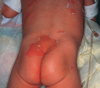
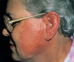
Figure 27-3. Early staphylococcal scalded-skin syndrome demonstrating diffuse erythema and early desquamation.
[/not-level-membership-for-dermatology-category]
