B
bacitracin An antibiotic drug with similar properties to penicillin and effective principally against gram-positive bacteria, such as Staphylococci and Streptococci. It is mainly used in combination with other agents (e.g. polymyxin B) for treating external eye infections (e.g. blepharoconjunctivitis).
back haptic size See haptic size, back.
back of a lens Relating to that surface nearer to the eye (British Standard).
back optic zone diameter See optic zone diameter.
back optic zone radius See optic zone radius, back.
back vertex focal length See vertex focal length.
back vertex power See power, back vertex.
backward masking See metacontrast.
baclofen An analogue of gamma-aminobutyric acid (GABA) used orally to treat skeletal muscle spasm and in the management of nystagmus, particularly periodic alternating nystagmus.
bacteria Microscopic unicellular organisms that commonly reproduce by cell division (fission) and contained within a cell wall. They are a natural component of the human body, particularly on the skin, mouth and intestinal tract. Many are beneficial to the environment and living organisms, but some are the cause of many infectious diseases. Infectious bacteria enter the body through torn tissues or by its orifices (e.g. nose, mouth, lungs) and can provoke inflammation. Many bacterial infections may spread from host to host (e.g. contagious conjunctivitis). Infections caused by bacteria are treated with antibiotics. Singular: bacterium.
See antibiotics; Gram stain.
bacterial conjunctivitis See conjunctivitis, acute.
bacteriostatic A term describing substances such as sulfonamides and tetracycline which inhibit the growth and propagation of bacteria, but do not actually destroy bacteria.
See antibiotic.
Badal’s optometer See optometer, Badal’s.
‘bag’, capsular A sack-like structure remaining within the eye following extracapsular cataract extraction or phacoemulsification. The implanted intraocular lens is placed within this structure to recreate the usual phakic state.
See cataract extraction; lens, intraocular; phacoemulsification.
Bagolini’s glass; test See glass, Bagolini’s.
Bailey–Lovie acuity chart See chart, Bailey–Lovie.
Bailliart’s ophthalmodynanometer See ophthalmodynanometer.
balance, binocular Condition characterized by the two eyes being simultaneously in focus or equally out of focus.
balance, muscle The status of the eye muscle function as represented by the phoria measurement.
See heterophoria.
balancing lens; test See lens, balancing.
Baldwin’s illusion See illusion, Baldwin’s visual.
Balint’s syndrome See syndrome, Balint’s.
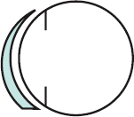
ballast Additional weight of material incorporated in a part of a contact lens to maintain it in a given orientation (Fig. B1). This is often provided by giving prismatic power to the lens (prism ballast lens).
band keratopathy See keratopathy, band.
band, retinoscopic A strip of light seen in the retinoscopic reflex of an astigmatic eye, especially when neutralizing one of the principal meridians.
band-shaped corneal dystrophy See keratopathy, band.
bandage lens See lens, therapeutic soft contact.
Bangerter foils Semitransparent membranes of various degrees of opacification which can be pressed onto a spectacle lens to reduce visual acuity. There are 10 degrees, graded from 1.0 (6/6 or 20/20) to complete occlusion. They are used in the treatment of amblyopia and intractable diplopia. Syn. Bangerter graded occluder; Bangerter occluder.
See occlusion treatment; penalization.
bar reading See test, bar reading.
Barany’s nystagmus See nystagmus.
Bardet–Biedl syndrome See syndrome, Laurence–Moon–Bardet–Biedl.
baring of the blind spot See spot, baring of the blind.
barrel-shaped distortion See distortion.
Barrer A unit of oxygen permeability of a contact lens material. Symbol: Dk. It is equal to the product of the diffusion coefficient D of oxygen through the material (i.e. the speed at which oxygen molecules pass through the material) and the solubility k of oxygen in the material (i.e. the number of oxygen molecules that can be absorbed in a given volume of material).
See oxygen permeability; oxygen transmissibility.
basal 1 . In anatomy, denoting a layer or cells farthest away from the surface. Example: the basal cells of the corneal epithelium nearest Bowman’s layer. 2. In optics, denoting the surface opposite to the apex of a prism.
base-apex direction See base setting.
base-apex line See base setting.
base curve See curve, base.
base of prism The edge of a prism at which the faces are separated by a maximum distance.
See base setting.
base setting The direction of the line from apex to base of a prism in a principal section (a section lying in a plane perpendicular to the refracting edge). The setting position for the base of a prism is normally specified by the direction ‘base-up’ (or base-down, in or out as the case may be) in which ‘up’ and ‘down’ have their ordinary meanings, ‘in’ means towards the nose and ‘out’ towards the temple. Base-apex line, base-apex meridian and base-apex direction are deprecated terms (British Standard). Alternatively, the TABO notation is used. Abbreviations for the placement of the base of the prism are BD (for base-down), BI (for base towards the nose), BO (for base towards the temple) and BU (for base-up).
See axis notation, standard; base of prism.
base of vitreous See humour, vitreous.
Bassen–Kornzweig syndrome See syndrome, Bassen–Kornzweig.
Batten–Mayou disease See disease, Batten–Mayou.
beam of light See light, beam of.
beam splitter An optical system which separates an incident beam of light into two beams of lesser intensity, one reflected and the other transmitted, e.g. a semi-silvered mirror. Some beam splitters are made of birefringent material, which splits the incident light beam into oppositely polarized beams. They are called polarizing beam splitters. Example: Wollaston prism.
‘bear tracks’ See retinal pigment epithelium, congenital hypertrophy of the.
bearing, apical An area of contact between the back surface of a rigid contact lens and the apex of the cornea. It is observed with the fluorescein test.
bedewing, endothelial A cluster of inflammatory cells deposited on the posterior surface of the corneal endothelium. They have been noted with anterior eye inflammation and contact lens wear. The symptoms may include slight stinging sensation, some interference with vision and intolerance to contact lens wear. Reduction of wearing time is usually indicated and in severe cases contact lens wear must be ceased.
See blebs, endothelial; corneal endothelium.
Behçet’s disease See syndrome, Behçet’s.
belladonna See atropine.
Bell’s palsy See palsy, Bell’s.
Bell’s phenomenon See phenomenon, Bell’s.
Benedikt’s syndrome See syndrome, Benedikt’s.
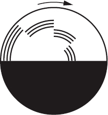
Benham’s top A disc, half black and half white with a number of concentric black bars on the white half which when rotated evokes a sensation of colour, called Fechner’s colours or Fechner–Benham colours (Fig. B2). Syn. Benham–Fechner top.
benoxinate hydrochloride See oxybuprocaine hydrochloride.
Benson’s disease See asteroid hyalosis.
benzalkonium chloride See antiseptic.
Berger’s loupe See loupe, Berger’s.
Berger’s postlenticular space See space, Berger’s postlenticular.
Bergmeister’s papilla See papilla, Bergmeister’s.
Berlin’s disease See disease, Berlin’s.
Bernell clip See clipover.
Best’s disease See disease, Best’s.
best vision sphere See method, fogging.
Best’s vitelliform macular dystrophy See disease, Best’s.
best-form lens See lens, best-form.
beta-adrenergic blocking agent See beta-blocker.
beta-blocker A drug that blocks or reduces the action of neurotransmitters on beta-adrenergic receptors. It reduces secretion of aqueous humour and consequently intraocular pressure and it is used in the treatment of glaucoma. Common beta-blockers include timolol maleate, betaxolol hydrochloride, carteolol hydrochloride, levobunolol hydrochloride and metipranolol. Timolol is often used together with another agent (combination drugs), e.g. timolol and brimonidine, timolol and dorzolamide, timolol and latanoprost. Syn. beta-adrenergic antagonist; beta-adrenergic blocking agent.
See adrenergic receptors; miotics; sympatholytic drugs.
betamethasone See antiinflammatory drugs.
betaxolol hydrochloride See adrenergic receptors; beta-blocker.
bethanechol chloride See pilocarpine.
bevacizumab See anti-VEGF drugs; macular degeneration, age-related.
Bezold–Brücke phenomenon See phenomenon, Bezold–Brücke.
Bianchi’s valve See lacrimal apparatus; valve of Hasner.
bichrome test See test, duochrome.
biconcave lens See lens, biconcave.
biconvex lens See lens, biconvex.
Bidwell’s experiment See experiment, Bidwell’s.
Bidwell’s ghost A special case of a moving positive after-image occurring behind a moving spot of light. This after-image seems like a ghost light trailing behind. Syn. Purkinje after-image.
See after-image.
Bielschowsky’s head tilt test; phenomenon; phenomenon test See under the nouns.
Bietti’s band-shaped corneal dystrophy See keratopathy, actinic.
bifixation Imaging of an object on the fovea of each eye simultaneously. Syn. bifoveal fixation.
bilateral strip See nasotemporal overlap.
billiards spectacles See spectacles, billiards.
bimatoprost See prostaglandin analogues.
binasal hemianopia See hemianopia, binasal.
binocular Pertaining to both eyes.
binocular balance See balance, binocular.
binocular disparity See acuity, stereoscopic visual; disparity, retinal; perception, depth.
binocular fusion See fusion, sensory.
binocular indirect ophthalmoscope; lock; lustre; parallax; rivalry See under the nouns.
binocular single vision See vision, binocular single.
binocular vision See vision, binocular.
binocular visual field See field, binocular visual.
binoculars A set of two identical telescopes, one for each eye, which gives binocular vision of magnified distant objects. The images are erected using either an eyepiece of negative power, or prisms, or very occasionally, an additional lens system placed between objective and eyepiece. On binoculars, the magnification M and the diameter D of the objective or entrance pupil are shown as M × D (e.g. 8 × 30). Syn. field glasses; prism binoculars (for those which use prisms as erectors).
See erector; telescope, galilean; telescope, terrestrial.
binoculars, prism See binoculars.
biocular Pertaining to the use of the two eyes but without fusion or stereopsis. The term is primarily used in clinical testing and vision therapy in which different prisms are placed in front of each eye.
biofeedback A technique whereby visual (or bodily) processes normally under involuntary control (e.g. accommodation) are displayed to the subject, enabling voluntary control to be learnt. It has been used in myopia control and in acuity improvement but the value of the technique in these conditions is still unproven.
Biological–statistical theory See theory, biological–statistical.
bioluminescence Emission of light by living organisms, e.g. firefly, certain fungi, etc.
See luminescence.
biometry of the eye The measurement of the various dimensions of the eye and of its components and their interrelationships. The axial length and the corneal curvature are essential measurements to predict the correct lens power of an intraocular lens. There are several biometers which are used prior to cataract surgery some based on ultrasound, others on optical systems. A commonly used optical biometry method called partial coherence interferometry (PCI) e.g. IOL Master, uses infrared laser light and provides a measurement of axial length, lens thickness, anterior chamber depth and corneal curvature. It also includes software for the calculation of an intraocular lens power using a selection of formulae. It is not appropriate for eyes with dense cataracts or severe corneal oedema, in which case ultrasonography is preferable.
See constants of the eye; phakometer; SRK; ultrasonography.
biomicroscope 1. An instrument designed for detailed examination of ocular tissues containing a magnifying system and usually used in conjunction with a slit-lamp. Biomicroscopy can be used to examine both the anterior segment using various illumination techniques and the posterior segment of the eye. 2. Term commonly used to describe a slit-lamp (although this is not strictly correct).
biomicroscopy, fundus Observation of the fundus of the eye with a biomicroscope. It requires an additional, usually hand-held, lens (+90 D, +78 D, +60 D, etc.) placed between the patient’s eye (with the pupil usually dilated) and the slit-lamp, which is adjusted to be coaxial with the eye. This method provides a real, inverted and reversed stereoscopic view of the fundus.
See illumination; lens, gonioscopic; lens, Hruby; slit-lamp.
biomimetic contact lens See lens, biomimetic contact.
bioptic telescope See telescope, bioptic.
bipolar cell See cell, bipolar.
bi-prism, Fresnel’s Optical device consisting of two prisms of very small refracting power, set base to base and which forms two images of a single source. It is often used to produce interference fringes. It may also be used to measure cyclophoria. Syn. double prism; Fresnel’s double prism; Maddox bi-prism; Maddox double prism.
See interference fringes; prism, Wollaston; test, double prism.
birdshot retinochoroidopathy See retinochoroidopathy, birdshot.
birefringence Property of anisotropic media such as crystals, whereby an incident light beam is split up into two beams, each plane polarized at right angles to the other. One beam, called ordinary, obeys Snell’s law, while the other, called extraordinary, does not. Syn. double refraction.
See anisotropic; law of refraction; prism, Nicol; prism, Wollaston.
bitemporal hemianopia See hemianopia, bitemporal.
Bitot’s spot See spot, Bitot’s.
Bjerrum screen See screen, tangent.
Bjerrum’s scotoma; sign See scotoma, Bjerrum’s.
black A visual sensation having no colour and being of extremely low luminosity.
black body; eye See under the nouns.
blackout Synonym for amaurosis fugax. It also includes the temporary loss of vision and consciousness occurring in unprotected pilots, due to a reduction of blood supply to the eye and brain at high acceleration.
See amaurosis fugax.
blanching, limbal A whitening of the limbal area due to pressure from the edge of a soft lens which fits too tightly.
See lens, steep; limbus; test, push-up.
bleaching 1. The process of changing colour from the pink of a dark-adapted retina to a pale yellow colour after it has been exposed to light. This is due to the reaction of the rhodopsin pigment. The process is reversible if the healthy retina is allowed to remain in the dark. 2. Process to remove a tint from organic lenses.
blebs, endothelial Oedema of some cells of the corneal endothelium which bulge towards the aqueous humour. With specular microscopy or with high magnification biomicroscopy the cells appear as black areas as they do not reflect light towards the observer. Blebs occur within minutes of inserting a contact lens on the eye and disappear within hours after insertion. They may result from a local acidic pH shift at the endothelium.
See bedewing, endothelial; corneal endothelium; illumination, specular reflection.
blending The process by which the different curvatures of a contact lens or of a bifocal lens are made to merge in a transition zone, with the purpose of eliminating the dividing line.
See optic zone diameter; transition.
blennorrhoea neonatorum See ophthalmia neonatorum.
blephara The eyelids. Singular: blepharon.
blepharitis Inflammation of the eyelids. The most common of these is marginal blepharitis.
See glands, meibomian; hordeolum, external.
angular b. Inflammation of the canthi, affecting especially the inner canthus.
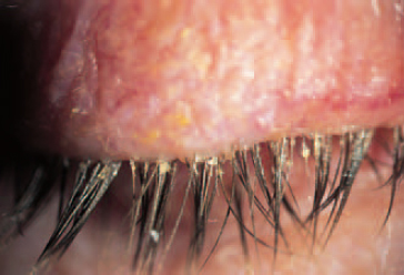
marginal b. Chronic inflammation of the eyelid margin accompanied by crusts or scales usually due to a bacterial infection (e.g. Staphylococcus aureus), an allergy, or to excessive secretion of lipid by the meibomian glands and the glands of Zeis (seborrhoeic blepharitis). The condition is commonly associated with keratoconjunctivitis sicca. Symptoms and signs include burning, itching, grittiness, and the eyelid is hyperaemic and crusted and usually worse in the morning. Treatment consists mainly of frequent cleaning of the lid margins with a cotton-tipped applicator (or face cloth or cotton ball) dipped in a diluted solution of baby shampoo; warm compresses and an antibiotic ointment (e.g. erythromycin) and occasionally systemic antibiotics such as tetracycline, especially in seborrhoeic blepharitis. In complicated cases, corticosteroids will also be used (Fig. B3).
See acne rosacea; glands, meibomian; glands of Zeis; meibomianitis; trichiasis.
posterior b. Chronic inflammation resulting from dysfunction of the meibomian glands characterized either by excessive meibomian secretion (seborrhoeic blepharitis), which is frequently associated with seborrhoeic dermatitis involving the scalp, brows and ears, or inflammation and obstruction of the meibomian glands (meibomianitis).
seborrhoeic b. See blepharitis, marginal.
ulcerative b. Inflammation of the eyelid margin characterized by small ulcers.
blepharochalasis An atrophy of the upper eyelids causing a fold of tissue which often hangs over the eyelid margins. The condition follows recurrent episodes of oedema and inflammation, usually in young people. Treatment is surgical.
See dermatochalasis; epiblepharon.
blepharoconjunctivitis Inflammation of the conjunctiva and eyelids.
See keratitis, herpes simplex.
blepharon See blephara.
blepharophimosis A congenital condition characterized by a generalized narrowing of the palpebral fissure. It produces a pseudoptosis but it commonly forms part of the blepharophimosis syndrome.
blepharoplasty Any operation of the eyelid. It may be done for cosmetic reasons (e.g. to erase the signs of ageing) or for medical reasons (e.g. ptosis, entropion, ectropion).
See blepharochalasis; dermatochalasis.
blepharoplegia Paralysis of an eyelid.
blepharoptosis See ptosis.
blepharorrhaphy suturing of eyelids or of a lacerated lid.
See tarsorrhaphy.
blepharospasm Tonic or chronic spasm of the orbicularis oculi muscle which involves involuntary closure of the eyelids. It is often provoked by a foreign body in the eye, an abrasion or inflammation of the cornea or conjunctiva, or by excessive exposure to ultraviolet light (e.g. actinic keratoconjunctivitis). Treatment consists chiefly of injection into the muscles around the eyelids of botulinum toxin.
See botulinum toxin; chemodenervation; keratoconjunctivitis, actinic; muscle, orbicularis.
blepharostat See eye speculum.
blepharosynechia Adhesion of the eyelids to each other or to the eyeball.
blind Totally or partially unable to see.
b. test See study, single-blind; study, double-blind.
blindness 1 . Inability to see. 2. Absence or severe loss of vision so as to be unable to perform any work for which eyesight is essential. The World Health Organization (WHO) defines blindness as the best corrected visual acuity of 3/60 (20/400) or less, in the better eye. Syn. ablepsia; ablepsy; amaurosis.
blue b. See tritanopia.
colour b. Sometimes this term is incorrectly used to cover all forms of colour vision deficiency, however mild or severe.
See achromatopsia; colour vision, defective; deuteranopia; monochromat; protanopia; tritanopia.
congenital stationary night b. Night blindness (nyctalopia) inherited as either autosomal dominant with non-progressive nyctalopia but normal daylight visual acuity and visual fields and presumed to be due to a defect in neural transmission between the rods and the bipolars in the retina, or autosomal recessive or X-linked with congenital nyctalopia, myopia, nystagmus and reduced visual acuity.
See disease, Oguchi’s; fundus albipunctatus; hemeralopia; retinitis pigmentosa.
cortical b. Loss of vision due to lesions in the areas of both occipital lobes of the brain associated with visual functions. It may result from trauma or from a vascular disease (e.g. a circulatory occlusion caused by a stroke). A lesion in one occipital lobe may result in homonymous hemianopia, often with macular sparing.
day b. See hemeralopia.
eclipse b. Partial or complete loss of central vision due to a foveal lesion caused by fixating the sun without adequate eye protection. This condition is caused mainly by the infrared radiations from the sun.
See actinic.
flash b. See keratoconjunctivitis, actinic.
green b. See deuteranopia.
hysterical b. Blindness associated with an emotional shock, which occurs without a physical or organic cause. The patient has normal blink and pupillary responses and the fundus appears normal. A placebo therapy and/or psychological counselling may be required.
legal b. The definition varies from country to country. In the UK it is equal to either 3/60 (20/400) or worse; or 6/60 (20/200) or worse, with markedly restricted fields.
motion b. A very rare condition in which a patient is unable to process information about motion, although other visual functions are unimpaired. This is believed to be the result of damage to the middle temporal cortex (V5).
See areas, visual association.
night b. See hemeralopia.
perceptual b. See agnosia.
red b. See protanopia.
river b. See onchocerciasis.
snow b. See keratoconjunctivitis, actinic.
word b. See alexia.
blindsight A term used to indicate someone who is totally blind but yet is able, unconsciously, to locate an object on the basis of visual cues. It indicates a lesion which has destroyed the visual cortex but in which the retinotectal pathway to the superior colliculus remains unaffected. This pathway is not involved in conscious vision but receives some information from the retina.
See pathway, retinotectal.
blink A temporary closure of the eyelids (usually of both eyes). Blinks are usually involuntary but may be voluntary. The frequency of blinking is conditioned by a number of external and internal factors, e.g. glare, wind, emotion, attention, tiredness, etc. Normal blink rate is about 10 blinks per minute, although there are wide variations. The duration of a full blink is approximately 0.3–0.4 s. Blink rates are often altered with contact lens wear and in some diseased states (e.g. chalazion, Graves’ disease).
Blivet figure See figure, Blivet.
blobs Cluster of cells found in each hypercolumn of the primary visual cortex (V1), which are, in most instances, colour-opponent but insensitive to orientation, shape or movement. They are about 0.2 mm in diameter and separated by an area about 0.5 mm wide called ‘interblob’. The reason why these cells are called blobs is because they are easily revealed when stained with the enzyme cytochrome oxidase, as these cells derive energy from the oxidase metabolism, whereas the interblobs stain very lightly.
See column, cortical; hypercolumn.
blocking The mounting of one or a number of lens blanks on a holder to form a unit (termed a ‘block’) ready for surfacing. The lens blanks are cemented with pitch, wax, etc.
See surfacing.
blood–brain barrier A mechanism that prevents some substances in the blood from reaching the brain. It is achieved by brain capillaries, which unlike other capillaries elsewhere in the body, are composed of endothelial cells sealed together in continuous tight junctions and surrounded by astrocytes that contribute to the selective passage of substances. Lipid-soluble substances such as alcohol, caffeine, nicotine and most anaesthetics, as well as glucose, oxygen and water, pass rapidly into brain cells, whereas proteins, most antibiotics and ions do not enter or enter very slowly. The mechanism protects brain cells against harmful substances and pathogens.
See system, central nervous.
blood–retina barrier See retina.
blood pressure See sphygmomanometer.
bloomed lens See lens, coated.
blooming See coating.
blot haemorrhage See haemorrhage, blot.
‘blow-out fracture’ See fracture, orbital.
blue Visual sensation evoked by radiations within the waveband 450–490 nm. It is a primary colour and the complementary of yellow.
See colour, complementary; colours, primary.
blue blindness See tritanopia.
blue field entoptoscope See entoptoscope, blue field.
blue-yellow blindness See tritanopia.
blur 1. Degradation of an image formed by an optical system as a result of lack of focusing, aberrations, diffusion of light, etc. 2. A pattern in which the border is indistinct.
See lens flare; vision, blurred.
b. back test See test, plus 1.00 D blur.
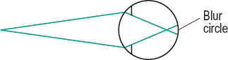
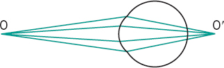
b. circle A circular patch of light formed on the retina resulting from a point object whose image is focused either in front of, or behind the retina, or due to excessive aberrations of the optical system of the eye. The size of the blur circle increases with the distance of the ocular image from the retina and with the diameter of the pupil. Its diameter can be expressed in angular terms (in min arc) as
< ?xml:namespace prefix = "mml" />
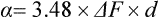
where ΔF is the defocus (in dioptres) with respect to the object point, and d the pupil diameter (in mm) (Figs. B4 and B5). Example: An object at infinity is viewed by a 2 D uncorrected myope with a 4.0 mm pupil diameter, i.e. α = 3.48 × 2 × 4 = 28 min arc. Syn. circle of confusion; circle of diffusion.
See aberration; depth of field.
plus 1.00D b. test See test, plus 1.00 D blur.
spectacle b. Reduction in visual acuity noticed with spectacles after removal of hard contact lenses (PMMA). This may be due to corneal oedema, alteration of the corneal index of refraction, surface distortion of the cornea, etc. Refitting the patient with gas permeable lenses usually relieves this symptom.
blurred vision See vision, blurred.
bobbing, ocular Spontaneous, rapid downward movements of both eyes followed by a slow drift to the straight-ahead position. It occurs in patients, usually comatose, who have lesions of the brainstem.
body 1. Any discrete mass. 2. The main and largest part of a structure. 3. A substance of any kind.
black b. Thermal radiator which absorbs completely all incident radiation, whatever the wavelength, the direction of incidence or the polarization. This radiator has, for any wavelength, the maximum spectral concentration of radiant flux at a given temperature (CIE). Syn. full radiator; planckian radiator.
See absorption; colour temperature; law, Planck’s.
colloid b’s. See drusen.
cytoid b’s. Small, swollen white spots found on the retina resembling cells. They are due to degenerated retinal nerve fibres in which cellular components become trapped in the peripheral axons of the optic nerve blocking axonal flow. Collection of cytoid bodies are thought to represent the ‘cotton-wool’ spots found on or around the optic disc in papilloedema, retinal trauma, diabetic retinopathy, AIDS, systemic lupus erythematosus, etc.
See exudate.
lateral geniculate b’s. See geniculate bodies, lateral.
vitreous b. See humour, vitreous.
white b. A sample exhibiting diffuse reflection and having a reflectance of approximately 100%. Examples: coating of magnesium oxide; sandblasted opal glass surface; plaster of Paris.
Bommarito clip See clipover.
bones of the orbit See orbit.
book retinoscopy See retinoscopy, dynamic.
botulinum toxin A poisonous substance which paralyses muscles and leads to inhibition of the release of acetylcholine from presynaptic neuromuscular terminals. The effect can last for weeks after being injected into a muscle. It is used as an alternative or addition to extraocular muscle surgery in the management of strabismus. It is also sometimes used in the management of blepharospasm. Example: In esotropia, the medial rectus muscle is injected to paralyse its action.
See chemodenervation.
Bowen’s disease See disease, Bowen’s.
Bowman’s layer See layer, Bowman’s.
boxing centre; system See under the nouns.
brachium of the superior colliculus A bundle of nerve fibres that leaves the optic tract below the pulvinar of the thalamus to enter the pretectal nucleus near the superior colliculus. Some fibres also connect the superior colliculus to the lateral geniculate body. Damage to the brachium results in a reduced pupil reflex to light, but not to near objects.
See nucleus, pretectal; reflex, pupil light.
brachymetropia Term proposed by Donders for myopia.
bracketing A procedure used in subjective refraction in which large and equal steps of dioptric changes are made above and below the presumed correct answer, and then reducing the size of the dioptric changes and shifting the centre of the range, until the finest and just detectable blur is induced by equal steps above and below the refractive error. It is commonly used with patients with low vision and also to check the range of clear vision provided by a near addition.
Braille System of printing for blind persons, consisting of points raised above the surface of the paper used as symbols to indicate the letters of the alphabet. Reading is accomplished by touching the points with the fingertips.
break-up time test See test, break-up time.
Brewster’s angle See angle of polarization.
Brewster’s stereoscope See stereoscope, Brewster’s.
bridge That part of a spectacle frame which forms the main connection between the lenses or rims. The bridge assembly is generally taken to include the pads, if any (British Standard).
See spectacles.
flush b. The bridge of a spectacle frame with zero projection.
inset b. A spectacle frame so shaped that the bearing surface of the bridge is behind the plane of the lenses.
keyhole b. Bridge of a spectacle frame with pads, looking like the outline of the upper part of a keyhole.
pad b. A bridge of a spectacle frame with two pads acting as the resting surface on the nose.
saddle b. A bridge so shaped as to rest on the nose over a continuous area, but in which the ends of the bearing surface are extended to lie behind the back plane of the front (British Standard).
brightness Attribute of visual sensation according to which an area appears to emit more or less light. Syn. luminosity. Note 1: In British recommended practice, the term brightness is now reserved to describe brightness of colour (i.e. the opposite of dullness) as used in the dyeing industry. Note 2: This attribute is the psychosensorial correlate, or nearly so, of the photometric quantity luminance (CIE).
Brightness Acuity Tester (BAT) See glare tester.
b. constancy See constancy, brightness.
b. enhancement See effect, Brücke– Bartley.
brimonidine tartrate See alpha-adrenergic agonist.
brinzolamide See carbonic anhydrase inhibitors.
Broca’s pupillometer See pupillometer, Broca’s.
Broca–Sulzer phenomenon See effect, Broca–Sulzer.
Brock’s after-image test See test, after-image transfer.
Brock’s string A white string used to demonstrate physiological diplopia. One end of the string is placed against the bridge of the nose and the other end against a distant object (e.g. a doorknob). The subject should see two strings intersecting wherever the horizontal components of the visual axes meet. Red and green filters, one before each eye, enhance or facilitate the observation of the two strings. Several beads, each of a different colour, are usually threaded on the string so that they can be moved at will. One bead may be used for fixation while the other/s appear double; in crossed diplopia for the one closer to the eyes than the fixation bead, and in uncrossed diplopia for the one further away than the fixation bead. Brock’s string is commonly used in visual training. The observation of physiological diplopia with Brock’s string is often referred to as Brock’s string test. Syn. bead on string.
Brodmann’s areas See areas, Brodmann’s.
Brown’s superior oblique tendon sheath syndrome See syndrome, Brown’s superior oblique tendon sheath.
Bruch’s membrane See membrane, Bruch’s.
Brücke’s muscle See muscle, ciliary.
Brücke–Bartley effect See effect, Brücke–Bartley.
Bruckner’s method See method, Bruckner’s.
bruit A sound heard on auscultation of the heart, lungs, large arteries or veins, or any large cavity (e.g. the orbit). The auscultation is carried out with a stethoscope. Example: An occlusive disease of the carotid artery caused by atherosclerosis leads to a reduction in blood flow through the carotid arteries (and a concomitant reduction in blood flow through vessels of the eye and orbit). It gives rise to a swishing sound with the chest piece of the stethoscope on the neck over the carotid artery. See amaurosis fugax.
brunescent cataract See cataract, nuclear.
brushes, Haidinger’s See Haidinger’s brushes.
Brushfield’s small white or yellow iris spots associated with Down’s syndrome.
B-scan See ultrasonography.
bulbar Pertaining to the eyeball.
bulbar conjunctiva See conjunctiva.
bull’s eye maculopathy See maculopathy, bull’s eye.
bulla A fluid-filled blister appearing on the surface of the cornea when it is severely oedematous (increased thickness of more than 25%). It gives rise to a reduction of visual acuity and pain on rupturing. Example: bullous keratopathy. Plural: bullae.
See keratopathy, bullous.
bullous keratopathy See keratopathy, bullous.
bundle of light See light, beam of.
Bunsen–Roscoe law See law, Bunsen–Roscoe.
buphthalmos See glaucoma, congenital.
bupivacaine hydrochloride A local anaesthetic of the amide type used in eye surgery. It is used in 0.25–0.75% solution. It is often mixed with lidocaine hydrochloride. Its action starts after about 5 minutes and lasts for about 10 hours.
Busacca’s nodules See Koeppe’s nodules.
button The preformed piece of glass which will become the segment of a fused bifocal or multifocal lens. It is ground and polished on one side to the appropriate curvature for fusing to the main lens (British Standard).
See lens, bifocal.


