Autoimmune (IgG4) Pancreatitis

Published on 19/07/2015 by admin

Filed under Radiology

Last modified 22/04/2025

Print this page

rate 1 star rate 2 star rate 3 star rate 4 star rate 5 star
Your rating: none, Average: 0 (0 votes)

This article have been viewed 2201 times

 Sausage-like enlargement of pancreas (with smooth contour) and loss of normal pancreatic lobulations

image Hypoattenuating halo or capsule around pancreas
image Absence of retroperitoneal fluid, fluid collections/pseudocysts, or inflammation
image Less enhancement than expected in arterial phase; parenchyma/capsule may show delayed enhancement
image Diffuse or segmental narrowing of pancreatic duct
image MRCP: Multiple discontiguous MPD/bile duct strictures which resolve after secretin (duct penetrating sign)
• Focal form

image Focal mass or localized enlargement of pancreas (usually head/uncinate) with delayed enhancement
image Lack of biliary or pancreatic ductal dilatation

TOP DIFFERENTIAL DIAGNOSES

• Pancreatic ductal adenocarcinoma
• Chronic pancreatitis
• Acute edematous pancreatitis

PATHOLOGY

• Two distinct histologic subtypes

image Type I: Lymphoplasmacytic sclerosing pancreatitis 

– Positive IgG4 tissue staining; serum IgG4 elevated
– Extrapancreatic organ involvement common (∼ 60%); inflammatory bowel disease in only 2-6%
– Older patients (usually > age 60) with M > F
image Type II: Idiopathic duct-centric pancreatitis

– No IgG4 tissue staining; serum IgG4 not elevated
– No extrapancreatic organ involvement; inflammatory bowel disease in 30%
– Younger patients (mean age 43) with M=F
image
(Left) Axial CECT shows diffuse infiltration and enlargement of the pancreas with loss of normal fatty lobulation. There is a hypodense halo or capsule image around the pancreas, with relatively little spread into adjacent tissues, compatible with autoimmune pancreatitis. All symptoms and signs resolved with steroid therapy.

image
(Right) Transhepatic cholangiogram in a patient with autoimmune pancreatitis shows multifocal strictures image indistinguishable from those of primary sclerosing cholangitis.
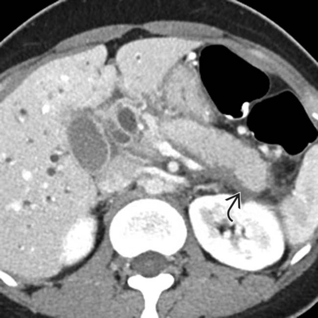
image
(Left) Axial CECT demonstrates a diffusely enlarged pancreas with a low attenuation halo image around its margin.

image
(Right) Coronal CECT from the same patient shows similar findings with a low attenuation capsule image 
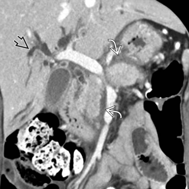
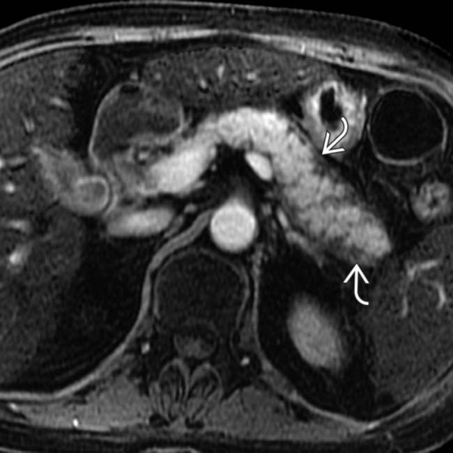
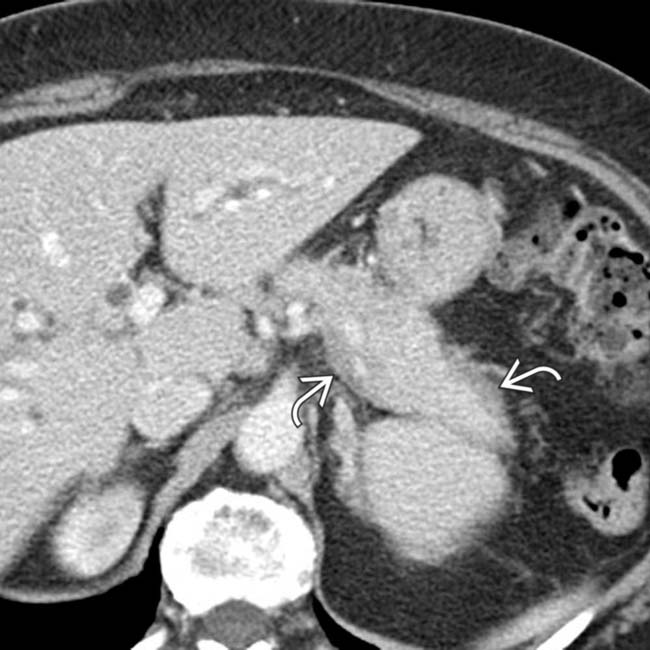
around the enlarged pancreatic margin. Note the presence of biliary dilatation image in this patient with a history of biliary strictures, often associated with autoimmune pancreatitis.

TERMINOLOGY

Abbreviations

• Autoimmune pancreatitis (AIP)

Synonyms

• Lymphoplasmacytic sclerosing pancreatitis; primary sclerosing pancreatitis; tumefactive pancreatitis; non-alcoholic destructive pancreatitis

Definitions

• Immune-mediated fibroinflammatory disease primarily involving pancreas responding to steroid therapy

IMAGING

General Features

• Best diagnostic clue

image Diffusely/focally enlarged pancreas with hypodense halo
image No vascular involvement, calcifications, or pseudocysts
image Lack of significant dilatation of main pancreatic duct
• Location

image May be diffuse, multifocal, or focal/mass-forming
• Morphology

image Sausage-shaped appearance of pancreas

Imaging Recommendations

• Best imaging tool

image MRCP and gadolinium-enhanced MR

CT Findings

• Diffuse form

image Diffuse sausage-like enlargement of pancreas (with smooth contour) and loss of pancreatic lobulations
image Hypoattenuating halo or capsule around pancreas
image Often less enhancement than expected in arterial phase; delayed enhancement of involved parenchyma/capsule
image No retroperitoneal fluid collections or inflammation
image Lymphadenopathy common (20%) with similar halo or capsule
• Focal form

image Focal mass or localized enlargement of pancreas (usually head/uncinate) with delayed enhancement
• Diffuse or segmental narrowing of pancreatic duct
• Extrapancreatic imaging findings

image IgG4 cholangitis in 90%: May be indistinguishable from primary sclerosing cholangitis

– Stricture of common bile duct (CBD) ± intrahepatic ducts with hyperenhancement of duct wall
image Renal involvement in 35% of patients with AIP

– Round or wedge-shaped low attenuation parenchymal lesions
– Diffuse renal enlargement
– Perirenal soft tissue rind (mimicking lymphoma)
– Urothelial thickening in renal pelvis
image Retroperitoneal fibrosis, IgG4-related lung disease, and enlarged salivary glands or salivary gland mass
image IgG4-related lung disease

– Solid nodules, ground glass opacities, interstitial opacities

MR Findings

• Diffuse enlargement of pancreas (T1WI hypointense and T2WI hyperintense)
• Capsule of peripheral hypoenhancement and low T2WI signal with delayed enhancement
• Delayed enhancement of involved parenchyma
• MR cholangiopancreatography (MRCP): Multiple discontiguous main pancreatic duct/bile duct strictures

image Strictures resolve after secretin (duct-penetrating sign)
• DWI: Mildly restricted diffusion of affected tissue

image Cannot use DWI to differentiate focal AIP from pancreatic cancer

Ultrasonographic Findings

• Transcutaneous ultrasound is of limited value; may have normal appearance
• Endoscopic ultrasound

image Enlarged hypoechoic gland with sausage-like appearance, narrowed MPD, and thickening of CBD wall
image Echogenic interlobular septa

Nuclear Medicine Findings

• PET/CT

image FDG uptake very common (83-100%) compared to traditional chronic pancreatitis (13%)
image Diffuse uptake throughout pancreas (unlike malignancy)
image Extrapancreatic uptake (salivary glands, kidneys) suggests AIP
image FDG uptake may decrease after therapy with steroids

Other Modality Findings

• ERCP: Diffuse or segmental narrowing (often with multiple discontinuous strictures) of main pancreatic duct (MPD)

image Long, narrow strictures typically greater than 1/3 length of pancreatic duct
image Lack of upstream dilatation of pancreatic duct proximal to strictures
image Side branch ducts arising from stricture

DIFFERENTIAL DIAGNOSIS

Pancreatic Ductal Carcinoma

• Focal AIP difficult to differentiate from pancreatic cancer
• Upstream MPD dilatation and glandular atrophy (unlike AIP)
• Multiple strictures (as with AIP) uncommon
• Encasement of major peripancreatic vessels

Chronic Pancreatitis

• Atrophic gland with beaded, dilated pancreatic duct and parenchymal/intraductal calcifications
• History of alcoholism and pain symptoms (unlike AIP)

Acute Edematous Pancreatitis

• Enlarged, inflamed pancreas with retroperitoneal fluid and inflammation (uncommon in AIP)

PATHOLOGY

General Features

• Etiology

image Exact pathogenesis unknown, but autoimmune etiology likely given steroid response/elevated antibody titers
image Variety of autoantibodies have been identified in association with AIP, as well as alterations in regulatory T-cells and cytokines
image Unclear whether IgG4 plays a pathogenic role in type I or is an epiphenomenon
• Genetics

image Genetic studies suggest association with specific human leukocyte antigen (HLA) types
• Associated abnormalities

image Often associated with other autoimmune diseases (Sjögren syndrome, IgG4-associated cholangitis, autoimmune thyroiditis, interstitial nephritis, primary biliary cirrhosis, ulcerative colitis, SLE)

Staging, Grading, & Classification

• Type I: Lymphoplasmacytic sclerosing pancreatitis

image More common than Type II (80% of cases in US)
image Older patients (usually > age 60) with M > F
image Symptoms can mimic pancreatic cancer (obstructive jaundice in 75%, acute pancreatitis in 15%, diabetes in 85%, chronic abdominal pain, weight loss, steatorrhea)
image Positive IgG4 tissue staining; serum IgG4 elevated
image Extrapancreatic organs often involved (∼60%)
image Inflammatory bowel disease in 2-6%
image Relapses after steroid therapy frequent (up to 40%)
image Imaging: Diffuse (60%) or focal lesions (40%)
• Type II: Idiopathic duct-centric pancreatitis

image Younger patients (mean age 43); no M/F predominance
image No IgG4 tissue staining; serum IgG4 normal
image No extrapancreatic organ involvement; IBD in 30%
image Presentation: Obstructive jaundice 50%, acute pancreatitis 33%; abdominal pain
image Rarely relapses after steroid therapy
image Imaging: Focal pancreatic lesions (85%)

Gross Pathologic & Surgical Features

• Firm or rock-hard in palpation
• Inflammatory process involving either entire pancreas or only a portion of it (most likely head)

Microscopic Features

• Type 1: Dense lymphoplasmacytic infiltration of parenchyma with scattered eosinophils

image Inflammatory cells aggregated around interlobular pancreatic ducts
image Storiform fibrosis and obliterative phlebitis common
image Pancreatic ductal architecture preserved
image Prominent lymphoid follicles
image Positive IgG4 staining
• Type 2: Neutrophilic infiltration of parenchyma associated with characteristic granulocyte-epithelial lesions

image Negative staining for IgG4
image Pancreatic duct destruction
image Rarely storiform fibrosis and obliterative phlebitis
image Dense periductal inflammation with neutrophils
• Both types 1 and 2 lack intraductal protein plugs, calcifications and pseudocysts seen with other forms of chronic pancreatitis such as alcoholic pancreatitis

CLINICAL ISSUES

Presentation

• Most common signs/symptoms

image Type I AIP: Obstructive jaundice 75%, acute pancreatitis 15%, diabetes 85%, chronic abdominal pain, weight loss
image Type II AIP: Obstructive jaundice 50%, acute pancreatitis 33%, abdominal pain
image Much less pain than other forms of pancreatitis
image If pancreatic atrophy develops, patients may have steatorrhea
• Other signs/symptoms

image Type II AIP patients may have symptoms of IBD
image Type I AIP often has extrapancreatic disease, including thyroiditis, dry eyes and mouth from Sjögren-like syndrome, peripheral lymphadenopathy, and renal impairment from IgG4 nephropathy and retroperitoneal fibrosis
• Clinical profile

image Lab findings: Type I: ↑ IgG4 (sensitive and specific) in 80%, normal or ↑ serum amylase/lipase, ↑ ALP/GGT, ↑ bilirubin, ↑ ANA, ↑ autoantibodies, ↑ CA 19-9
image Lab findings: Type II: No elevation of IgG4
image Of note is potential for false positive elevation of IgG4 in 10% of patients with pancreatic cancer

Demographics

• Age

image Middle-aged to elderly (Type I in 6th decade; type II in 4th decade)
• Gender

image Type I: Male > female; type II: Male = female
• Epidemiology

image 5-6% of chronic pancreatitis cases
image 6-8% of pancreatic resections for presumed carcinoma had autoimmune pancreatitis
image Incidence in Japan estimated at 0.82% per 100,000
image No history of alcohol abuse, smoking, or biliary stones

– No traditional risk factors for chronic pancreatitis

Natural History & Prognosis

• Slowly progresses to end-stage disease unless treated
• Type I may have frequent relapses after steroid therapy
• 50% have diabetes that may improve with steroid therapy
• No acute exacerbation (unlike EtOH chronic pancreatitis)
• Dramatic response to steroids, with immunomodulators utilized if steroid resistant

Treatment

• Some cases may resolve spontaneously without treatment
• Prednisone 40 mg; immunomodulators if steroid resistant

DIAGNOSTIC CHECKLIST

Consider

• Pancreatic carcinoma if focal mass with significant upstream pancreatic ductal dilatation

Image Interpretation Pearls

• Sausage-like enlargement of pancreas without pancreatic ductal dilatation
image
(Left) Axial T1 C+ MR image in the arterial phase demonstrates diffuse pancreatic enlargement with a low signal rim or capsule image around the margin of the pancreas.

image
(Right) Axial T1 C+ MR in a more delayed phase demonstrates that the capsule image 
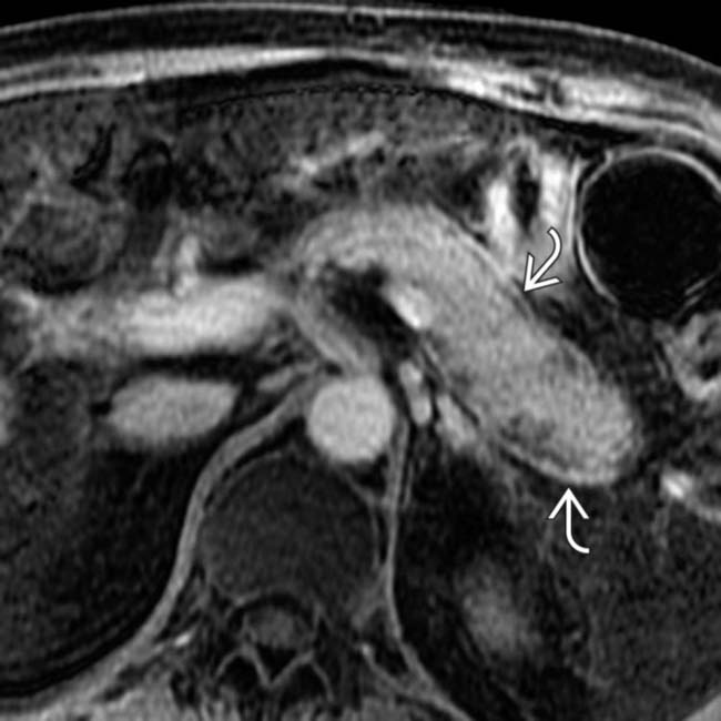
now shows avid delayed enhancement, a characteristic feature of autoimmune pancreatitis.
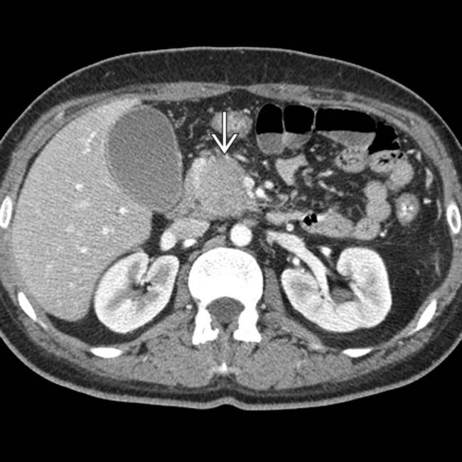
image
(Left) Axial CECT shows a hypodense mass in the head of the pancreas mimicking a ductal adenocarcinoma image.

image
(Right) Coronal reformatted CECT in the same patient shows the focal mass image 
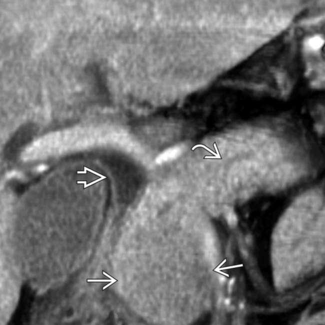
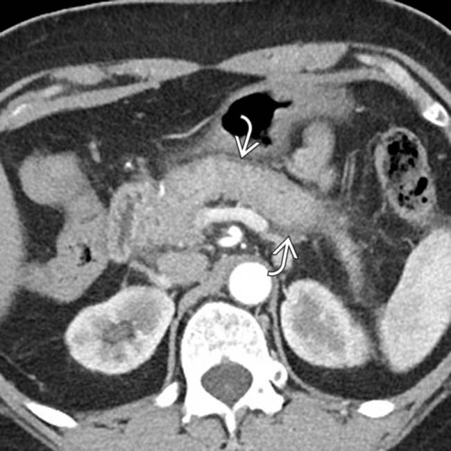
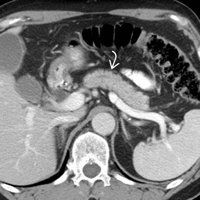
in the pancreatic head, a tapered stricture of the common bile duct image, and a normal pancreatic duct image. Endoscopic ultrasound-guided biopsy of the pancreatic head mass revealed autoimmune pancreatitis. The lack of dilatation of the pancreatic duct was an important clue to the diagnosis.
image
(Left) Axial CECT shows diffuse enlargement and loss of fatty lobulation of the entire tail of the pancreas image
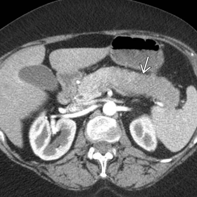
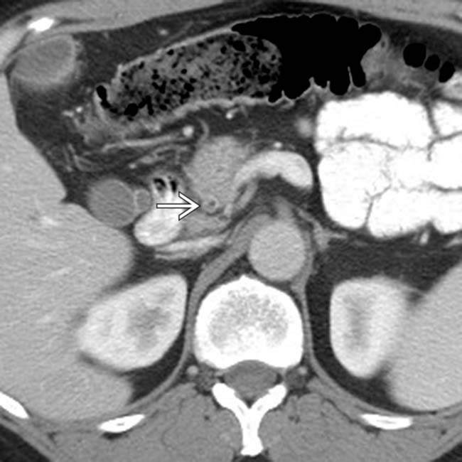
. The pancreatic duct in this segment is not identified and there is little surrounding inflammation.

image
(Right) Axial CECT at a more caudal level in the same patient shows that the pancreas has a subtle surrounding low-attenuation halo image
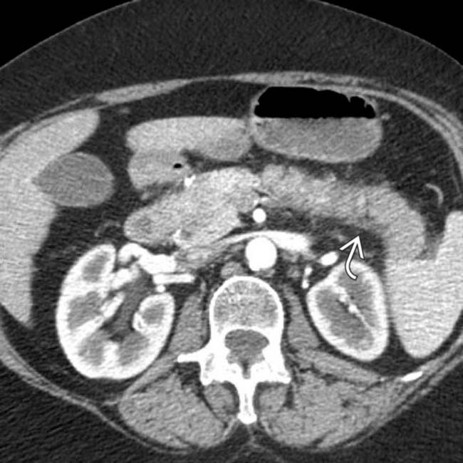
, typical of autoimmune pancreatitis.
image
(Left) Initial axial CECT shows a focal hypodense mass image 
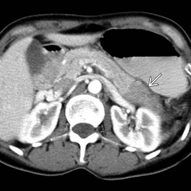
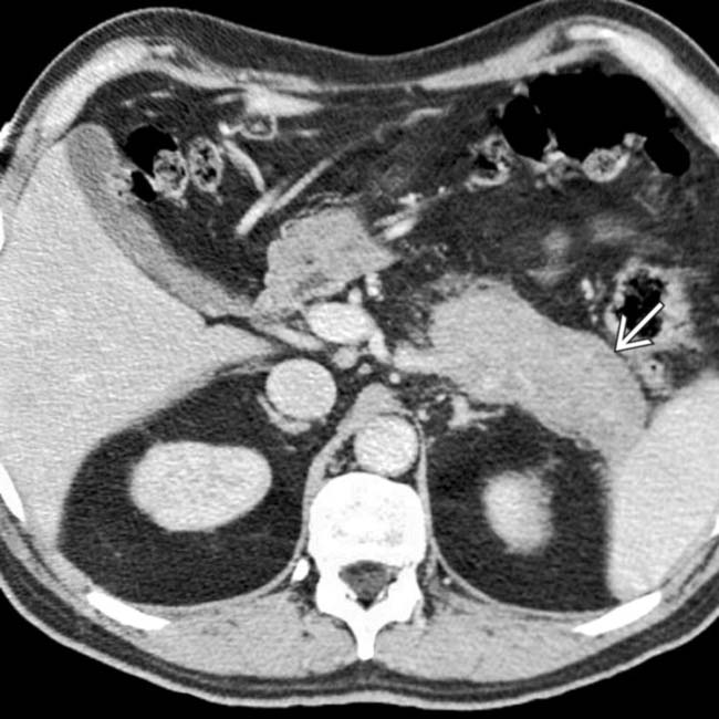
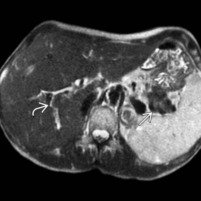
in the tail of the pancreas. Serologic testing (IgG4) suggested the diagnosis of autoimmune pancreatitis and steroid therapy was started.

image
(Right) Repeat axial CECT in the same patient 10 months later shows substantial decrease in size of the inflammatory mass image 
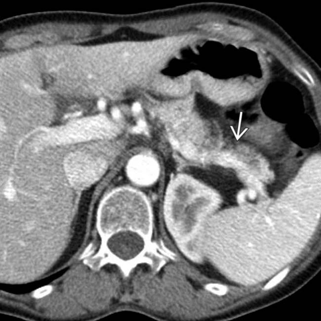
and slight atrophy of the affected pancreatic tail segment.
image
(Left) Axial CECT demonstrates mild enlargement of the pancreas with a subtle low-density capsule image 
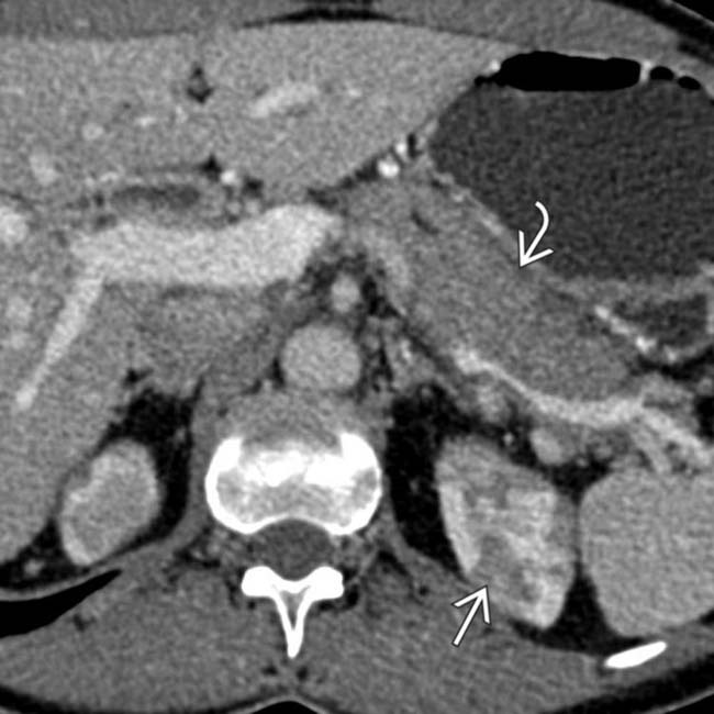
in a patient with an elevated IgG4, compatible with autoimmune pancreatitis. Note the low-density wedge-shaped lesions image in the left kidney.

image
(Right) Coronal CECT in the same patient better demonstrates the full extent of the wedge-shaped low density lesions in both kidneys. Renal lesions, likely a manifestation of autoimmune disease, are a frequent associated finding in patients with AIP.
image
(Left) Axial CECT in a patient with chronic abdominal pain demonstrates an enlarged pancreas with a subtle low-attenuation capsule image
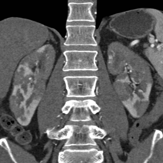
. This was thought to likely represent autoimmune pancreatitis, and the patient was found to have elevated IgG4.

image
(Right) Axial CECT in the same patient following treatment with steroids demonstrates that the pancreas now appears normal in size with resolution of the previously seen low-attenuation halo.
image
Axial CECT in a patient with autoimmune pancreatitis demonstrates diffuse enlargement of the pancreas with a low-attenuation halo image 
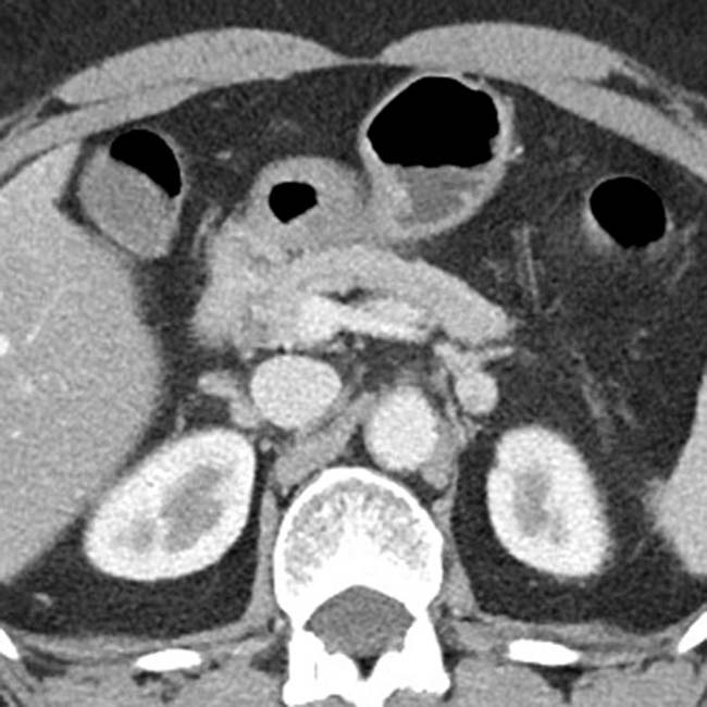
surrounding the gland.
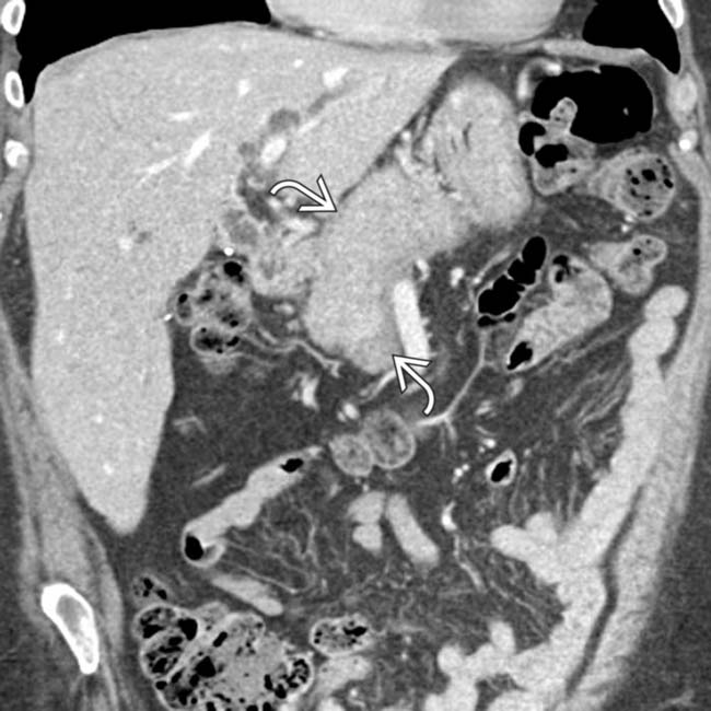
image
Coronal CECT from the same patient again demonstrates a diffusely sausage-shaped, enlarged gland with a low-attenuation halo, highly suggestive of autoimmune pancreatitis.
image
Axial CECT demonstrates a mildly enlarged pancreas with a subtle low-attenuation halo image.
image
Axial CECT in the same patient demonstrates hyperenhancement of the wall of the distal CBD image. This constellation of findings is highly suggestive of autoimmune pancreatitis.
image
Axial T2WI in the same patient shows diffuse enlargement and loss of normal fatty lobulation of the pancreas, along with a hypointense halo image 
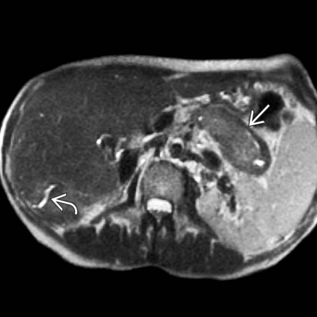
around the pancreas, typical features of autoimmune pancreatitis. Multifocal, irregular dilation of the intrahepatic bile ducts is also seen image.
image
Axial T2WI in a 43-year-old woman with thyroiditis, cholangitis, and pancreatitis shows irregular dilation of the intrahepatic bile ducts image and a hypointense halo image around the tail of the pancreas.

Share this: