136
Atypical mole syndrome (dysplastic nevus syndrome)
Atypical mole syndrome consists of multiple clinically atypical nevi, together with an increased risk of melanoma. It occurs as a familial syndrome.
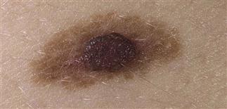
The fried egg pattern for an atypical nevus. The center is dark and raised with a lighter, less distinct scalloped border.
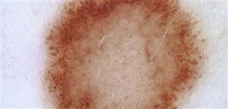
The pigmentation of atypical nevi has several distinct patterns. Here, there is a broad homogeneous white center with a hyperpigmented rim.
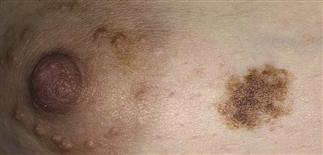
Here, the hypopigmented and hyperpigmented components are intermixed. Magnification with dermoscopy showed that the pigmentation was uniform.
DESCRIPTION
Multiple clinically atypical nevi, with increased melanoma risk. Familial syndrome or sporadic.
HISTORY
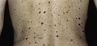
• As familial syndrome, affected members have many irregular nevi, multiple family members with melanoma. Inheritance appears autosomal dominant with variable penetrance. • Nevi were considered marker for risk of developing melanoma. • Syndrome is uncommon. • Solitary atypical nevi common, prevalence of 5–20%. • Males, females equally affected. • Atypical nevi not present at birth; begin appearing in childhood. Unlike common acquired nevi, which stop appearing after age 30, atypical nevi continue to appear in adulthood. • While sun exposure favors appearance of nevi, lesions also develop in sun-protected areas. • Most affected people have > 50 nevi, some of which appear atypical. • Striking heterogeneity from one nevus to another. • Atypical nevi regarded as along continuum between benign and malignant melanocytic neoplasms. Likelihood of an individual atypical nevus subsequently developing into melanoma cannot be estimated. Lifetime melanoma risk estimated at 1.3%. Risk for persons with atypical nevi, but without family history of melanoma, estimated at 6%. Risk increases to 15% in patients with atypical nevi and family history of melanoma. Persons with familial atypical nevi have 150-fold increased risk of developing melanoma by age 70, 500-fold increased risk if patient has already had melanoma.
PHYSICAL FINDINGS
• Atypical nevi usually larger (6–15 mm in diameter), with irregularly outlined, indistinct border. Color varies: pink, tan, brown, black. Surface irregular, may contain central or eccentric papule surrounded by a prominent macular component. • Affected persons often have nevi in sun-protected areas. • Pathologists often use terms such as mild to moderate atypia when describing these lesions. This does not mean that they are or would have evolved into a melanoma.
TREATMENT
• Patients with atypical nevi should have routine full-skin examinations after puberty, with follow-up every 3–12 months. Consider dermatologic referral for regular monitoring of nevi. Examine entire cutaneous surface. Also examine family members. • Changing lesions should be biopsied. • Emphasize patient education and awareness. Teach patients to perform regular skin self-examinations. • Consider photomapping to assist patient in self-monitoring between visits.