20
Atopic dermatitis
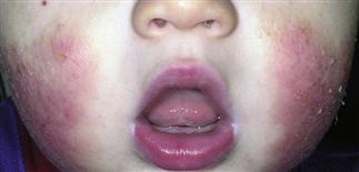
Atopic dermatitis in a young toddler. The child scratches the face by rubbing. Eczematous patches are evident on both cheeks. Use of emollients daily is helpful.
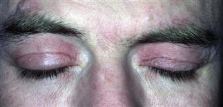
Atopic dermatitis has a predilection for the eyelids. Affected eyelids are red and scaling, and develop lichenification over time. Avoidance of irritants and allergens is a good management strategy.
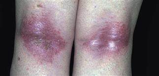
Eczematous dermatitis on dorsal forearm. The prominent finding is lichenification; this indicates a long-standing problem. Ointment-based treatments will penetrate this thickened skin better.
A typical site for atopic dermatitis in childhood is the flexure popliteal fossae and the antecubital fossae. These erythematous scaling patches are very pruritic and sometimes weeping.
DESCRIPTION
An eczematous dermatitis, distressingly pruritic, recurrent, often flexural, and symmetric.
HISTORY
• Major diagnostic criteria: pruritus; young age at onset; flexural lichenification and linearity in adults; facial and extensor involvement in infants; chronic relapsing course; personal or family history of asthma, allergic rhinoconjunctivitis, atopic dermatitis. • Minor, less specific features: xerosis, ichthyosis, palmar hyperlinearity, keratosis pilaris, immediate type 1 skin test responses, dermatitis of hands and feet, cheilitis, nipple eczema, increased susceptibility to cutaneous infections, perifollicular accentuation. Most children outgrow, but some develop chronic relapsing disease, especially retroauricular, eyelids, and hands.
PHYSICAL FINDINGS
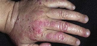
• Often begins abruptly with erythema, severe pruritus. • Red pruritic papules, patches of erythema, scaling. • Acute lesions oozing and vesicular; subacute lesions scaly and crusted; chronic lesions dull red, lichenified, very pruritic. • Infant distribution: cheeks, perioral, scalp, dorsal feet, elbows. • Children: flexural; antecubital, popliteal, neck, wrists, ankles. • Adults: flexural. Hand dermatitis may be sole manifestation. Upper eyelids common site. Can be diffuse and patchy. • Secondary infection with Staphylococcus aureus resulting in flare or rash persistence. Increased susceptibility to herpes simplex, molluscum contagiosum, S. aureus, and fungal infections. • Hypopigmentation and hyperpigmentation may result from inflammation. • Patch test if dermatitis pattern changes or is refractory to treatment. • Children with severe disease may have food allergies. Rarely a problem for adults.
TREATMENT
• Frequent application of a plain, thick, greasy moisturizer (Vaseline, Aquaphor, Aveeno, Vanicream) without fragrances or common sensitizing preservatives is ideal. • Advise soft, light cotton clothing; avoid wool, coarse fibers. • Keep environment cool. • Oral antihistamines with sedative effects for pruritus—such as diphenhydramine (Benadryl), hydroxyzine (Atarax), and doxepin—especially at night. • Control or eliminate inflammation and infection. • Topical corticosteroids of appropriate strength for patient’s age and affected area are applied twice daily to inflamed skin for 10–21 days (see Topical therapy). • Second-line treatment: tacrolimus (Protopic) ointment 0.03% for children aged 2–15 years, and 0.03% and 0.1% for adults, or pimecrolimus (Elidel) cream 1% twice daily for short-term and intermittent long-term therapy. Helpful for atopic dermatitis of face, eyelids, or limited involvement. • Staph aureus is the usual flare provoking bacteria, but sensitivity patterns are changing. Oral antibiotics may be helpful; choice should be guided by culture and sensitivity results. Topical mupirocin (Bactroban) cream twice daily for 5 days if infection is limited. • For acute lesions and severe flares, wet dressings with Burow’s solution applied for 20 min 2–3 times a day, followed by a topical steroid. • Consult dermatologist for severe, refractory disease.


