Atelectasis

Published on 22/03/2015 by admin

Filed under Pediatrics

Last modified 22/04/2025

Print this page

rate 1 star rate 2 star rate 3 star rate 4 star rate 5 star
Your rating: none, Average: 0 (0 votes)

This article have been viewed 1428 times

Chapter 402 Atelectasis

Atelectasis, the incomplete expansion or complete collapse of air-bearing tissue, results from obstruction of air intake into the alveolar sacs. Segmental, lobar, or whole lung collapse is associated with the absorption of air contained in the alveoli, which are no longer ventilated.

Pathophysiology

The causes of atelectasis can be divided into five groups (Table 402-1). Respiratory syncytial virus (RSV) and other viral infections in young children can cause multiple areas of atelectasis. Mucous plugs are a common predisposing factor to atelectasis. Massive collapse of one or both lungs is most often a postoperative complication but occasionally results from other causes, such as trauma, asthma, pneumonia, tension pneumothorax (Chapter 405), aspiration of foreign material (Chapters 379 and 389), and paralysis, or after extubation. Massive atelectasis is usually produced by a combination of factors, including immobilization or decreased use of the diaphragm and the respiratory muscles, obstruction of the bronchial tree, and abolition of the cough reflex.

Table 402-1 ANATOMIC CAUSES OF ATELECTASIS

CAUSE CLINICAL EXAMPLES
External compression on the pulmonary parenchyma Pleural effusion, pneumothorax, intrathoracic tumors, diaphragmatic hernia
Endobronchial obstruction completely obstructing the ingress of air Enlarged lymph node, tumor, cardiac enlargement, foreign body, mucoid plug, broncholithiasis
Intraluminal obstruction of a bronchus Foreign body, granulomatous tissue, tumor, secretions, including mucous plugs, bronchiectasis, pulmonary abscess, asthma, chronic bronchitis, acute laryngotracheobronchitis
Intrabronchiolar obstruction Bronchiolitis, interstitial pneumonitis, asthma
Respiratory compromise or paralysis Neuromuscular abnormalities, osseous deformities, overly restrictive casts and surgical dressings, defective movement of the diaphragm, or restriction of respiratory effort

Clinical Manifestations

Symptoms vary with the cause and extent of the atelectasis. A small area is likely to be asymptomatic. When a large area of previously normal lung becomes atelectatic, especially when it does so suddenly, dyspnea accompanied by rapid shallow respirations, tachycardia, cough, and often cyanosis occurs. If the obstruction is removed, the symptoms disappear rapidly. Although it was once believed that atelectasis alone can cause fever, studies have shown no association between atelectasis and fever. Physical findings include limitation of chest excursion, decreased breath sound intensity, and coarse crackles. Breath sounds are decreased or absent over extensive atelectatic areas.

Massive pulmonary atelectasis usually presents with dyspnea, cyanosis, and tachycardia. An affected child is extremely anxious and, if old enough, complains of chest pain. The chest appears flat on the affected side, where decreased respiratory excursion, dullness to percussion, and feeble or absent of breath sounds are also noted. Postoperative atelectasis usually manifests within 24 hr of operation but may not occur for several days.

Acute lobar collapse is a frequent occurrence in patients receiving intensive care. If undetected, it can lead to impaired gas exchange, secondary infection, and subsequent pulmonary fibrosis. Initially, hypoxemia may result from ventilation-perfusion mismatch. In contrast to atelectasis in adult patients, in whom the lower lobes and, in particular, the left lower lobe are most often involved, 90% of cases in children involve the upper lobes and 63% involve the right upper lobe. There is also a high incidence of upper lobe atelectasis and, especially, right upper lobe collapse in patients with atelectasis being treated in neonatal intensive care units. This high incidence may be due to movement of the endotracheal tube into the right mainstem bronchus, where it obstructs or causes inflammation of the bronchus to the right upper lobe.

Diagnosis

The diagnosis of atelectasis can usually be established by chest radiographic examination. Typical findings include volume loss and displacement of fissures. Atypical presentations include atelectasis manifesting as a masslike opacity and atelectasis in an unusual location. Lobar atelectasis may be associated with pneumothorax.

In asthmatic children, chest radiography demonstrates an abnormality rate of 44%, compared with a thorax high-resolution CT (HRCT) scan abnormality rate of 75%. Children with asthma and atelectasis have an increased incidence of right middle lobe syndrome, acute asthma exacerbations, pneumonia, and upper airway infections.

In foreign body aspiration, atelectasis is one of the most common radiographic findings. The site of atelectasis usually indicates the site of the foreign body (Chapter 369.1). Atelectasis is more common when diagnosis of foreign body aspiration is >2 wk.

Bronchoscopic examination reveals a collapsed main bronchus when the obstruction is at the tracheobronchial junction and may also disclose the nature of the obstruction.

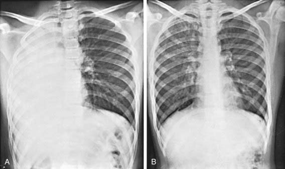
Massive pulmonary atelectasis is generally diagnosed on chest radiograph. Typical findings include elevation of the diaphragm, narrowing of the intercostal spaces, and displacement of the mediastinal structures and heart toward the affected side (Fig. 402-1).

Treatment

Treatment depends on the cause of the collapse. If effusion or pneumothorax is responsible, the external compression must first be removed. Often vigorous efforts at cough, deep breathing, and percussion will facilitate expansion. Aspiration with sterile tracheal catheters may facilitate removal of mucous plugs. Continuous positive airway pressure (CPAP) may improve atelectasis.

Bronchoscopic examination is immediately indicated if atelectasis is the result of a foreign body or any other bronchial obstruction that can be relieved. For bilateral atelectasis, bronchoscopic aspiration should also be performed immediately. It is also indicated when an isolated area of atelectasis persists for several weeks. If no anatomic basis for atelectasis is found and no material can be obtained by suctioning, the introduction of a small amount of saline followed by suctioning allows recovery of bronchial secretions for culture and, possibly, for cytologic examination. Frequent changes in the child’s position, deep breathing, and chest physiotherapy may be beneficial. Intrapulmonary percussive ventilation is a chest physiotherapy technique that has been shown to be safe and effective. Oxygen therapy is indicated when there is dyspnea or desaturation. Intermittent positive pressure breathing and incentive spirometry are recommended when atelectasis does not improve after chest physiotherapy.

In some conditions, such as asthma, bronchodilator and corticosteroid treatment may accelerate atelectasis clearance. Recombinant human DNase (rhDNase), which is approved only for the treatment of cystic fibrosis, has been used off label for patients without cystic fibrosis who have persistent atelectasis. This product reduces the viscosity of purulent bronchial debris. In patients with acute severe asthma, diffuse airway plugging with thick viscous secretions frequently occurs, with the resulting atelectasis often refractory to conventional therapy. rhDNase has been used in both nebulized form for nonintubated patients with acute asthma as well as intratracheally for atelectasis in intubated asthmatics, with resolution of atelectasis unresponsive to conventional asthma therapies.

Lobar atelectasis in cystic fibrosis is discussed in Chapter 395.

Atelectasis can occur in patients with neuromuscular diseases. These patients tend to have ineffective cough and difficulty expelling respiratory tract secretions, which lead to pneumonia and atelectasis. Several devices and treatments are available to assist these patients, including intermittent positive pressure breathing, a mechanical insufflator-exsufflator, and noninvasive bilevel positive pressure ventilation via nasal mask or full-face mask. Patients with neuromuscular disease who have undergone surgery are at substantial risk for postoperative atelectasis and subsequent pneumonia. Migrating atelectasis in the newborn infant, a rare and unique presentation, may be secondary to neuromuscular disease.

There is an association between the development of lobar collapse and the requirement for mechanical ventilation. Although lobar collapse is rarely a cause of long-term morbidity, its occurrence may necessitate the prolongation of mechanical ventilation or re-intubation. In ventilated patients, positive end-expiratory pressure (PEEP) or CPAP is generally indicated.

Airway clearance therapies utilized for adults are often recommended and/or utilized in pediatric populations. However, given the differences in respiratory physiology and anatomy between children and adults, practices applicable to one may or may not apply to the other. Atelectasis due to cystic fibrosis is the only pediatric entity that clearly benefits from airway clearance therapy, although that due to neuromuscular disease, cerebral palsy, or mechanical ventilation probably benefits from such therapy (Table 402-2). Thus far no specific airway clearance therapy has been demonstrated to be superior.

Table 402-2 BENEFIT OF AIRWAY CLEARANCE THERAPIES IN PEDIATRIC CONDITIONS

CLEAR AND PROVEN BENEFIT

Cystic fibrosis

PROBABLE BENEFIT

POSSIBLE BENEFIT

Prevention of post-extubation atelectasis in neonates

MINIMAL TO NO BENEFIT

From Schechter MS: Airway clearance applications in infants and children, Resp Care 52:1382–1390, 2007.

Bibliography

Ashizawa K, Hayashi K, Aso N, et al. Lobar atelectasis: diagnostic pitfalls on chest radiography. Br J Radiol. 2001;74:89-97.

Birnkrant DJ. The assessment and management of the respiratory complications of pediatric neuromuscular diseases. Clin Pediatr. 2002;41:301-308.

Engoren M. Lack of association between atelectasis and fever. Chest. 1995;107:81-84.

Girardi G, Contador AM, Castro-Rodriguez JA. Two new radiological findings to improve the diagnosis of bronchial foreign-body aspiration in children. Pediatr Pulmonol. 2004;38:261-264.

Hendriks T, de Hoog M, Lequin MH, et al. DNase and atelectasis in non-cystic fibrosis pediatric patients. Critical Care. 2005;9:R351-R356.

Miske LJ, Hickey EM, Kolb SM, et al. Use of the mechanical In-Exsufflator in pediatric patients with neuromuscular disease and impaired cough. Chest. 2004;125:1406-1412.

Scolieri P, Adappa ND, Coticchia JM. Value of rigid bronchoscopy in the management of critically ill children with acute lung collapse. Pediatr Emerg Care. 2004;20:384-386.

Thomas K, Habibi P, Britto J, et al. Distribution and pathophysiology of acute lobar collapse in the pediatric intensive care unit. Crit Care Med. 1999;27:1594-1597.

Tokar B, Ozkan R, Ilhan H. Tracheobronchial foreign bodies in children: importance of accurate history and plain chest radiography in delayed presentation. Clin Radiol. 2004;59:609-615.

Tsai SL, Crain EF, Silver EJ, et al. What can we learn from chest radiographs in hypoxemic asthmatics? Pediatr Radiol. 2002;32:498-504.

Yen Ha TK, Bui TD, Tran AT, et al. Atelectatic children treated with intrapulmonary percussive ventilation via a face mask: clinical trial and literature overview. Pediatr Int. 2007;49:502-507.