Asthma: Epidemiology, Pathophysiology, and Risk Factors

Published on 23/05/2015 by admin

Filed under Pulmolory and Respiratory

Last modified 22/04/2025

Print this page

rate 1 star rate 2 star rate 3 star rate 4 star rate 5 star
Your rating: none, Average: 2 (1 votes)

This article have been viewed 4744 times

Chapter 38 Asthma

Epidemiology, Pathophysiology, and Risk Factors

Asthma has been described throughout time, beginning with ancient Egyptians. The Georg Ebers Papyrus, found in Egypt in the 1870s, contains prescriptions written in hieroglyphics for more than 700 treatments for the disorder. The inhalation of fumes extracted from the heating of herbs in bricks was one of the remedies available. The term asthma comes from the Greek word aazein, meaning “to pant, or breathe with the mouth open.” Between the 1930s and 1950s, asthma was considered to be a psychosomatic illness. During the 1960s, the emergence of the inflammation theory refuted a psychological origin and proved that asthma is a physical disease. Nonetheless, an association between psychological conditions (e.g., anxiety, depression) and difficult-to-treat asthma has been recognized since that time.

Nowadays, asthma is defined as a chronic inflammatory process characterized by reversible and variable airflow obstruction due to bronchial responsiveness secondary to multiple external stimuli in which genetic factors interact with environmental factors. The Global Initiative for Asthma (GINA) gives an operational description of asthma, as follows:

At present, much research is directed at elucidation of the underlying causes of asthma, which remain unknown. The risk for development of asthma is supported by evidence for a mixture of genetic, environmental, and lifestyle factors. Asthma does not respect age or gender, affecting both children and adults from kindergarten and school through work to retirement. For the moment, there is no cure for this condition, so preventive strategies are being effectively applied at the community level to prevent onset of the condition and to control worsening of asthma symptoms in the future. Nevertheless, some of these approaches have failed to reach the entire spectrum of the community population, for various reasons, such as limited access to information, personal beliefs, religious practices, and ethnicity within emigrational cohorts. Moreover, clinical practice guidelines and their distribution are basic for acute asthma diagnosis, treatment, and control.

Epidemiology

The World Health Organization (WHO) estimates that 300 million people have asthma worldwide, producing significant morbidity and interfering with daily activities and quality of life. During the year 2009, 250,000 people died from this condition in low- to middle-income countries. Furthermore, asthma is the most frequent chronic respiratory condition among children worldwide, and its prevalence is increasing. The Centers for Disease Control and Prevention (CDC) has estimated a significant rise in asthmatic patients of 12.3% since 2001 in the United States. Statistical figures for 2009 show that 24.6 million people had asthma, compared with 20.3 million at the beginning of the decade. This increase has come with a cost to society of $56 billion dollars in medical expenses and lost productivity. No clear explanation for this increased prevalence, however, has emerged, especially in view of the overall reduction in smoking and in second-hand smoke exposure with the implementation of laws banning smoking. The same situation is described outside the United States. From a metaanalysis of data obtained through routine statistics and population surveys, Anderson and co-workers concluded that asthma prevalence increased in the United Kingdom from 1955 to 2004.

Despite a large number of research studies, the reasons for why some people develop asthma and others do not and why asthma has emerged as a public health problem in some populations earlier than in others are not well understand. The main problem in focusing on the epidemiology of asthma is to address a proper operational definition in an attempt to unify the large conclusions of many epidemiologic studies from different areas of the world. The need to derive a practical and tangible definition of asthma for use in large-scale questionnaire-based epidemiologic research has led to a focus on asthma symptoms and their highly subjective expressions as part of the results. In questionnaire surveys, the presence of asthma often is defined on the basis of responses to questions about symptoms of wheeze in the past weeks or months, “wheeze ever,” and doctor-diagnosed asthma. This approach has been shown to have good short-term repeatability but may lack specificity, particularly in children, because other causes of wheezing illness, such as viral infection, may be misdiagnosed as asthma. With a focus on symptoms, the most common clinical presentation is one of breathlessness, wheezing, chest tightness, and cough, especially at night, coexisting with asymptomatic periods. Because of the nonspecificity and variability inherent in personal perception of respiratory difficulty, implementation of vital patient educational programs and pharmacologic treatment of asthma symptoms may be delayed. Clinical symptoms vary from one person to another, so a severe asthma exacerbation that necessitates urgent medical attention may not be recognized at first, and consequent delay in getting required treatment may lead to a poorer outcome.

Reported prevalence rates for asthma are widely variable. Despite the difficulty in obtaining a consensus on epidemiologic data, several studies have shown that asthma prevalence is increasing worldwide, with at least 7% to 10% of the population affected. Inclusion of data obtained using nonstandardized methods (questionnaires versus definition agreement) in a majority of the epidemiologic studies to determine asthma prevalence underlies the notable differences in global rates published in contemporary literature. This particular finding has no significance for geographic distribution of asthma, rates for which remain very low in many rural villages, in contrast with data reported for Western populations. Pollution in industrialized civilizations acts like a silent predator on the susceptible airway, leading to asthma in persons with certain respiratory diseases. A “hygiene hypothesis” has been proposed to explain an increased risk in children for the development of asthma that may reflect reduced microbe exposure in early life.

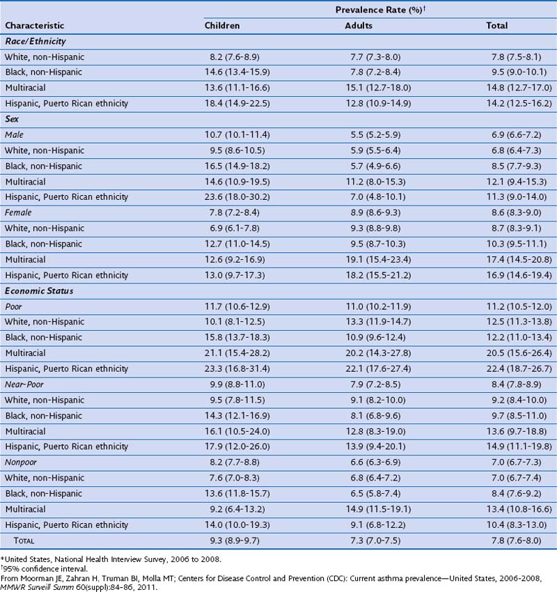
In a morbidity and mortality report from the CDC for the period 2006 to 2008, asthma prevalence was estimated as 7.8% for the U.S. population (Table 38-1). Current asthma prevalence was higher among the multiracial (14.8%), Puerto Rican Hispanics (14.2%), and non-Hispanic blacks (9.5%) than among non-Hispanic whites (7.8%). Current asthma prevalence also was higher among children (9.3%) than among adults (7.3%), among females (8.6%) than among males (6.9%), and among the poor (11.2%) than among the near-poor (8.4%) and nonpoor (7.0%).

With respect to the worldwide variability in reported prevalence rates for asthma, different studies have examined the impact on asthma care in two of the most ethnically diverse nations, such as the United Kingdom and the United States. Emigrational cohorts of Mexican Hispanics have demonstrated a lower rate of asthma than that in U.S.-born Hispanics. Moreover, in a comparison of U.K.-born persons of the same ethnic group with those already settled after emigration, the second group demonstrated a lower rate of physician visits for asthmatic symptoms. Nevertheless, Westernization does not explain this variance, and the inequalities also may be influenced by differences in genetics, environmental risk factors, and social activities.

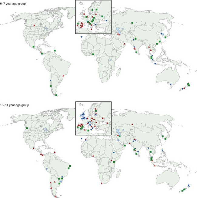
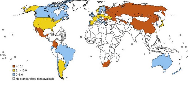
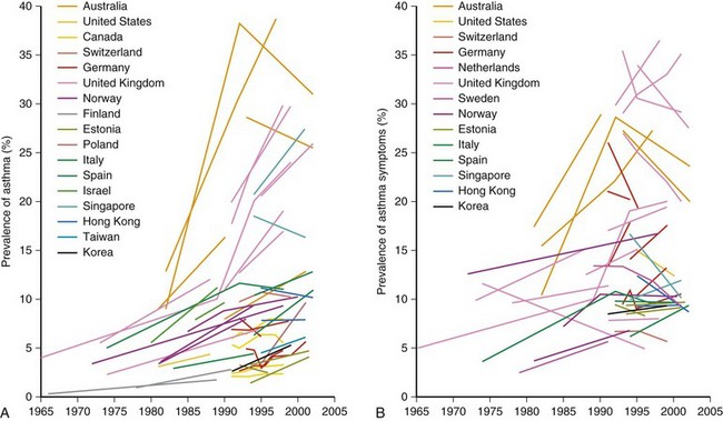
One of the main objectives of the European Community Respiratory Health Survey (ECRHS) was to estimate the variation in prevalence of asthma, asthma-like symptoms, atopic sensitization, and bronchial hyperreactivity, predominantly in Western Europe and many other countries (Figure 38-1). The highest rates in Europe were in the United Kingdom (15.2%), and the lowest were in Georgia (0.28%). Another important ongoing multicenter study, the International Study of Asthma and Allergies in Children (ISAAC), had as its main aim to determine the asthma prevalence in children. A comparative survey conducted in Canadian children between 2 and 7 years of age showed an asthma prevalence of 9.8% overall, with a 4.6% higher rate in boys than in girls (Figure 38-2). GINA embraced the results of both of these valuable studies in the Global Burden of Asthma report and estimated that by 2025, 400 million people around the world will have a diagnosis of asthma. In the same way, this increase in prevalence is directly related to the augmented rates of rhinitis, eczema, and other atopic disorders.

image

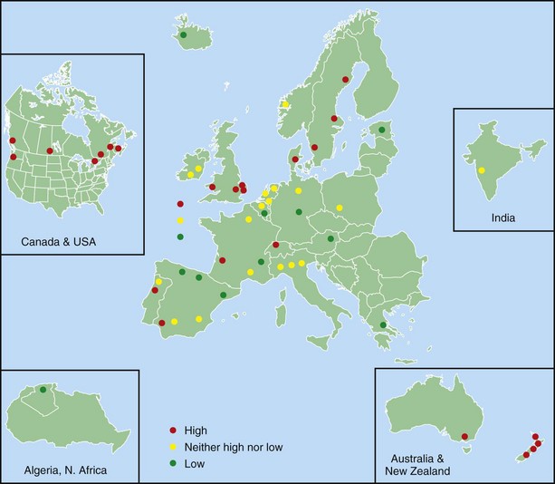
Figure 38-1 Prevalence of asthma in Europe.

(Courtesy European Community Respiratory Health Survey I Centers. Available at: http://www.ecrhs.org/ECRHS%20I.htm.)

In relation to mortality, asthma accounts for an estimated 250,000 annual deaths worldwide. Statistical data show large differences between countries, and asthma death rates do not parallel prevalence rates (Figure 38-3). Mortality seems to be high in countries where access to essential drugs is low. According to the WHO, many asthma-related deaths are preventable, being a result of suboptimal long-term medical care and delay in obtaining help during the final, fatal attack. In many areas of the world, people with asthma do not have access to basic asthma medications and health care. Nations with the highest death rates are those in which controller medications are not available. In many countries, deaths due to asthma have declined recently as a result of better asthma management. Considerable evidence points to an overall trend of decreasing mortality. For instance, mortality rates in Australia registered by the Australian Institute of Health and Welfare decreased by approximately 70% between 1989 and 2006. Persons older than 65 years of age and people of lower socioeconomic status were at higher risk for dying from asthma, mainly secondary to respiratory infections during winter. Overall, asthma-related mortality in Australia remains uncommon, accounting for 402 (0.30%) in the year 2006 of all deaths (Australian Centre for Asthma Monitoring: Asthma in Australia 2008, AIHW Asthma Series no. 3, Cat. no. ACM 14, Canberra, Australian Institute of Health and Welfare, 2008) (Figure 38-4).

image

Figure 38-3 World map of asthma case-fatality rates: asthma deaths per 100,000 people with asthma in the 5- to 34-year-old age group.

(From Masoli M, Fabian D, Holt S, Beasley R; Global Initiative for Asthma [GINA] Program: The global burden of asthma: executive summary of the GINA Dissemination Committee Report, Allergy 59:469–478, 2004.)

image

Figure 38-4 Changes in prevalence of diagnosed asthma (A) and asthma symptoms (B) over time among children and young adults.

(From Eder W, Ege MJ, von Mutius E: The asthma epidemic, N Engl J Med 355:2226–2235, 2006.)

It is worth emphasizing that many of the deaths secondary to asthma are preventable, and that the high economic costs attributable to this disease can be diminished. The elevated prevalence and mortality rates are associated with not inconsideable consumption of health-related resources, placing an additional economic load on health care services.

Pathogenesis

Asthma is an inflammatory disorder of the airways in which multiple mediators and several types of inflammatory cells are involved. This pattern of inflammation is strongly associated with airway hyperresponsiveness and classic asthma symptoms of wheezing, breathlessness, chest tightness, and coughing.

A genetic predisposition to develop specific immunoglobulin E (IgE) antibodies directed against common environmental allergens, or atopy, is the strongest identifiable risk factor for the development of asthma. Intrinsic abnormalities in airway smooth muscle and airway remodeling in response to injury and inflammation add to the effects of airway inflammation in creating the clinical presentation of asthma (Figure 38-5).

image

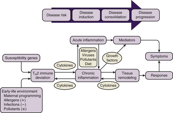
Figure 38-5 Natural history of asthma.

(From Szefler SJ: The natural history of asthma and early intervention, J Allergy Clin Immunol 109:S550, 2002. Adapted from Holgate ST: The cellular and mediator basis of asthma in relation to natural history, Lancet 350[Suppl 2]:5–9, 1997.)

Airway Inflammation in Asthma

Despite the highly heterogeneous clinical expression of asthma, the presence of airway inflammation remains a consistent feature. Although airway inflammation in asthma is persistent even though symptoms are episodic, no clear relationship between the severity of asthma and the intensity of inflammation has been discovered. The inflammation affects all airways, including in most patients the upper respiratory tract and nose, but its pathophysiologic effects are most pronounced in medium-sized bronchi. The pattern of inflammation in the airways appears to be essentially the same in all clinical forms of asthma, whether allergic, nonallergic, or aspirin-induced, and at all ages.

Inflammatory Cells

Studies using bronchoalveolar lavage and bronchial biopsies have demonstrated that a variety of cells and mediators are involved, with IgE and mast cells implicated in the acute response and eosinophils and eosinophil granule proteins in the late response, with T cells, particularly TH2 cells, orchestrating these responses through their production of cytokines such as the interleukins IL-4, IL-5, IL-9, and IL-13. Stromal and epithelial cells also are involved in the inflammatory response, as shown by the ability of these cells to respond to TH2 cytokines with the production of chemokines that initiate and perpetuate tissue inflammatory reactions. As a result, a pathogenetic construct for atopic asthma (and possibly other forms of asthma) has been proposed in which (1) TH2 cells play a central role in the recognition of antigens and the initiation and perpetuation of inflammation; (2) eosinophils are important proinflammatory and epithelium-damaging cells; and (3) a variety of cells, including mast cells, epithelial cells, basophils, fibroblasts, smooth muscle cells, and macrophages, contribute through the secretion of cytokines that generate tissue inflammation or influence TH2 lymphocyte or eosinophil function.

Implicit in the “inflammation theory” of asthma is the belief that inflammation is both necessary and sufficient to account for the complex features of asthma. Although inflammation is undoubtedly a cornerstone of asthma, it is now clear that the asthmatic response is more complex. Also clear from pathologic investigations is that structural alterations exist in the asthmatic airway. Mathematical modeling studies have provided evidence that these alterations contribute to the symptoms and physiologic dysregulation seen in asthma. As a result, it has been proposed that the chronic inflammation that is characteristic of the asthmatic airway leads to a remodeling response, and that the structural alterations induced by this response play an important role in generating the manifestations of the disorder. This conceptual evolution predicts that an enhanced understanding of asthma pathogenesis can be expected when asthma is studied in the context of paradigms of injury and wound healing, as well as the traditional paradigms relevant to the interface of inflammation and airway physiology. It also suggests that studies utilizing this new perspective will identify novel targets against which therapies can be directed and will help to elucidate the biologic basis for the patient-to-patient variability encountered in clinical practice.

Inflammatory Mediators

More than 100 different mediators are now recognized to be involved in asthma and to mediate the complex inflammatory response in the airways. Chemokines are expressed mainly in airway epithelial cells and are important in the recruitment of inflammatory cells into the airways. Eotaxin is relatively selective for eosinophils, whereas macrophage-derived chemokines (MDCs) recruit TH2 cells.

Cytokines such as IL-1β and TNF-α, which amplify the inflammatory response, and granulocyte-macrophage colony-stimulating factor (GM-CSF), which prolongs eosinophil survival in the airways, orchestrate the inflammatory response in asthma and determine its severity. TH2-derived cytokines include IL-5, which is required for eosinophil differentiation and survival; IL-4, which is important for TH2 cell differentiation; and IL-13, needed for IgE formation.

Cysteinyl leukotrienes are potent bronchoconstrictors that act as proinflammatory mediators mainly derived from mast cells and eosinophils. Their inhibition has been associated with an improvement in lung function and asthma symptoms.

The prostaglandin PGD2 is a bronchoconstrictor derived predominantly from mast cells and is involved in TH2 cell recruitment to the airways.

Histamine is released from mast cells and contributes to bronchoconstriction and to the inflammatory response.

Nitric oxide (NO) has been associated with the presence of eosinophilic inflammation in asthma. It is produced predominantly from the action of inducible nitric oxide synthase (iNOS) in airway epithelial cells. Exhaled NO concentration is increasingly being used to diagnose asthma in the context of a compatible clinical history and to monitor the effectiveness of asthma treatment.

Pathophysiology

Airway narrowing is the final common pathway leading to symptoms and physiologic changes in asthma. As discussed next, several factors contribute to the development of airway narrowing in asthma (Figure 38-6).

image

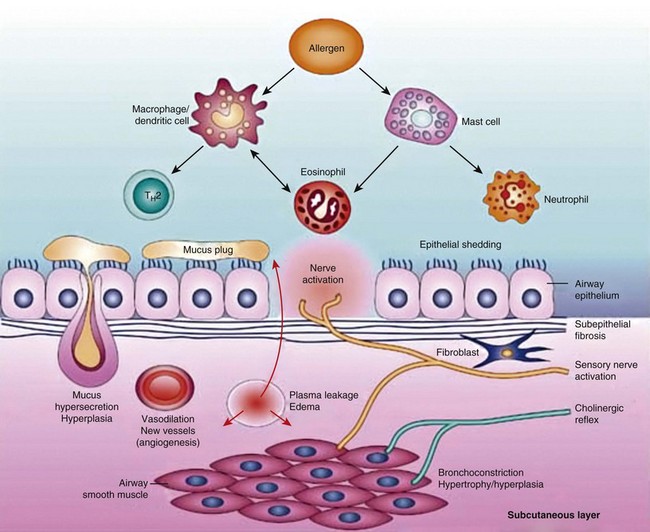
Figure 38-6 Pathobiology of asthma.

(From Barnes PJ: New drugs for asthma, Nat Rev Drug Discov 3:831–844, 2004.)

Airway Remodeling

In addition to the inflammatory response, characteristic structural changes, often described as airway remodeling, are observed in the airways of asthma patients. Some of these changes are related to the severity of the disease and may result in relatively irreversible narrowing of the airways. These changes may represent repair in response to chronic inflammation.

Airway remodeling can be defined as an airflow obstruction despite aggressive antiinflammatory therapies, including regimens of inhaled corticosteroids and systemic corticosteroids. It was first appreciated over 75 years ago by Huber and Koessler in their classic description of fatal asthma. The natural history of airway remodeling is poorly understood, and it does not appear to be a universal finding or a modifiable factor with early treatment.

The effect of bronchoconstriction on airway remodeling has been studied by exposing subjects to a dust mite allergen or repeated methacholine inhalation challenge. Both agents cause bronchoconstriction, although dust mite allergen induces an eosinophilic inflammation. The repetition of the exposure leading to bronchoconstriction after 4 days, observed in bronchial biopsy specimens, established that both the allergen and methacholine groups developed airway remodeling that was independent of inflammation for the second group.

On histopathologic examination, the airway remodeling is associated with a variety of features including airway wall thickening, mucous gland hyperplasia, persistence of inflammatory cellular infiltrates, release of fibrogenic growth factors along with collagen deposition, and elastolysis. Increase in the number and size of vessels in the airway wall is one of the most consistent features of asthma remodeling occurring in the lungs of patients with mild, moderate, and severe asthma. Airway wall thickening is the result of a dense fibrotic response that occurs in the lamina reticularis. The pathologic significance of subepithelial fibrosis is not clear, but this process has been associated with disease severity and correlated with a decline in forced expiratory volume in 1 second (FEV1).

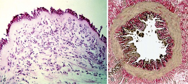
All components of the airway wall have been reported to be thickened in asthma (Figure 38-7). Elements involved in this response are increase in airway smooth muscle, increased myocyte muscle mass, edema, inflammatory cell infiltration, glandular hypertrophy and hyperplasia, and connective tissue deposition. The recently popularized “perturbed equilibrium hypothesis” also suggests that wall thickening can destabilize the dynamic forces that control airway caliber, leading to airway collapse.

Special Circumstances

Risk Factors

Asthma is a condition characterized by lung airway inflammation initiated and perpetuated by an inappropriate immune response, increased airway responsiveness, and variable airflow obstruction. These results come from complex interactions between multiple genetics and environmental influences. Although a clear cause-and-effect association is not always possible to demonstrate, numerous risk factors have been identified (Box 38-1).

The asthma risk factors can be divided into genetic and environment factors, including in the latter allergens, nutritional factors, perinatal factors, and tobacco smoke (active and secondhand smoking). In addition, a series of trigger factors, either direct or indirect, also can worsen asthma symptoms. Among the direct factors are infections, nonspecific irritants such as tobacco smoke, outdoor air pollution, industrial spills, bonfire smoke, and meteorologic changes (low temperatures, high humidity). Indirect factors are exercise, extremes of emotion, food allergens, medications, menstruation, gastroesophageal reflux disease (GERD), and pregnancy.

Genetics

Certain components of the asthma phenotype appear to be strongly heritable, such as atopy, airway hyperresponsiveness, obesity, and gender. In fact, a family history of asthma is a clear risk factor for developing asthma. Current data show that multiple genes may be involved in the pathogenesis of asthma, although the specific genes responsible for these inherited components have not yet been identified.

The relative risk for asthma in a first-degree relative of the patient is between 2.5% and 6%; in monozygotic twins, the risk is 60% and in dizygotic twins, 25%. In contrast with other pathologic conditions caused by a unique genetic abnormality (e.g., cystic fibrosis) in which a hereditary pattern can be explained with a simple mendelian pattern, asthma is a heterogeneous disease in which multiple genes interact. The final phenotype depends on the smaller additive actions of these particular genes, combined with and modulated by environmental factors.

One of the most commonly studied genetic factors is atopy. Atopy can be defined as the genetic predisposition to develop IgE antibodies against specific allergens. Atopy is considered the main factor contributing to development of asthma. The atopic pattern is characterized by atopic dermatitis, allergic rhinitis, and asthma. Not all atopic patients, however, develop all three conditions. The incidence of atopy in the general population is approximately 30%, whereas that of asthma is much lower—the implication being that not all atopic persons develop asthma, but that almost all asthmatic patients have atopy.

Other areas of the asthmatic phenotype in which a search for genetic factors has been focused are airway hyperresponsiveness, inflammatory responses, and TH1-TH2 immune responses. In some cases, a number of chromosomal regions associated with asthma susceptibility have been identified, such as the major locus near the long arm of chromosome 5 (5q), although with inconsistent results.

Obesity is another factor that has been associated with asthma. Asthma is more frequently observed in obese people (body mass index greater than 30) and is more difficult to control in obese asthmatics. The influence of obesity on lung mechanics and the generation of a proinflammatory state in addition to hormonal or neurogenic influences are likely to play a role. This risk may be greater with nonallergic asthma than with allergic asthma.

A clear-cut genetic factor also has been established—asthma is twice as frequent in male children as in females. This predilection reverses by adulthood, when the prevalence of asthma is greater in women than in men, and it reaches its maximal differences when related to severe asthma.

Approximately 20 genome-wide linkage screening studies have been reported in different populations for investigating chromosomal regions that are linked to asthma and atopy, or related phenotypical features such as elevated IgE levels, wheezing, and bronchial hyperresponsiveness. A number of chromosomal regions have been repeatedly identified, across multiple studies, that contain genes of biologic relevance to asthma and allergic disease, including the cytokine cluster on 5q (containing interleukin-3 [IL-3], IL-5, and GM-CSF), FCER1B on 11q, IFNG (interferon-γ) and STAT6 on 12q, and IL4R (the IL-4Rα chain, also part of the IL-13 receptor) on 16p. Linkage studies followed by positional cloning approaches have resulted in the identification of a handful of novel asthma susceptibility genes, including CYFIP2, DPP10, HLAG, PHF11, GPRA, and ADAM33. GPRA (G protein—coupled receptor for asthma) and ADAM33 (a disintegrin and metalloproteinase domain–containing protein 33) have generated considerable interest, because their expression in bronchial smooth muscle cells suggests roles in the pathobiology of asthma and pulmonary allergic disease.

Environmental Factors

The environment plays a pivotal role in the development of asthma because it interacts with the genetic susceptibility of the subject to develop an asthmatic phenotype. Environmental factors, however, frequently overlap with trigger factors that can precipitate asthma symptoms, and their influence can be additive.

Risk Factors in Childhood

Lung Function

Decreased airway caliber in infancy has been reported as a risk factor for transient wheezing, perhaps related to prenatal and postnatal exposure to environmental tobacco smoke. Furthermore, the presence of airways with decreased caliber has been associated with increased bronchial responsiveness and increased symptoms of wheeze. Several studies have suggested an association between reduced airway function in the first few weeks of life and asthma in later life. The magnitude of the effect of this risk factor in isolation (i.e., without concomitant allergy) is unclear; perhaps persons with smaller airways require less stimulus (i.e., airway inflammation) before symptoms become apparent. Children with wheezing (and diagnosed asthma) persisting to adulthood have a fixed decrement in lung function as early as the age of 7 or 9 years. Recent studies of preschool children have documented abnormal lung function in those with persistent wheezing as early as the age of 3 years. However, some infants in whom persistent wheezing develops have normal lung function shortly after birth, which suggests a critical period of exposures within the first few years of life, before the development of these persistent abnormalities in expiratory flow. By contrast, infants who exhibit early transient wheezing exhibit decreased airflow shortly after birth. Maternal smoking with in utero nicotine exposure has been correlated with this type of lung dysfunction, but the effects of other exposures have been less well studied.

Antibiotics and Infections

The use of antibiotics has been associated with early wheezing and asthma in several studies. One suggested mechanism for this association is immunologic stimulation through changes in the bowel flora, but other studies found no coincident increase in eczema or atopy, despite increased wheezing rates—an observation that argues against this mechanism. Greater antibiotic use also may potentially represent a surrogate marker for a higher numbers of infections (perhaps viral) in early life.

Viral infections of the lower respiratory tract contribute to early childhood wheezing. Whether lower respiratory tract infection promotes sensitization to aeroallergens causing persistent asthma is controversial: Childhood viral infections may be pathogenic in some children but protective in others. Infants of mothers with allergy or asthma have a relatively persistent maturational defect in TH1 cytokine synthesis in the first year of life, which may play a role in the development of persistent or severe viral infections. Severe viral infection of the lower respiratory tract in genetically susceptible infants who are already sensitized to inhalant allergens may lead to deviation toward TH2 responses promoting asthma. It is unclear whether these effects of lower respiratory tract infection are virus-specific (e.g., RSV, rhinovirus) or whether synergistic exposures to allergens can induce asthma even in children (or adults) who are not genetically susceptible. Interactions of genes with environmental exposures (including allergens, air pollution, environmental tobacco smoke, and diet) modulate the host response to infections. It remains controversial whether the occurrence or timing of childhood infection is pathogenic or protective for the development and long-term outcome of asthma and allergy and of nonallergic wheeze phenotypes. This controversy relates in part to small sample size, cross-sectional analysis, lack of precise case definition, and incomplete microbial assessment in studies of this phenomenon. Respiratory infections in early childhood are associated with early wheezing, but it is unclear whether infection alone has a role in the development of persistent asthma. Repeated lower respiratory tract infection may affect infants who are already at risk for asthma because of family history or atopy. Severe infection with certain viruses such as RSV and rhinovirus may play a role in persistent wheezing, although other studies have suggested no effect. Considered as a proxy for viral infections, day care attendance is associated with higher incidence of early wheeze but lower incidence of persistent wheeze.

Allergens

Allergens are well known to trigger asthma symptoms and also are considered to be the main environmental risk factors. The exposure to allergens can be outdoor (pollens, fungi, molds, yeasts) or indoor (house dust mite, animal proteins [particularly dog and cat dander], cockroaches, and fungi). Most of the allergens associated with asthma are in the air and to induce a condition of hypersensitivity must be present in abundance over considerable periods of time. As soon as the sensitization occurs, the patient can show extreme reactivity, to the point that minimum quantities of the antigen produce significant exacerbations of the disease.

Total serum IgE level, a surrogate for allergen sensitivity, has been associated with the incidence of asthma. High levels of IgE at birth were associated with greater incidence of both atopy and aeroallergen sensitivity but not necessarily asthma. However, sensitization to aeroallergens, particularly house dust mite, cat, and cockroach allergens, is well documented as being associated with asthma. Immune responses in the developing infant and young child may affect the development of asthma. For example, impairment in interferon-γ production at 3 months was associated with a greater risk for development of wheeze. Immaturity in neonatal immune responses may promote the persistence of the TH2 immune phenotype and development of atopy, but an association with persistent asthma is as yet unproved. More recent work has focused on the role of the innate immune system in handling and presentation of antigens and suggests that polymorphisms in Toll-like receptors (TLRs) may play a greater role than has been previously recognized in the development of the skewed immune responses associated with persistent asthma.

Perinatal Factors

Several prenatal and perinatal factors are proposed to be associated with the development of asthma. Risk factors in the prenatal period are multifactorial. Assessment is complicated by the variety of wheezing conditions that may occur in infancy and childhood, only some of which evolve to classic asthma. The principal prenatal factor is exposure to maternal smoking. The exposure to smoking during pregnancy increases the risk of acquiring asthma up to 37% at the age of 6 and up to 13% after that age. The exposure is clearly associated with fetus growth retardation with disproportionately small airways relative to the size of the lung parenchyma, which in turn is associated with increased airway hyperreactivity, low pulmonary function, asthma, and increased frequency of emergency department visits for treatment of acute exacerbations.

The perinatal factors studied have been maternal age, diet during pregnancy, prematurity, mode of delivery, and in utero exposure to antibiotics. The incidence of wheezing illnesses during childhood is inversely related to maternal age. In one study evaluating young maternal age as a risk factor for childhood asthma, children born to mothers younger than 20 years of age had the highest risk for development of asthma.

Diet and Nutrition

Observational studies examining prenatal nutrient levels or dietary interventions and the subsequent development of atopic disease have focused on foods with antiinflammatory properties (e.g., omega-3 fatty acids) and antioxidants such as vitamin E and zinc. Several studies have demonstrated that higher intake of fish or fish oil during pregnancy is associated with lower risk of atopic disease (specifically eczema and atopic wheeze) up to the age of 6 years. Similarly, higher prenatal vitamin E and zinc levels have been associated with lower risk of development of wheeze up to the age of 5 years. However, no protective effect against the development of atopic disease in infants has been shown for maternal diets that excluded certain foods (e.g., cow’s milk, eggs) during pregnancy. Two recent studies reported an inverse relation of maternal vitamin D levels with onset of wheeze in early life, but no relation with atopy or symptoms in later life. The Mediterranean diet during pregnancy is associated with reduced risk of atopy and asthma.

Prematurity and mode of delivery also have been proposed to be associated with asthma. Delivery by cesarean section has been reported to increase the risk of childhood asthma over that associated with vaginal delivery. The possible explanation—an extension of the hygiene hypothesis—is that microbial exposure and infections during early childhood protect against allergic disease; however, data in support of this extended hypothesis are conflicting. Development of atopy was two to three times more likely among infants delivered by emergency cesarean section, although no such association occurred with elective cesarean section. Potential reasons for these findings include maternal stress and differences in the infant’s gut microflora associated with specific modes of delivery.

Adult-Onset Asthma

Asthma in adults may have persisted from childhood, may have occurred as a relapse of earlier childhood asthma (whether or not recalled by the affected patient) or may be true adult-onset asthma, with history of no symptoms earlier in life. New-onset asthma in adulthood may have environmental (especially occupational) causes with or without allergen sensitization. Although adult asthma may develop in relation to treatment with specific drugs (e.g., beta blockers, nonsteroidal antiinflammatory drugs) or, in women, the use of hormone replacement therapy, occupational exposure to sensitizing agents or irritants is more common.

Occupational Exposure

Occupational asthma is found predominantly in adults, and workplace sensitization is responsible for 1 in 10 cases of asthma among adults of working age (see Chapter 40). High-risk occupations for the development of asthma include farming and agricultural work, painting, plastic manufacturing, and cleaning. IgE- and cell-mediated allergic reactions are involved in its pathobiology.

Trigger Factors

As mentioned earlier, a number of factors are associated with worsening of symptoms and the development of asthma exacerbations.

Indirect Triggers

Suggested Readings

Akinbami LJ, Moorman JE, Liu X. Asthma prevalence, health care use, and mortality: United States, 2005-2009. Natl Health Stat Rep. 2011;32:1–14.

Asher MI, Montefort S, Bjorksten B, et al. Worldwide time trends in the prevalence of symptoms of asthma, allergic rhinoconjunctivitis, and eczema in childhood: ISAAC phases one and three repeat multicountry cross-sectional surveys. Lancet. 2006;368:733–743.

Bara I, Ozier A, Tunon de Lara JM, et al. Pathophysiology of bronchial smooth muscle remodelling in asthma. Eur Respir J. 2010;36:1174–1184.

Bisgaard H, Bønnelykke K. Long-term studies of the natural history of asthma in childhood. J Allergy Clin Immunol. 2010;126:187–197.

Breysse PN, Diette GB, Matsui EC, et al. Indoor air pollution and asthma in children. Proc Am Thorac Soc. 2010;7:102–106.

Busse WW, Lemanske RFJr, Gern JE. Role of viral respiratory infections in asthma and asthma exacerbations. Lancet. 2010;376:826–834.

D’Amato G, Cecchi L, D’Amato M, Liccardi G. Urban air pollution and climate change as environmental risk factors of respiratory allergy: an update. J Investig Allergol Clin Immunol. 2010;20:95–102.

Eder W, Ege MJ, von Mutius E. The asthma epidemic. N Engl J Med. 2006;355:2226–2235.

Global Initiative for Asthma. Global strategy for asthma management and prevention. HHLBI/WHO Workshop report, updated 2011 (article online). http://www.ginasthma.org/. accessed February 2, 2012

Grainge CL, Lau LC, Ward JA, et al. Effect of bronchoconstriction on airway remodeling in asthma. N Engl J Med. 2011;364:2006–2015.

Anderson HR, Gupta R, Strachan DP, Limb ES. 50 years of asthma: UK trends from 1955 to 2004. Thorax. 2007;62:85–90.

Lemanske RFJr, Busse WW. Asthma: clinical expression and molecular mechanisms. J Allergy Clin Immunol. 2010;125(2 Suppl 2):S95–S102.

Lloyd A, Price D, Brown R. The impact of asthma exacerbations on health-related quality of life in moderate to severe asthma patients in the UK. Prim Care Respir J. 2007;16:22–27.

Murphy DM, O’Byrne PM. Recent advances in the pathophysiology of asthma. Chest. 2010;137:1417–1426.

Subbarao P, Mandhane PJ, Sears MR. Asthma: epidemiology, etiology and risk factors. CMAJ. 2009;181:E181–E190.

Xepapadaki P, Papadopoulos NG. Childhood asthma and infection: virus-induced exacerbations as determinants and modifiers. Eur Respir J. 2010;36:438–445.

Share this: