Ascites and pleural fluid
Ascites
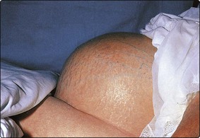
Ascites is fluid in the peritoneal space. It can usually be detected by clinical examination (Fig 64.1). Laboratory analysis of ascitic fluid may provide answers to important clinical questions, as its composition varies depending on the underlying cause.
Serum-ascites albumin gradient
The serum-ascites albumin gradient (SAAG) is defined as the serum albumin concentration minus the ascitic fluid albumin concentration. The SAAG correlates directly with the portal pressure. Patients with a wide SAAG (defined as ≥11 g/L) have portal hypertension, whereas patients with a narrow SAAG (<11 g/L) do not (Table 64.1).
Table 64.1
Serum-ascites albumin gradient
| Wide (>11 g/L) | Narrow (<11 g/L) |
| Chronic liver disease (cirrhosis) | Peritoneal carcinomatosis |
| Veno-occlusive disease | Reduced plasma oncotic pressure (e.g. nephrotic syndrome) |
| Massive hepatic metastases | Secondary peritonitis |
| Congestive cardiac failure | Tuberculous peritonitis |
| Spontaneous bacterial peritonitis |
Peritonitis
SBP or secondary infection?
Secondary peritonitis tends to be more severe than SBP, probably because of the heavier bacterial load. This severity is reflected in the ascitic fluid biochemistry (Table 64.2). Bacteria and neutrophils consume glucose, anaerobic metabolism of which results in the production of lactate, which correlates inversely with pH. Lysis of stimulated neutrophils results in the release of LDH and other cellular proteins, with a consequent rise in the ascitic fluid total protein concentration.
Table 64.2
Spontaneous bacterial peritonitis compared with secondary infection
| Ascitic fluid parameter | Spontaneous bacterial peritonitis | Secondary |
| Glucose (mmol/L) | >2.8 | <2.8 |
| Lactate dehydrogenase | <upper limit of reference interval | >upper limit of reference interval |
| Total protein (g/L) | <10 | >10 |
Pleural fluid
Transudate or exudate?
Clinicians usually request pleural fluid analysis because they want to know what is causing an effusion. In some cases, a specific cause is suspected, but much more frequently the question is posed in more general terms, by asking if the effusion is a transudate or an exudate. The underlying assumption is that fluid formed by ‘exudation’ from inflamed or tumour-infiltrated pleura is likely to be high in protein, whereas fluid formed by ‘transudation’ from normal pleura due to an imbalance in hydrostatic and oncotic forces is likely to be low in protein; in general terms, exudates are more likely to reflect local pathology, and to warrant further investigation. The criteria established by Light and colleagues in 1972, and subsequently modified, continue to be applied widely in classifying pleural fluids as exudates or transudates. They are summarized in Table 64.3.
Table 64.3
Modified Light’s criteria for identification of an exudate
Pleural fluid is classified as an exudate if any of the following criteria are met:


Other questions
 Malignant pleural effusions. As with ascites, finding malignant cells in pleural fluid indicates the presence of malignancy, although only 70% of patients with malignant effusions will have positive cytology. Again, measurement of tumour markers in pleural fluid is rarely indicated. This in part reflects the utility of other modalities of investigation in the diagnosis of malignancy, e.g. imaging.

Pleural fluid glucose
Ascites and pleural fluid


