Ascites
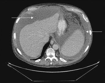
Ascites is the accumulation of excess free fluid in the peritoneal cavity (Fig. 5).
History
Symptoms
Patients with ascites can present with abdominal discomfort, increasing abdominal girth, weight gain and ankle or sacral swelling.
Onset
The development of ascites is usually gradual, but sudden onset can result from acute decompensation of liver cirrhosis, malignancy, portal or splenic vein thrombosis and Budd–Chiari syndrome.
Past medical history
Any history of malignancy in the abdomen or pelvis is relevant; however, abdominal metastasis can also result in ascites, especially from breast, ovarian, prostatic, testicular and haematological malignancies. Cirrhosis of the liver may result from alcoholism, previous hepatitis, Wilson’s disease, primary and secondary biliary cirrhosis and haemochromatosis. A previous history of TB should raise suspicion of secondary disease.
Examination
Classical findings are shifting dullness and a fluid thrill. As liver disease and carcinomatosis account for 90% of the cases presenting with ascites, a detailed examination of all systems is required. The next commonest causes are cardiac failure and nephrotic syndrome.
Inspection
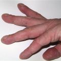
Evidence of liver disease would be suggested by the presence of jaundice, spider naevi, loss of body hair, gynaecomastia, palmar erythema and caput medusae. The jugular venous pressure (JVP) is elevated in the presence of cardiac failure, and a prominent ν wave is seen with tricuspid regurgitation. Further elevation of the JVP on inspiration (Kussmaul’s sign) may be observed with a pericardial effusion.
Palpation
Careful abdominal examination should be performed to look for possible abdominal or pelvic tumours. ‘Dipping’ may be required to feel masses in the presence of gross ascites. Hepatomegaly and splenomegaly can occur with portal hypertension and hepatic and haematological malignancies. Displacement of the apex beat with cardiomegaly may result from cardiac failure. Pedal oedema occurs with cirrhosis, cardiac failure, malabsorption, obstruction of lymphatic flow due to intra-abdominal or pelvic tumours, and nephrotic syndrome.
Auscultation
A third heart sound, systolic murmurs (functional tricuspid and mitral regurgitation) and crepitations (pulmonary oedema) may be audible with cardiac failure. Heart sounds are muffled with pericardial effusions. A pericardial friction rub or knock is occasionally audible in pericarditis.
Percussion
Dullness of the lung bases occurs with pulmonary oedema and pleural effusions (which may occur secondary to ascites).
Internal examination
A rectal examination may reveal ulceration or a fixed mass suggestive of a carcinoma. In women, an adnexal mass PV may be the first indication of a pelvic tumour.
General Investigations
History and examination findings may reveal the underlying cause. General investigations can be used to confirm or indicate the possible aetiology.
■ Urine dipstick
Will be strongly positive for protein in nephrotic syndrome. If so, a 24-hour urine collection for protein should be undertaken: more than 3.5 g/24 h is indicative of nephrotic syndrome.
■ FBC
A raised white count may indicate an infective aetiology but a differential white count is more specific.
■ U&Es
Elevated urea and creatinine implies severe renal impairment, and may occur as a component of hepatorenal syndrome, which is renal impairment secondary to liver failure.
■ LFTs
May be deranged in the presence of liver disease. The serum albumin will be able to indicate hypoalbuminaemia but the underlying cause must still be sought.
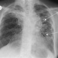
■ CXR
Findings suggestive of cardiac failure are cardiomegaly, upper venous diversion of blood, the presence of Kerley B lines, pulmonary oedema and pleural effusions. Occasionally, the presence of carcinoma may be suggested by a lesion in the lung fields.
■ US abdomen/pelvis
In addition to confirming the presence of ascites, US will detect any intra-abdominal masses that are not palpable on clinical examination, and visualise fatty deposits in the liver in the presence of cirrhosis. Dilated collateral veins may be seen in conditions that result in obstruction of the venous outflow of the liver, including cirrhosis and Budd–Chiari syndrome.
■ Abdominal paracentesis
Aspiration of the ascitic fluid is very useful to help determine the underlying cause. A sample should be sent to microbiology, clinical chemistry and pathology.
Ascitic fluid
Appearance
Chylous
The milky white appearance of chylous ascites is due to obstruction of the lymphatic ducts.
Bile-stained
Is suggestive of bile peritonitis.
Haemorrhagic
Can occur in malignancy, trauma and TB.
Straw-coloured
The usual appearance with most other causes.
Biochemistry
Protein
The ascitic protein levels are often used to classify the ascites as a transudate or an exudate; however this may not always be reliable. A transudate would be ascitic fluid with the protein concentration of less than 25 g/L in the sample or 11 g/L lower than the serum protein level, while an exudate is the opposite.
Amylase
Usually elevated in pancreatic ascites.
Glucose
Low in bacterial infections.
Triglyceride
Elevated in chylous ascites; may indicate obstruction of drainage of the thoracic duct.
Bilirubin
Elevated in bile ascites.
Microbiology
Gram and Ziehl–Neelsen staining with cultures may be positive with a bacterial aetiology.
Cytology
WCC
Neutrophilia is suggestive of a bacterial peritonitis, while tuberculous peritonitis usually results in a lymphocytosis. Malignant cells may be identified and a primary source may possibly be located.
Specific Investigations
■ Echocardiography
Cardiac failure will manifest as a dilated and poorly contracting ventricle with a reduced ejection fraction. A pericardial effusion is visible as an echo-free space between the left ventricle and the pericardium. Tricuspid regurgitation can be visualised with colour-flow Doppler imaging.
■ Liver biopsy
Will be able to confirm and may be able to ascertain the underlying cause of liver cirrhosis.
■ Renal biopsy
Reveals the underlying cause of nephrotic syndrome.
■ Portal venography
Is indicated if obstruction to the venous outflow of the liver is suspected and can confirm Budd–Chiari syndrome and veno-occlusive disease.