115 Ascites
Salient features
Examination
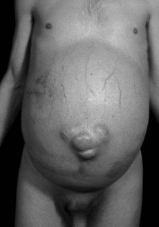
• Full flanks and umbilicus (Fig. 115.1)
• Presence of shifting dullness (always percuss with your finger parallel to the level of fluid)
• If the ascites is gross, use the ‘dipping’ method of palpation to feel the liver and spleen
• Look for stigmata of underlying disease (e.g. signs of cirrhosis, cardiac failure, renal failure or malignancy).
Questions
Advanced-level questions
What are the mechanisms of ascites formation in cirrhosis?
• It is caused by a combination of liver failure and portal hypertension. Liver failure decreases renal blood flow, resulting in retention of salt and water
• Secondary hyperaldosteronism caused by increased renin release and decreased metabolism of aldosterone by the liver
• Decreased metabolism of aldosterone by the liver
• Decreased metabolism of antidiuretic hormone
• Hypoalbuminaemia, which decreases colloid oncotic pressure
• Lymphatic obstruction, resulting in a ‘weeping’ liver
• Overproduction of nitric oxide has been proposed to be important in the pathogenesis of ascites sodium and water retention and hemodynamic abnormalities in cirrhosis (N Engl J Med 1998;339:533–41). Inhibition of nitric oxide synthesis improves sodium and water excretion in cirrhotic rats with ascities.
What is the relation between ascites and chronic liver disease?
• Ascites indicates decompensation of previously asymptomatic chronic liver disease.
• Ascites occurs in about half of the patients within 10 years of a diagnosis of compensated cirrhosis.
• The development of fluid retention in patients with chronic liver disease is a poor prognostic sign: only half of these patients survive beyond 2 years.
How would you manage a patient with cirrhosis and ascites?
The most important treatments are sodium restriction and diuretics (N Engl J Med 1994;330:337–42):
• Sodium restriction to 88 mmol/day; only 15% of these patients lose weight or have a reduction in ascitic fluid with this therapy alone.
• Fluid restriction is usually not necessary unless the serum sodium concentration falls below 120 mmol/l.
• When the patient has tense ascites, 5 litres or more of ascitic fluid should be removed to relieve shortness of breath, to diminish early satiety and to prevent pressure-related leakage of fluid from the site of a previous paracentesis.
• Diuretic therapy should be initiated immediately, before which the serum sodium concentration of a random urine sample should be measured. Serial monitoring of urinary sodium concentration helps to determine the optimal dose of diuretic; doses are increased until a negative sodium balance is achieved. The most effective diuretic regimen is a combination of spironolactone and furosemide. More than 90% of patients respond to this therapy.
In patients with cirrhosis and refractory or recurrent ascites would you prefer paracentesis or transjugular intrahepatic portasystemic stent shunt?
What are the complications of ascites?
• Respiratory embarrassment may complicate large amounts of ascites
• Spontaneous bacterial peritonitis is seen in cirrhotics: suspect when there is an ascitic fluid leukocyte count of 500 × 106 cells/l, or polymorphonuclear cell count of >250 × 106 cells/l. Empirical therapy with a non-nephrotoxic broad-spectrum antibiotic should be initiated immediately.
What do you know about the pathogenesis of ascites in cirrhotics?
Two theories have been proposed (N Engl J Med 1982;307:1577).
Underfilling theory. Primary abnormality is inappropriate sequestration of fluid within the splanchnic vascular bed caused by portal hypertension, resulting in a decrease in intravascular volume. The kidney responds by retaining salt and water.
Overflow theory. Primary abnormality is inappropriate retention of salt and water by the kidney in the absence of volume depletion.
G Budd (1808–1882), Professor of Medicine at King’s College, London.
H Chiari (1851–1916), Professor of Pathology at the German University in Prague.
JV Meigs (1892–1963), Professor of Gynaecology at Harvard, Massachusetts General Hospital.
Le Veen, gastroenterologist at the Veterans Administration Hospital in New York.


