Asbestosis

Published on 29/05/2015 by admin

Filed under Pulmolory and Respiratory

Last modified 22/04/2025

Print this page

rate 1 star rate 2 star rate 3 star rate 4 star rate 5 star
Your rating: none, Average: 0 (0 votes)

This article have been viewed 1565 times

Chapter 52 Asbestosis

Inhalational exposure to asbestos produces both malignant and nonmalignant diseases of the chest. The focus of this chapter is on the two major categories of nonmalignant disease—asbestosis and asbestos-related pleural disorders, listed in Table 52-1. These conditions have received a great deal of attention from the scientific and medical communities because of the ubiquitous use of asbestos in modern society and its diverse and pernicious toxicities. Despite major progress in awareness of the issues and in control of exposure, a large burden of asbestos-related disease will continue to accrue as a consequence of ongoing exposure and the characteristic disease latency.

Table 52-1 Nonmalignant Asbestos-Related Diseases

Condition Locus of Pathologic Change Description
Asbestosis Parenchyma Interstitial pulmonary fibrosis
Benign nodules Parenchyma Lymphoid or fibrotic nodular scars
Benign pleural effusion Pleura Exudative, transient effusion
Pleural plaques Pleura Collagenous, hyalinized masses; circumscribed, avascular; usually involving the parietal pleura
Diffuse pleural thickening Pleura Collagenous, hyalinized masses; diffuse, avascular; involving the parietal and visceral pleura and interlobular space
Rounded atelectasis Combined pleura and parenchyma Scarring of pleura and adjacent lung tissue, resulting in retraction, entrapment, and local partial collapse of lung

Epidemiology, Risk Factors, and Pathophysiology

Epidemiology

The first well-documented cases of asbestosis were reported in the early 1900s among asbestos textile workers. Through the 1920s and 1930s, reports emerged of asbestosis, pleural thickening, pleural calcification, and right ventricular failure in asbestos-exposed workers. Radiographic studies that began in the 1930s documented an asbestosis prevalence of 25% to 55% in these workers, especially among those with greater cumulative exposure. Early evidence from these studies suggested that exposure to higher concentrations of asbestos over longer periods of time resulted in increased risk for development of pulmonary fibrosis.

Asbestos use increased extensively in the early 1940s, and this mineral substance was used widely in the United States for the next 30 years. Asbestos use began to diminish when the Occupational Safety and Health Administration (OSHA) regulated workplace asbestos exposure in 1972. It is estimated that between 1940 and 1979, more than 27 million people had potential exposure in the United States alone. Data from a study of sheet metal workers examined between 1986 and 2004, reported by Welch and colleagues, found that asbestosis is continuing to occur even 50 years after first exposure. The study also found that the strongest predictor of nonmalignant asbestos-related disease in the workers was the cutoff year in which employment commenced: Prevalence was lowest among those who began working in the sheet metal industry after 1970 and highest among those who began work before 1949. With thousands of commercial applications and the mineral’s resistance to degradation, asbestos remains ubiquitous. Efforts have been made to limit ongoing exposure through abatement programs of asbestos removal from buildings and/or on-site encapsulation. Nevertheless, asbestos-related disability and mortality will continue well into the next decade.

Although the use of asbestos has been curtailed in many developed nations, in less-developed countries this inexpensive but hazardous material continues to be used widely. Many developed countries are now fast replacing the developed ones in the production and use of asbestos. Currently, Russia is the leading producer of asbestos worldwide, followed by China, Brazil, Kazakhstan, and Canada. More than 85% of the world production of asbestos is used to manufacture products in Asia and Eastern Europe. Although asbestos usage in developing countries is increasing, information about asbestos-related disease remains largely obscure. Epidemiologic questions have not been systematically researched.

In keeping with the long latency required before asbestosis becomes clinically apparent, all past and current asbestos workers must be considered to be at risk for development of this fibrosing lung disorder.

Etiology and Risk Factors

The minerals referred to herein as “asbestos” are a family of naturally occurring, flexible, fibrous hydrous silicates found in soil worldwide. Mined asbestos fibers are categorized as either long and curly (serpentine) or straight and rodlike (amphibole). The serpentine fiber, chrysotile, accounts for most of the commercially used asbestos, favored for its properties of heat resistance, flexibility, and ease of spinning for textiles. Five categories of amphiboles are recognized: crocidolite, amosite, anthophyllite, tremolite, and actinolite. These more rigid fibers are less commonly used but are still pathogenic. All major commercial forms have been associated with development of nonmalignant respiratory disorders and of lung cancer and mesothelioma, as discussed in Chapters 47, 65, and 70.

Asbestosis is the result of either direct or “bystander” exposure to asbestos-containing materials. Major sources of exposure are summarized in Table 52-2. During the first half of the 20th century, high-level exposures to asbestos dust occurred in the manufacturing of asbestos textiles and construction materials and in the construction and ship-building trades. Potential exposures still occur in the construction trades and in the process of asbestos abatement. Although the use of asbestos has been curtailed in many developed nations since the 1970s, in less developed countries this inexpensive but hazardous material continues to be used widely. High cumulative, occupational exposures in these settings are still commonplace.

Table 52-2 Major Asbestos Uses and Sources of Exposure

Environment Type of Exposure Source of Exposure
Occupational Asbestos-cement products Construction industry (sheeting used in roofing and cladding of structural materials, molded into roof tiles, pipes, gutters; filler for wall cracks, cement, joint compound, adhesive, caulking putty)
Floor tiling Filler and reinforcing agent in asphalt flooring, vinyl tile, adhesive
Insulation, fireproofing Insulators, pipefitters
Construction industry (pipes, boiler covers, ship bulkheads, sprayed on walls and ceilings as fireproofing, soundproofing)
Textiles Fireproof textiles used in clothing, blankets
Paper products Roofing felt, wall coverings, mill board, insulating paper
Friction materials Brake linings
Rubber, plastic manufacture Filler in rubber and plastics
Building trades, secondhand exposure Building maintenance activities, pipefitting, electrical repair, boiler tending and secondhand exposure repair, boiler tending and repair, power station maintenance
Carpenters, plumbers, welders
Domestic “Fouling the nest” Carrying home asbestos in hair and clothes of exposed workers results in exposure to family members
Secondhand exposure Residential remodeling, removal, handling of frayed, friable asbestos in homes can cause environmental exposure
General Contaminated buildings Found in low levels in buildings under normal use
Elevated exposures from remodeling, renovation, asbestos removal, disturbance of contaminated materials such as acoustic ceiling tiles, vinyl floor tiles, paints, plaster, pipes, boilers, steel beams
Geologic exposure Living near asbestos mines or cement factories, or in geographic areas in which naturally occurring asbestos is found in ambient air
Urban environment Ambient air levels slightly higher in cities, perhaps because of asbestos shed from automotive brakes in denser traffic, and high concentration of industry and construction

Environmental exposure to asbestiform fibers also is well described as the cause of nonmalignant and malignant asbestos-related lung disease in countries including Turkey, Greece, Japan, and China and the territory of New Caledonia. Exposure in these settings usually occurs when villagers in rural areas disturb natural soil deposits while working in fields or when applying whitewash prepared from these outcrops to their dwellings. In the United States, asbestos-related lung disease caused by nonoccupational exposures is a recently recognized problem, mostly in current and former residents of Libby, Montana. Amphibole asbestos–contaminated vermiculite was mined, milled, and processed near this small town for many years. Personal and commercial use of the contaminated mineral was widespread among residents. During a health screening in 2000 to 2001, nearly 18% of 6668 participants were noted to have pleural thickening on chest radiographs. Less than 1% had findings compatible with asbestosis.

A clear dose-response relationship between asbestos exposure and asbestosis has been recognized, although controversy remains concerning risks at low-level exposure. Risk for asbestosis varies widely among industries, with more disease seen in textile and construction workers than with those in mining. Development of the disease is associated with factors such as respirability of the fiber type, the cumulative dose of exposure, the capacity of the lung to clear the fibers, and the biopersistence of the asbestos. In general, the relative risk for development of asbestosis for asbestos workers increases in proportion to the asbestos exposure levels in the workplace. More severe disease has been associated with higher retention of asbestos fiber in the lungs. Typical asbestos fibers found in the lungs are 20 to 50 µm long and initially are deposited at the bifurcations of conducting airways. Thin fibers, of diameters less than 3 µm, translocate readily into the alveolar space, interstitium, and pleural space. Thicker fibers tend to be incompletely phagocytosed by alveolar macrophages and are retained in the lung, where they can trigger the inflammatory events that lead to fibrosis, as discussed next.

Histopathology and grading

Asbestosis is defined histopathologically as bilateral, diffuse interstitial fibrosis of the lungs caused by the inhalation of asbestos fibers (Figure 52-1). Gray streaks of fibrosis can be seen in the parenchyma along interlobar and interlobular septa. Later, the pleural surface becomes more nodular in appearance, and the parenchyma loses volume and elasticity and forms more fibrotic scars and honeycombing. The gross pathologic changes are most obvious in the lower lung zones bilaterally, with the worst disease nearest to the pleura.
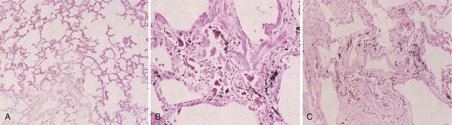
The College of American Pathologists and Pulmonary Pathology Society has modified a 12-point grading scheme that has been shown to be consistently reproducible. The modified grading scheme, based on histologic criteria presented in the 2010 update on the original diagnostic criteria, consists of the following categories of disease severity:

Although histologic evidence of pulmonary fibrosis may occasionally be obtained in the course of clinical evaluation, routine lung biopsy and lavage are not recommended, and microscopic evidence is rarely required to diagnose asbestosis.

Occasionally, the determination of asbestos fibers in lung tissue, bronchoalveolar lavage, or sputum may be used to document past exposure, although these measurements are neither usually relied on nor required for the clinical diagnosis of asbestosis. Under light microscopy or by use of transmission electron microscopy, uncoated fibers or fibers coated with proteinaceous material may be detected. These coated fibers—so-called asbestos bodies or ferruginous bodies—are nonspecific, because they can be found in occupationally unexposed people, in occupationally exposed people who have no asbestos-related lung disease, and in workers who have asbestosis. Generally, the most exposed and most severely affected people have higher asbestos fiber counts, but a significant interlaboratory variability is found in these measures.

Pathogenesis

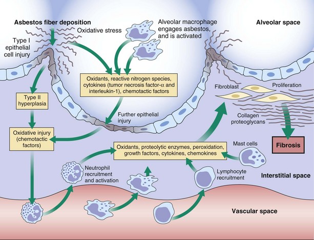
Some of the major events thought to be involved in the pathogenesis of asbestosis are summarized in Figure 52-2. Within minutes after asbestos fibers have been inhaled, a local tissue response is initiated at the bifurcations of terminal bronchioles and alveolar ducts. The first changes occur in epithelial cells and then in alveolar macrophages as they attempt to engulf and are pierced by the fibers. In addition to cell death, which leads to the release of macrophage contents, asbestos-activated macrophages release reactive oxygen species that directly damage the tissue through peroxidation and direct cytotoxicity. Asbestos also can induce toxicity by mechanisms independent of its ability to promote formation of reactive oxygen species.

Increasing numbers of alveolar macrophages accumulate within 48 hours of first exposure. With chronic inhalation, a localized fibrosing alveolitis in the peribronchiolar region develops, followed by diffuse fibrotic scarring. Increasing the dose of asbestos increases the cellular response. A cascade of events ensues in which the macrophages and neutrophils release various cytokines (such as interleukin-8 and interferon-γ), chemokines, oxidants, and growth factors (such as fibronectin, platelet-derived growth factor, insulin-like growth factor, transforming growth factor-β, tumor necrosis factor-α, and fibroblast growth factor). These attract and alter the function of other inflammatory cells and resident cells, thereby promoting inflammation and fibrosis. The response of fibroblasts to these signals is to proliferate and produce the constituents of extracellular matrix (e.g., collagen, proteoglycans) in the pulmonary interstitium. Resident cells themselves are both targets and perpetrators of the fibrotic response. The pathogenesis of the chronic fibrotic response is complex and remains to be fully elucidated. It is clear, however, that this chronic response is progressive and that, apart from macrophage, neutrophil, and epithelial cells, a number of other cell types, such as lymphocytes and mast cells, contribute to the cycle of lung remodeling and fibrosis. Multiple, functionally overlapping, redundant inflammatory events occur in the lung simultaneously during the period of fibrogenesis. The consequence is an irreversible alteration in the structure and function of the lung.

Clinical Features

Asbestosis is the pulmonary fibrotic disease that results from asbestos exposure. It affects the lungs symmetrically and typically is diagnosed on the basis of a consistent occupational or environmental history of asbestos exposure plus evidence of pulmonary fibrosis, usually obtained by chest radiography. The latency period from first exposure to clinical disease is 10 to 20 years but can be up to 40 years or more, with shorter latency and more severe disease seen in those workers with the highest inhalational exposures. In some situations, the exposure history may be difficult to document. If needed, further evidence of occupational exposure can be verified by identifying high numbers of asbestos bodies in bronchoalveolar lavage fluid, sputum, or lung tissue, as discussed previously. Evidence of bilateral pleural plaques is pathognomonic for previous asbestos exposure. The radiographic finding of bilateral interstitial markings in the lower lung zone is sufficient radiographic evidence of asbestosis, although (as discussed in the following section) other tools such as computed tomography (CT) and measures of lung physiology also aid diagnosis.

The most common signs and symptoms of asbestosis are the insidious onset of dyspnea on exertion (and eventually at rest), dry cough that can be paroxysmal, and fatigue. Hemoptysis, chest pain, and weight loss are not common and should raise suspicion for asbestos-related malignancy. Although physical abnormalities are uncommon at the early stages of asbestosis, patients may exhibit such signs later in the clinical course, such as dry, bilateral basilar rales evident on end inspiration, digital clubbing, cyanosis, and signs of cor pulmonale.

Radiologic Findings

Radiographically, asbestosis typically manifests in the lower lobes with irregular “reticular” markings toward the lung periphery and costophrenic angles (Figure 52-3). Linear opacities that resemble extensions of vascular markings may assume a netlike appearance. In early or less severe disease, the middle and upper lung zones may appear relatively spared. With progression, the linear and irregular opacities thicken and spread to the midlung zones, but rarely to the apex. The International Labour Organization (ILO) International Classification of Radiographs of Pneumoconioses characterizes each type of irregular opacity on the basis of increasing size and thickness as either s, t, or u, and on a scale of profusion (number) of opacities from normal profusion (0/−, 0/0, 0/1) to severe (3/2, 3/3, 3/+) (see Chapter 51, Table 51-2, for classification details). When irregular opacities are seen on the chest radiograph in conjunction with pleural thickening, the radiograph can be considered virtually pathognomonic for asbestosis. However, the chest radiograph lacks sensitivity, because 15% to 18% of symptomatic, biopsy-proven asbestosis cases are associated with a normal appearance on chest radiographs. Focal masses are uncommon except for those caused by rounded atelectasis.

image

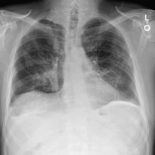
Figure 52-3 Chest radiograph illustrating parenchymal abnormalities in asbestosis. Descriptive designations are from the International Labour Organization (ILO) classification for the radiographic appearance of pneumoconioses (see Chapter 51, Table 51-2). In this case, fine linear opacities of profusion category 1/0 can be seen in the middle and lower lung zones. Calcified pleural plaques are seen along the diaphragm bilaterally.

Computed Tomography

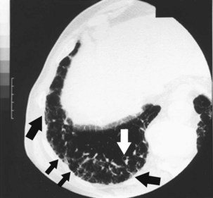
Conventional CT and high-resolution CT (HRCT) are more sensitive and specific than radiography for the diagnosis of pleural disease related to asbestos. HRCT, with use of 1- to 3-mm slices, is superior in sensitivity to plain chest radiography for detection of the fine reticular opacities in this disease. Of asbestos-exposed workers who have normal-appearing chest radiographs, 10% to 30% have HRCT scans suggestive of underlying interstitial disease. Thus, the HRCT scan can prove useful when the clinical index of suspicion for asbestosis is high but the chest radiograph appears normal. The most common HRCT findings in asbestosis are short, peripheral septal lines; subpleural curvilinear lines; ground glass attenuation; peripheral cystic lesions (honeycombing); parenchymal bands adjacent to areas of pleural thickening; and bronchiolar thickening (Figure 52-4). The density of interstitial abnormalities found on HRCT has been shown to correlate with the symptoms and with physiologic and inflammatory indicators of asbestosis, although in general, findings on both the chest radiograph and the HRCT scan demonstrate only a limited correlation with disease severity measured by assessment of lung physiology.

Diagnosis

The long latency between asbestos exposure and development of asbestosis and the gradually progressive nature of the symptoms mean that this disease has a tendency to remain undetected until fairly late in its course. Efforts to conduct workplace surveillance by use of the chest radiograph and ILO readings of these films have improved disease detection. The diagnosis is based on a consistent history of exposure to asbestos, with sufficient duration of latency, and evidence of interstitial fibrosis. A careful work and environmental history holds the key to determining that past exposure has occurred. As discussed previously, the presence of bilateral pleural plaques or demonstration of asbestos bodies in lavage or on biopsy also can aid in the assessment of exposure. Although most algorithms for diagnosis of asbestosis suggest that the combination of histologic analysis plus mineralogic assessment is the most sensitive and specific method of diagnosis, frequently the required biopsy material is unavailable, and such studies are unnecessary in making a probable determination of disease. Even the lung pathophysiologic findings should be considered in context with clinical data. The main considerations in the histologic differential diagnosis include the other pneumoconioses and other causes and agents of pulmonary fibrosis such as pharmaceutical drugs, metal dusts, infectious agents, autoimmune disorders, and idiopathic pulmonary fibrosis.

In the absence of lung histologic and mineralogic analysis, the clinical diagnosis of asbestosis can be made with reasonable confidence on the basis of the following criteria:

Helpful, but less essential criteria include evidence of restrictive impairment of lung function, abnormalities of gas exchange, bilateral inspiratory crackles (rales), and digital clubbing.

Treatment

At present, no cure exists for asbestosis, and no benefit from the use of corticosteroids or other immunosuppressive therapy has been documented. After exposure and early disease have occurred, no prophylactic measures are available.

Medical management in cases of asbestosis focuses on the following interventions:

In the United States, patients with a diagnosis of asbestosis and occupational exposure to asbestos may apply for workers’ compensation benefits. Compensation may be granted on appropriate medical documentation of the degree of physical impairment in combination with employment and exposure requirements. Workers who have evidence of pleural plaques, also referred to as pleural asbestosis, also may be eligible for compensation in some programs, with or without evidence of coexisting pulmonary asbestosis.

Clinical Course

The prognosis for patients with asbestosis varies widely. It is dependent in part on the magnitude of exposure. In 1906, the disease was almost uniformly fatal by the third decade of life. Today, however, with fewer exposures and shorter exposure times, and with superior detection and supportive care, few patients demonstrate such severe progression of their disease. After removal from exposure, progression usually is slow and occurs in 5% to 40% of patients over approximately a decade of follow-up. Thus, if clinical deterioration occurs over a period of days or weeks, the clinician must look first for other explanations, such as infection or malignancy. Many patients may remain mildly symptomatic for many years and show little or no objective signs of disease progression, whereas others show steady, inexorable decline in lung function, gas exchange, worsening symptoms, development of end-stage respiratory insufficiency, and cor pulmonale with right ventricular failure.

Patients with asbestosis are at increased risk for intercurrent lung infections and lung cancer. The best prognosis is found in those workers who have the lowest ILO profusion scores (i.e., chest radiographs that show the fewest irregular opacities) at the time of termination of exposure. Multiple studies demonstrate that those with the greatest average and cumulative dust exposures tend to have higher initial profusions of small opacities on chest radiographs and more rapid disease progression.

Tobacco smoking also contributes to radiographic evidence of disease severity. Tobacco smoking has been shown to increase risk for development of asbestos-related fibrosis and asbestos-induced lung cancer. Several studies have proposed that smoking enhances the development of interstitial fibrosis in workers exposed to asbestos.

On the basis of National Center for Health Statistics data through 2005, for U.S. residents the age-adjusted mortality rate attributable to asbestosis began to plateau after 2000, but only after having risen from an age-adjusted mortality rate of 0.54 per 1 million population in 1968 to 6.03 per 1 million in 2005. Mortality rates are much higher among men than among women, and the age at which people die of asbestosis has risen from a median of 60 years in 1968 to approximately 79 years in 2005. In 2005, asbestosis resulted in more than 14,000 years of potential lives lost due to reduced life expectancy. Lung cancer is a significant contributing cause of increased mortality rates among patients with asbestosis.

Asbestos-Related, Nonmalignant Pleural Disorders

The most common pleural changes caused by asbestos are pleural plaques, with or without pleural calcification and diffuse pleural thickening. Presence of pleural thickening is a marker of exposure. Pleural changes are now known to contribute to the lung function abnormalities seen in asbestos-exposed workers. Both types of pleural alteration contribute independently to restrictive lung physiology (reduced vital capacity), reduced lung compliance, and diminished diffusing capacity. Of asbestos-exposed construction workers, 20% to 60% demonstrate chest radiographic evidence of pleural disease, which is remarkable in light of the insensitivity of the radiograph.

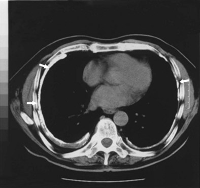
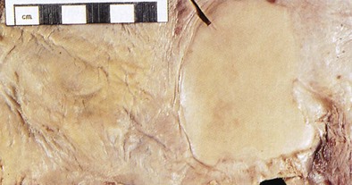
Circumscribed pleural plaques that involve the parietal pleura usually are symmetric and bilateral, most commonly between the fifth and eighth ribs toward the posterolateral aspects of the thorax (Figure 52-5); they also frequently involve the diaphragmatic pleura (Figure 52-6). These lesions remain discrete. Thus, if radiographic evidence of more diffuse thickening is found, either mesothelioma or diffuse pleural thickening must be considered. On histologic examination, pleural plaques are seen to be hyalinized, acellular, avascular masses, and they rarely contain asbestos bodies. They have a tendency to calcify, which can be mistaken for nodular infiltrates on the chest radiograph. Although the ILO classification system has an elaborate section devoted to characterization of pleural abnormalities on the chest radiograph, interreader agreement is relatively low. Because HRCT is more sensitive than chest radiography in the detection of pleural plaques, it helps to determine past asbestos inhalation, because these plaques are pathognomonic for that exposure. Also, HRCT helps to differentiate plaques from extrapleural fat pads. Asbestosis can occur in the absence of pleural disease, and inversely, pleural disease can occur without underlying pulmonary fibrosis, although autopsy studies suggest that when pleural changes are seen, histopathologic features of asbestosis often are evident even if the radiograph is normal in appearance. Pleural plaques rarely, if ever, undergo malignant transformation.

Diffuse pleural thickening involves both parietal and visceral pleura and is strongly associated with previous, benign asbestos-related pleural effusions. It also may develop when subpleural parenchymal fibrosis extends to the visceral pleura. Diffuse pleural thickening most commonly is located in the lower thorax, can blunt the costophrenic angles, and may be either unilateral or bilateral. Because it is so diffuse, this form of pleural thickening can produce dyspnea on exertion and dry cough, as well as loss of lung function. Other conditions that can induce similar diffuse thickening include past tuberculosis, thoracic surgery, chest trauma with hemorrhage, adverse drug reactions, and infection.

After direct contact of asbestos fibers pleura, an inflammatory, exudative, and often hemorrhagic effusion can develop. It is asymptomatic in two thirds of cases but can be associated with acute chest pain, with or without fever. It can occur in the presence or absence of asbestosis. Its incidence in asbestos-exposed workers has been estimated to be less than 5%. Although it can be the first manifestation of asbestos-related disease, the mean latency period for benign, asbestos-related pleural effusions is 30 years. These effusions often resolve spontaneously but recur in approximately one third of cases. The regression may be associated with pain. The consequences include not only diffuse pleural thickening but also the formation of adhesive fibrothorax. Benign pleural effusion is considered a diagnosis of exclusion.

Rounded atelectasis, although uncommon, is important to recognize because of its tendency to mimic lung tumors. It is thought to occur when visceral pleural thickening invaginates and folds on the lung parenchyma, resulting in atelectasis; CT is the preferred method of detecting its typical cicatricial pattern. Malignancy has only rarely been described in areas of rounded atelectasis. A positron emission tomography scan usually shows no abnormality in rounded atelectasis and may help differentiate the lesion from a lung cancer.

Controversies and Pitfalls

The threshold of asbestos exposure below which asbestosis will not occur is unclear, so any asbestos exposure carries some potential risk of asbestos-related disease. Prevention is superior to treatment of disease, because there is no cure. The best preventive measure is to eliminate inhalational exposure by the following measures or precautions:

Substituting materials that have less toxicity must be considered in industrial applications. When asbestos substitutes are not available, appropriately designed and maintained engineering controls, such as local exhaust ventilation systems, must be in place. Personal respiratory protection is appropriate for short periods of exposure or when other controls are not feasible. Such respirators must be appropriately fitted to the person and tested for the degree of protection they afford the worker by quantitative fit testing. Showering and changing of work clothes at the end of work shifts will help to eliminate take-home exposures. Workers must be educated about the combined risks of asbestos exposure and smoking for lung cancer and must be counseled to avoid future asbestos exposure. Companies that use asbestos must maintain strict compliance with government regulations on permissible exposure limits and appropriate medical surveillance of workers.

Suggested Readings

American Thoracic Society. Diagnosis and initial management of nonmalignant diseases related to asbestos. Am J Respir Crit Care Med. 2004;170:691–715.

Burgess WA. Asbestos products. In: Burgess WA, ed. Recognition of health hazards in industry. New York: John Wiley & Sons; 1995:443–451.

Craighead JE, Abraham JL, Churg A, et al. The pathology of asbestos-associated disease of the lung and pleural cavities: diagnostic criteria and proposed grading scheme. Arch Pathol Lab Invest. 1982;106:544–595.

Division of Respiratory Disease Studies. National Institute for Occupational Safety and Health (NIOSH) Work-related lung disease surveillance report (DHHS [NIOSH] publication no. 2003–2111). Washington, DC: U.S. Government Printing Office; 2002.

Hillerdal G. Rounded atelectasis: clinical experience with 74 patients. Chest. 1989;9:836–841.

International Labor Office. International classification of radiographs of pneumoconiosis. Geneva, Switzerland: International Labor Organization; 2003.

Kamp DW, Weitzman SA. The molecular basis of asbestos induced lung injury. Thorax. 1999;54:638–652.

Peipins LA, Lewin M, Campolucci S, et al. Radiographic abnormalities and exposure to asbestos-contaminated vermiculite in the community of Libby, Montana, USA. Environ Health Perspectives. 2003;111:1753–1759.

Roggli VL, Gibbs AR, Attanoos R, et al. Pathology of asbestosis—an update of the diagnostic criteria: report of the Asbestosis Committee of the College of American Pathologists and Pulmonary Pathology Society. Arch Pathol Lab Med. 2010;134:462–480.

Tossavainen A. International expert meeting on new advances in the radiology and screening of asbestos-related diseases. Scand J Work Environ Health. 2000;26:449–454.

Welch LS, Haile E, Dement J, Michaels D. Change in prevalence of asbestos-related disease among sheet metal workers 1986 to 2004. Chest. 2007;131:863–869.

Yamamoto S. Histopathological features of pulmonary asbestosis with particular emphasis on the comparison with those of usual interstitial pneumonia. Osaka City Med J. 1997;43:225–242.

Zhao XH, Jia G, Liu YQ, et al. Association between polymorphisms of DNA repair gene XRCC1 and DNA damage in asbestos-exposed workers. Biomed Environ Sci. 2006;19:232–238.