Arthroscopic Resection of Arthrosis of the Proximal Hamate

Published on 11/04/2015 by admin

Filed under Orthopaedics

Last modified 22/04/2025

Print this page

rate 1 star rate 2 star rate 3 star rate 4 star rate 5 star
Your rating: none, Average: 5 (1 votes)

This article have been viewed 2653 times

CHAPTER 25 Arthroscopic Resection of Arthrosis of the Proximal Hamate

Rationale and Basic Science

Pain in the ulnar aspect of the wrist is a common symptom in the field of hand surgery. This presenting complaint has a large number of possible etiologies, however, and the treatment for any given etiology is largely dependent on the diagnosis. Although treatment options for isolated tears of the triangular fibrocartilage complex (TFCC) or the lunotriquetral (LT) ligament have been studied for many years, there is very little research on outcomes for patients with arthritic change of the proximal pole of the hamate. This is surprising, given that the proximal hamate is one of the most frequent sites of arthrosis in the wrist.1,2

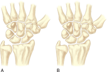
In 1990, two unrelated anatomical studies closely characterized the lunate articular anatomy.1,3 These descriptions referred to the distal joint morphology of the lunate in the midcarpal joint and two types were described (Figure 25.1). A type I lunate is one in which the distal lunate articulation makes contact with only the capitate. It was observed in approximately 30% of specimens. A type II lunate is one with an obvious distal lunate medial facet that articulates with the proximal pole of the hamate in addition to the regular facet that articulates with the capitate. The size of this medial facet ranges between 1 and 6 mm in width. This type II variant was observed in up to 70% of specimens. A type I lunate has no contact with the proximal pole of the hamate regardless of wrist position, whereas a type II lunate has a variable amount of contact between the hamate and medial lunate facet—primarily when the wrist is in an ulnarly deviated position.

Cadaver studies have revealed that cartilage erosion of the proximal pole of the hamate is the most common arthrotic condition within the wrist.1,2 An increasing rate of degeneration is noted with increased age. When proximal hamate arthrosis is present, it is noted almost exclusively in specimens with type II lunates. This appears to be directly related to the increased midcarpal motion observed in patients with type II lunates during radioulnar hand movements, and is presumed to be a result of overloading of the proximal hamate over the long term.4,5 Patients with symptomatic proximal hamate arthrosis typically complain of ulnar-sided wrist pain, especially with gripping. This appears to correlate with radiographic studies that clearly show increasing contact areas between the hamate and lunate with ulnar deviation. The larger the medial facet of the lunate the more likely arthrosis is to develop. The cartilage degeneration is most evident dorsally on the hamate, and matching degeneration on the medial facet of the lunate is observed up to 50% of the time.

Clinical and cadaveric studies have also demonstrated a strong association between arthrosis at the proximal hamate and tears in the lunotriquetral interosseous ligament (LTIL). Complete or partial disruption of the LTIL has been documented almost 90% of the time when arthrosis of the proximal hamate is identified. Because of this strong association, we have proposed the term HALT (hamate arthrosis lunotriquetral ligament tear) wrist to describe this common clinical condition. There has been little crossover of this anatomical and biomechanical data to the clinical arena, however. The role a torn LTIL may play in the development of hamate arthrosis has not been delineated. A prospective longitudinal study would be required to delineate whether LT instability predisposes wrists with type II lunates to increased or earlier degeneration of the proximal hamate.

Published clinical studies examining the treatment regimens for hamate arthrosis are limited. Thurston and Stanley reported on four patients with arthrosis of the proximal pole of the hamate who were treated by proximal hamate excision. All of the patients were reported to have done well after surgery.6 Harley et al. reviewed a much larger series of patients who were treated by arthroscopic excision of the proximal hamate and found favorable results in the majority of patients.7 A biomechanical arm of this latter study evaluated the unloading effect of proximal hamate resection. Cadaver wrists with type II lunates were mounted on a custom vice and testing stand.

Pressure-sensitive film was inserted into the ulnar half of the midcarpal joint to measure the joint pressures in the hamatolunate and hamatotriquetral articulations with the wrist loaded in 25 degrees of ulnar deviation. The proximal pole of the hamate was then excised and the joint loading was repeated. These studies revealed that on average resecting 2.4 mm of the proximal pole of the hamate fully unloads the hamatolunate articulation. This amount of bony resection was not observed to alter the load across the triquetrohamate articulation. This biomechanical data supports our clinical observation that an arthroscopic resection of the proximal pole of the hamate for the treatment of arthrosis is a sound surgical procedure.

Indications

The typical patient with a HALT wrist presents with a long history of increasing ulnar-sided wrist pain. The patient frequently presents after an acute exacerbation from a lifting or twisting injury, or in some cases a fall on the affected wrist. The pain typically worsens over time and has the typical arthritic qualities: pain with activities, reduced motion, post-activity ache, and variable response to anti-inflammatory medicines or splinting.

On the physical examination the patient will typically have diffuse nonlocalizing ulnar-sided wrist tenderness. In that the spectrum of pathological findings seen in patients with a HALT wrist encompasses LTIL tears, ulnocarpal and midcarpal synovitis, and chondromalacia of the proximal hamate, it is not surprising that these patients frequently do not specifically localize their pain to the hamate. In most cases, the LT “shuck” test and deep palpation of the hamate elicit a withdrawal reflex. However, most patients have little or no radial-sided tenderness. TFCC tears are noted frequently in patients with proximal hamate arthrosis. Ulnar-sided wrist pain attributable to these tears can further diminish the specificity of the physical exam.

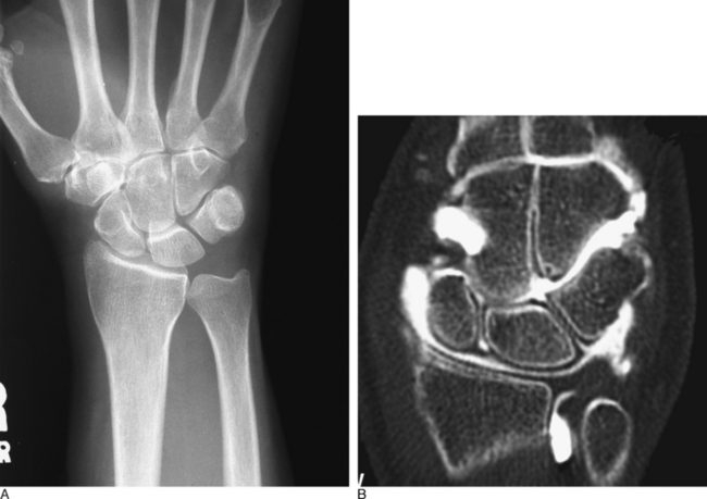
Plain radiographic studies of the affected wrist should be compared to the opposite asymptomatic wrist. Cystic change within the proximal hamate, or a sharp and sclerotic proximal hamate margin, can aid in the identification of this condition preoperatively. However, in most instances plain radiographic studies are noncontributory (Figure 25.2). Furthermore, it is important to realize that plain radiographs are not always diagnostic of a type II lunate morphology.8 MRI scans of the wrist that demonstrate edema within the proximal hamate are suggestive of this condition. Likewise, a wrist CT-arthrogram study that outlines a thinned proximal hamate cartilage surface and/or reveals a sclerotic subchondral ridge can also assist in making the diagnosis preoperatively. Unfortunately, neither of these two expensive diagnostic modalities consistently identifies this condition.

Surgical intervention for the HALT wrist should generally not be entertained until all nonoperative measures have been tried and have failed. A motivated, healthy, active patient with persistent ulnar-sided wrist pain represents the ideal surgical candidate. In our experience, advanced patient age has not precluded a successful surgical outcome. The preoperative discussion with a patient undergoing wrist arthroscopy for ulnar-sided wrist pain should always include the possibility of excision of an arthritic proximal hamate. If advanced chondromalacia is identified at the time of midcarpal arthroscopy, the appropriate treatment can be rendered at that time.

Contraindications

Contraindications to arthroscopic resection of the proximal hamate are few. Any symptomatic patient with advanced chondromalaciac changes identified at the time of arthroscopy can derive significant benefits from a proximal pole resection. Proximal hamate arthrosis represents a spectrum of pathological changes. Resection of the proximal hamate in a patient with grade I or II change is usually contraindicated (Table 25.1). A gray area exists for those patients with limited or focal grade III or IV chondromalacia.

Table 25.1 Chondromalacia Classification

Type I Localized softening, discoloration; no break in the surfaces
Type II Softening, discoloration; small areas of fibrillation or irregular surface
Type III Fibrillation and fissures extend to subchondral bone surface; “crabmeat” appearance
Type IV Areas with complete loss of articular cartilage; exposed subchondral bone

Although resection of the proximal hamate generally allows for marked symptom reduction in patients with isolated HALT wrist pathology, we have noted less impressive results from proximal pole resection in patients having multiple coincident interosseous ligament tears and/or cartilage surface degeneration elsewhere within the affected wrist. Similarly, proximal hamate excision in a patient with diffuse rheumatoid changes or an autoimmune disease that involves the wrist has a small likelihood of providing significant relief. A limited or total wrist fusion remains the treatment of choice in these more complex conditions.

Surgical Technique

The involved wrist and forearm are prepped and draped and placed in some type of traction device. A tourniquet is elevated after exsanguination. Antibiotic prophylaxis is not required for an isolated proximal hamate resection. A standard arthroscopic survey of the radiocarpal and midcarpal joints is then performed, and all cartilage surfaces and ligamentous structures are assessed. The usual working portals include the 3-/,4 and 4-/,5 portals as well as midcarpal-radial (MCR) and midcarpal-ulnar (MCU) portals. In the radiocarpal joint, close attention must be paid to the articular surface of the lunate as well as to the TFCC. If a TFCC perforation is present, the distal ulnar chondral surface of the ulnar head can also be assessed. The LTIL is best seen through the 4-/,5 portal. The 6-U portal is frequently utilized for instrumentation to help probe or debride a torn LTIL.

Once the radiocarpal joint pathology has been inventoried, the cartilaginous surfaces of the midcarpal are inspected. A midcarpal synovectomy is performed as needed to enhance visualization. The proximal pole of the hamate is most clearly evaluated with the arthroscope in the MCU portal. Instability of the LT interval is best assessed with a probe in the MCU portal and the arthroscope in the MCR portal. The cartilage surface of the proximal pole of the hamate should also be probed at this time to define the extent of any chondromalacia present. Seemingly minimal chondromalaciac change observed with the arthroscope is often found to extend much deeper upon probing. After a complete diagnostic arthroscopy of the midcarpal and radiocarpal joints, one can proceed with definitive treatment.

The TFCC, LTIL, and/or scapholunate interosseous ligament (SLIL) can be debrided or repaired as indicated—based on the acuity and severity of the pathology and on the patient’s preoperative symptoms. In our experience, the associated TFCC or LTIL pathology present in most patients with symptomatic arthrosis of the proximal hamate is chronic in nature and is hence usually treated with simple arthroscopic debridement.

Proximal hamate resection is carried out as follows. With the arthroscope in the MCR portal, a 2.7-mm full-radius resector and a 2.9-mm bur are placed alternately into the MCU portal. After the subchondral bone of the proximal hamate has been exposed with the shaver, 2 to 4 mm of the proximal pole of the hamate can be excised using the bur. Care must be taken to avoid damage to the cartilage surfaces of the proximal capitate or to the adjacent triquetrum. To allow adequate visualization during the procedure, frequent suctioning of the joint debris is required. The fat globules and bone debris arising from the resection must be adequately flushed out of the midcarpal joint to prevent leaving debris behind.

The most volar and distal portion of the proximal hamate resection is poorly visualized from the MCR portal. It is necessary to switch the arthroscope to the MCU portal to clearly visualize the full extent of the volar resection of the proximal hamate. Infrequently, the shaver or bur is placed into the MCR portal to complete the proximal hamate resection. The 2.9-mm bur can be used as a gauge to assess the adequacy of the bony resection. When placed in the area of resection, the exposed bur edge should lie nearly adjacent to the proximal capitate surface.

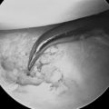
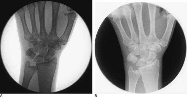
When the surgeon is satisfied with the arthroscopic decompression of the hamatolunate articulation, an intra operative fluoroscopic assessment is performed to confirm the adequacy of the proximal hamate resection (Figure 25.3). Whereas this often requires some maneuvering of both the surgeon and the instruments, the fluoroscopic pictures provide an important secondary means of evaluation. If the radiographs confirm that the resection is satisfactory, all portals are closed with skin tapes, local anesthetic is infiltrated into the joint and the surrounding subcutaneous tissues, and a light compressive bandage is applied for 10 days.

The postoperative regimen is determined primarily by the degree of intra-articular pathology and the amount of reconstructive work. In the setting of an isolated proximal hamate resection or a typical HALT wrist procedure (debridement of an LTIL tear followed by a proximal hamate resection), the patient is seen 10 days postoperatively and a hand therapy consultation is obtained. The therapist’s instructions usually include an early active assisted-motion protocol as well as edema control.

Authors’ Experience

A chart review of all patients undergoing wrist arthroscopy between 1991 and 2001 yielded 23 patients who had undergone arthroscopic resection of the proximal pole of the hamate. The mean age of the patients at the time of surgery was 43 years. Four patients were female and 19 were male. The symptomatic wrist was the patient’s dominant wrist in 13 cases. Seventeen patients related a specific distant injury to their wrist (fall or twist). All patients consented to arthroscopic treatment because of wrist pain unresponsive to nonoperative treatment. Preoperative X-rays revealed that 20 patients had an obvious type II lunate. Eight radiographs were suggestive of an erosive lesion of the proximal pole of the hamate. None of the patients had a static LT or SL carpal instability pattern.

At the time of arthroscopy, all wrists were found to have a variable amount of midcarpal synovitis and diffuse grade III or IV chondromalacia of the proximal pole of the hamate. Twenty-one wrists had pathology of the LTIL, seven had a TFCC tear, three were noted to have early radiocarpal arthritis, and two had a tear of the SLIL. In addition to resection of the proximal pole of the hamate, six patients underwent debridement of the proximal portion of the LTIL, five had a concurrent TFCC debridement, one had an SLIL debridement, one had a TFCC repair, and one underwent an open LTIL repair. The average bone resection of the proximal pole of the hamate, as measured by postoperative X-ray, was 2.2 mm (range 1 to 4 mm).

At final follow-up at 4.7 years, 18 of 23 patients had good or excellent results according to the modified Mayo scoring system (and one had a fair result). Ten patients were able to return to work within two weeks of their surgery, and 78% of patients returned to their preoperative employment or recreational status at an average of nine weeks post-surgery. At final follow-up, 18 patients reported minimal to no change in work or recreational status—whereas 5 either were not working or had to change jobs because of persistent wrist problems.

All patients with isolated HALT pathology (hamate arthrosis, LTIL tear, and midcarpal synovitis) had good or excellent results from hamate resection. Two-thirds of the patients with TFCC perforations, LTIL tears, and midcarpal synovitis had good or excellent results—whereas only half of the patients with LTIL, TFCC, and SLIL tears achieved these outcomes. No patients with preexistent radiocarpal arthritis (scaphoid or lunate facet of distal radius) had good or excellent outcomes.

Four patients had salvage surgery for poor outcomes. Of these four, two had salvage procedures within 18 months (one wrist fusion and one STT arthrodesis) and one underwent a four-corner fusion three years after the index procedure. The last patient underwent a proximal row carpectomy seven years after hamate resection.

We found that the majority of patients did well after surgery, and that the good results were generally sustained at a mean of 4.7 years from surgery. As a result of this study, it has become our practice to ensure that we resect the proximal hamate to the level necessary to completely unload the medial facet of the lunate. Other important observations from this study include (1) patients with increasing associated wrist pathology do not do as well as patients with pathology limited to the HALT condition, which should be discussed with patients, and (2) patients that developed the onset of hamate arthrosis after prior wrist surgical procedures for unrelated lesions were also more likely to have poorer results.

We have found that in the treatment of HALT wrists a fairly conservative approach to the LT instability appears warranted. We believe that most patients we encounter with hamate arthrosis and LTIL tears can be treated by arthroscopic excision of the proximal pole of the hamate and (at most) by debridement of the LTIL. We did not find that reconstruction of the LTIL with pinning and/or casting is necessary for symptomatic improvement. Perhaps the biggest advantage from this simplified approach is the institution of early wrist motion postoperatively, which contributed to a rapid return to work in 78% of patients.