188 Arterial leg ulcer
Salient features
History
Examination
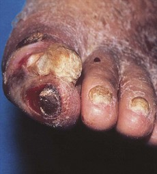
• Tender, punched-out ulcers on leg (usually away from the ankle region) (Fig. 188.1)
• The ulcer is usually in the plantar surface over the heads of first and fifth metatarsals (uncommon on the dorsum of the foot because the pressure is less sustained and perfusion is better)
• Peripheral pulses are not palpable or diminished (in diabetics and in chronic renal failure the arteries may be calcified, making the results of pulse examination less reliable)
Questions
How would you investigate such a patient?
• Radiographs may show calcification of the arteries of the leg
• Doppler ultrasonography to define the severity of arterial involvement
• Measurement of segmental limb pressures
• Pulse–volume waveform and transcutaneous oxygen are particularly useful in evaluating perfusion of the diabetic foot because they are not affected by vessel calcification
• Aortography shows narrowing and stenosis of the leg arteries.
How would you manage arterial ulcers?
• General: stop smoking; control diabetes, obesity and hypertension
• Limbs: should be kept warm (but avoid local heat)
• Feet: chiropodist and foot care, appropriate foot-wear
• Regular exercise to promote development of anastomotic collateral vessels
• Surgery: balloon dilatation for local iliac or femoral stenoses, aortoiliac bypass, amputation when complicated by gangrene
• Flow-limiting arterial lesions should be evaluated and reconstructed or bypassed: prosthetic vascular grafts or autologous vein grafts may be used.


