Approach to Diagnosis of Diffuse Lung Disease

Published on 23/05/2015 by admin

Filed under Pulmolory and Respiratory

Last modified 22/04/2025

Print this page

rate 1 star rate 2 star rate 3 star rate 4 star rate 5 star
Your rating: none, Average: 0 (0 votes)

This article have been viewed 2149 times

Chapter 46 Approach to Diagnosis of Diffuse Lung Disease

The term diffuse lung disease (DLD) includes infiltrative lung processes that involve the alveolar spaces or lung interstitium. This definition is fundamentally unsatisfactory because it groups together a variety of diverse disorders, some of which, such as cryptogenic organizing pneumonia, are not actually “diffuse,” but rather patchy and sometimes limited in extent. Furthermore, many secondary infiltrative abnormalities, including bacterial infection and malignancy, are excluded, whereas others, such as pulmonary involvement in connective tissue disease (CTD), are retained in most DLD classifications. The DLDs are grouped for historical reasons; in early series, DLDs presented most frequently with widespread clinical and chest radiographic abnormalities. However, with increasing clinician awareness of possible DLD, the diagnosis is often made when chest radiographic findings are limited, or disease is apparent only on high-resolution computed tomography (HRCT).

The diagnostic difficulties resulting from the multiple disorders within the DLDs are exacerbated by semantic confusion. Synonymous terms abound for some of the more frequently encountered DLDs, such as the following:

This problem has been partially addressed by the reclassification of the idiopathic interstitial pneumonias by a joint American Thoracic Society/European Respiratory Society (ATS/ERS) international consensus committee, discussed in detail in Chapter 47. However, the term cryptogenic fibrosing alveolitis (CFA) continues to cause difficulties. As defined in the ATS/ERS reclassification, CFA is strictly synonymous with idiopathic pulmonary fibrosis (IPF). The diagnosis of IPF/CFA now requires the presence of usual interstitial pneumonia (UIP) at surgical biopsy or typical appearances on HRCT, in association with a compatible clinical picture. This represents a radical change; in historical series, various disorders presenting with a clinical picture of IPF were grouped together as IPF/CFA. The entity of “clinical CFA syndrome” is still necessary for epidemiologic studies but should not be viewed as a final diagnosis in clinical practice.

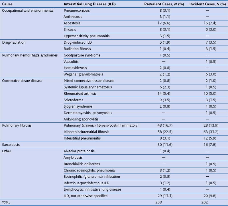
In routine practice, a simplified pragmatic approach to diagnosis of DLD is essential; consideration of a checklist of the more common diseases is often useful. The classification of DLD by their disease burden was addressed most definitively in a study from Bernalillo County, New Mexico, in which the incidence and prevalence of individual DLDs was quantified using a variety of methods (Table 46-1). New cases were estimated to occur in 32 : 100,000 years in males and 26 : 100,000 years in females; thus, although less common than lung infection, malignant disease or obstructive airways disease, the DLDs are responsible for a considerable disease burden. More recent evidence shows an increase in the prevalence of DLD, especially IPF, which inevitably means that the burden of disease has increased further in the previous one to two decades. Moreover, the workload for the respiratory medicine physician is disproportionate because the diagnosis of individual DLDs is often uncertain, despite more intensive investigation than is generally required in obstructive airways disease, malignancy, or chronic lung suppuration.

A consideration of the differential diagnosis of DLD, based on prevalence alone, is merely a starting point, for two reasons. First, clinical information at initial evaluation profoundly alters diagnostic probabilities; therefore a longer checklist of the DLDs, based on the possible underlying cause, is indispensable. Second, the length to which a specific diagnosis is pursued, with particular reference to surgical biopsy, is critically dependent on the importance of discriminating between likely differential diagnoses in individual cases. This crucial point is discussed in detail in the concluding section of this chapter.

Initial Clinical Evaluation

Even before chest radiography and HRCT findings are considered, a wealth of diagnostic information can be obtained from initial evaluation. A possible underlying cause is often apparent, although environmental and drug exposures occurring many years earlier and apparently limited exposures are often difficult to interpret in isolation. Much information can often be obtained on the longitudinal behavior of disease from the evolution of symptoms, serial chest radiographic data, and less frequently, serial spirometric volumes. Associated systemic disease and prominent airway-centered symptoms both provide useful diagnostic clues. Less frequently, physical examination may serve to broaden the differential diagnosis. In addition to chest radiography and HRCT, certain noninvasive ancillary tests should be performed in select patients, including autoimmune serology, measurement of precipitins, and echocardiography.

Clinical History

The identification of an underlying cause is the single most important contribution made by clinical evaluation. Table 46-2 provides a checklist of the more important causes of DLD. A careful occupational history is essential and should include details of all previous occupations, including short-term employment. Asbestos exposure is often extensive in railway rolling-stock construction, shipyard workers, power station construction and maintenance workers, naval boilermen, garage workers (involved in brake lining), and other occupations in which asbestos exposure is overt; generally, workers in these occupations are well aware of their asbestos exposure. However, other workers, including joiners, electricians, carpenters, and construction workers, who handle asbestos in the form of roofing and insulation material, are often unaware of significant exposure. Other occupations associated with DLD include coal mining (coal worker’s pneumoconiosis), metal polishing (hard metal disease), and sandblasting (silicosis).

Table 46-2 Frequently Encountered Diffuse Lung Diseases with Identifiable Underlying Cause

Cause Differential Diagnosis
Occupational-related or other inhalant–related
Inorganic

Organic

Collagen vascular disease–related Drug-related   Physical agents/toxins Neoplastic disease Vasculitis-related Disorders of circulation Chronic infection Smoking-induced

A careful history will also disclose exposure to organic antigens known to cause hypersensitivity pneumonitis. The two most prevalent forms of hypersensitivity pneumonitis are farmer’s (harvester’s, thresher’s) lung disease, in which the offending antigen, thermophilic actinomycetes (Thermoactinomyces vulgaris) is contained within moldy hay, and pigeon (bird) breeder’s lung disease (bird fancier’s lung), in which avian proteins are inhaled by those breeding birds, or more often, those who keep birds as domestic pets. However, a wide range of other exposures also cause hypersensitivity pneumonitis, and particular attention should be paid to molds (often arising in sites of water damage); bathroom molds (as in “basement shower syndrome” and “hot-tub lung” are easily overlooked. Hobbies should also be considered (e.g., cheese-maker’s lung, winemaker’s lung). There are now more than 100 known causes of hypersensitivity pneumonitis, and an up-to-date list (e.g., see Bertorelli et al.) of the 50 more frequent causes of hypersensitivity pneumonitis is highly useful.

A detailed drug history is also essential. The drugs most frequently causing DLD are probably amiodarone, methotrexate (at doses used in CTD), and antineoplastic agents, especially bleomycin. However, a wide variety of other agents (>200 at present) cause DLD, although often in only a small number of patients, and the list increases yearly. Fortunately, an international website is now devoted to drug-induced lung disease (www.pneumotox.com), through which all medications should be routinely checked in patients with DLD.

Other therapeutic modalities causing DLD include radiotherapy and exposure to high concentrations of oxygen (especially in those previously receiving bleomycin). Paraquat ingestion (causing acute or delayed proliferative bronchiolitis), inhalation of crack cocaine or heroin (causing eosinophilic pneumonia, diffuse alveolar hemorrhage, organizing pneumonia or pulmonary edema), and intravenous drug use (causing venoocclusive disease) are also relevant.

Smoking-related DLD is increasingly diagnosed; diseases other than chronic obstructive pulmonary disease caused by smoking include Langerhans cell histiocytosis, respiratory bronchiolitis associated with interstitial lung disease (RBILD), desquamative interstitial pneumonia, and nonspecific interstitial pneumonia. Recently, HRCT evaluation has shown that all these processes may coexist in the same patient. Further, both sarcoidosis and hypersensitivity pneumonitis are rare in current smokers. Because RBILD and hypersensitivity pneumonitis often have overlapping clinical and HRCT features, the smoking history is an important discriminator between these two disorders.

Information on likely longitudinal behavior often helps distinguish between acute and chronic disease, because acute infection, heart failure, and disseminated malignancy may all simulate DLD clinically and radiologically. The duration of dyspnea and cough, pattern of symptomatic progression, and previous responsiveness (or nonresponsiveness) to corticosteroid therapy may provide valuable diagnostic clues. Variable dyspnea and cough over several years, responding to steroid therapy, is compatible with hypersensitivity pneumonitis or sarcoidosis, whereas inexorably progressive dyspnea for 2 to 3 years, not responding to steroid therapy, is typical of IPF. Previous chest radiographs may be useful, with unchanging appearances over many years a frequent finding in sarcoidosis, but not in IPF. Previous full pulmonary function tests are seldom available, but serial spirometry may be performed in general practice, because asthma is often suspected when the first symptoms of DLD occur. Thus, useful conclusions may be drawn from the rapidity of decline, or conversely the duration of stability, of spirometric volumes.

Relevant systemic diseases associated with DLD include malignancy (lymphangitis carcinomatosis or multiple metastases) and CTDs complicated by DLD, especially rheumatoid arthritis, systemic sclerosis, systemic lupus erythematosus (SLE), polymyositis or dermatomyositis, and Sjögren syndrome. Lung disease may precede systemic manifestations in all the CTDs (most frequently in polymyositis/dermatomyositis) or may develop concurrently with systemic manifestations. Thus, a full history should include details of arthritis/arthralgia, myositis, skin disorders, Raynaud’s phenomenon, and dryness of the eyes or mouth. A subgroup of patients with autoimmune disease who fail to meet formal criteria for an individual disorder are considered to have “undifferentiated connective tissue disease” but may nonetheless develop DLD.

Airway-centered symptoms may help to refine the differential diagnosis. Cough occurs frequently in IPF, but prominent wheeze is more suggestive of hypersensitivity pneumonitis or, less frequently, sarcoidosis. Wheezing is also an important feature of some of the pulmonary vasculitides, especially Churg-Strauss syndrome and occasionally Wegener granulomatosis. Hemoptysis is the most frequent pulmonary symptom at presentation in Goodpasture syndrome; however, the volume is not a good guide to disease severity, because hemoptysis may be trivial or even absent, despite considerable alveolar hemorrhage.

Diagnostic Procedures

Blood Tests

In most cases, specific diagnostic tests for DLD are confined to autoimmune serology and precipitins against organic antigens. Serologic evidence of rheumatoid arthritis and the other major CTDs should be sought in any patient with apparently idiopathic DLD in whom the diagnosis is uncertain. This is particularly important when the diagnosis appears to be cryptogenic organizing pneumonia, because underlying connective disease is common; when present, prognosis is not always good, and prolonged treatment may be required. In the antisynthetase syndrome, characterized by Jo1 antibody positivity, polymyositis or dermatomyositis, and progressive pulmonary fibrosis, lung disease often precedes systemic manifestations. However, although antibodies to extractable nuclear antigens tend to be disease specific, levels of antinuclear antibodies and rheumatoid factor are often moderately increased in idiopathic pulmonary fibrosis and are less useful diagnostically unless titers are greatly elevated.

The presence of precipitins to organic antigens increases the likelihood of hypersensitivity pneumonitis but should never be considered diagnostic in isolation. Positive precipitins denote exposure to an antigen, with immune recognition, but alone are not indicative of clinically significant DLD; pigeon and other bird breeders often have avian precipitin positivity without overt lung disease. Moreover, in many patients with convincing exposure histories and a histologic diagnosis of hypersensitivity pneumonitis, the appropriate precipitins are not present; avian antigens, for example, vary between bird species and even between individual birds.

Chest Radiography

With recent attention focused on the diagnostic value of HRCT, it is often forgotten that the plain chest x-ray film provides invaluable information in diffuse lung disease. The chest radiograph points strongly toward a specific diagnosis in some patients. Sarcoidosis, the most prevalent DLD encountered in clinical practice, can be diagnosed with confidence from the clinical and chest radiographic features at presentation in many patients. HRCT seldom adds useful diagnostic information in this context.

Several radiographic features have useful positive predictive values (PPVs). The presence of hilar lymphadenopathy on chest radiography is particularly strongly predictive of sarcoidosis in the correct clinical context, although the radiographic differential diagnosis includes tuberculosis and malignancy, especially when hilar lymphadenopathy is asymmetric. Pleural effusions are an occasional feature of CTD, lymphangioleiomyomatosis, and asbestos-related disease, as well as disease processes that sometimes mimic DLD, including heart failure, infection, and malignancy. The distribution of disease on chest radiography is also diagnostically useful; granulomatous diseases (tuberculosis, sarcoidosis, hypersensitivity pneumonitis) tend to be more prominent in the middle to upper zones, whereas fibrotic diseases (IPF, fibrotic nonspecific interstitial pneumonia, asbestosis) have a predominantly lower-zone distribution.

However, apart from a large subgroup of patients with sarcoidosis, diagnoses based on chest radiography are seldom confident. Chest radiography is sometimes insensitive; in one often-quoted series (Epler et al.), 10% of patients with biopsy-proven DLD had normal chest radiographic appearances. The superior diagnostic accuracy of HRCT over chest radiography has been documented in numerous series, and the increased confidence associated with an HRCT diagnosis is a considerable aid to management. The classification of chest radiographic abnormalities as “nodular, reticulonodular, or reticular” provides relatively little diagnostic information. Predominantly basal honeycombing on chest radiography (invariably associated with honeycombing on HRCT) may be diagnostically useful in increasing the likelihood of IPF but is radiographically overt in surprisingly few IPF patients. It is now generally accepted that, except in patients with obvious sarcoidosis, routine HRCT is almost always warranted in DLD, although occasional exceptions exist, including elderly patients with obvious lower-zone honeycombing on chest radiography, indicative of IPF, and patients with long-standing pulmonary fibrosis on serial chest radiography and an obvious underlying cause, such as coal mining.

Two chest radiographic appearances pose particular diagnostic difficulties. Persistent unexplained multifocal consolidation has usually been treated unsuccessfully as for community-acquired pneumonia and has a wide differential diagnosis. Serial chest radiography tends to be more useful than HRCT in refining investigation, because the crucial diagnostic distinction lies between fixed infiltrates (nonbacterial infection, including tuberculosis, alveolar cell carcinoma, and other malignant processes) and changing infiltrates, in which these diagnoses are effectively excluded. However, immunologically mediated diseases, including eosinophilic pneumonia, cryptogenic organizing pneumonia, and vasculitic disorders, may give rise to either fixed or evanescent radiographic abnormalities, and a histologic diagnosis is often warranted. Diffuse alveolar filling processes giving rise to widespread air-space consolidation are generally indicative of life-threatening disease. Although this picture may represent DLD (e.g., acute interstitial pneumonitis, acute eosinophilic syndromes, drug-induced pulmonary infiltration), it is essential to broaden the differential diagnosis beyond DLD to include diffuse pulmonary infection, toxic inhalation, severe aspiration, opportunistic infection (especially Pneumocystis pneumonia), diffuse alveolar hemorrhage syndromes, mitral stenosis, and most importantly, heart failure. In both these radiographic presentations, successful management often depends on consideration of a wide differential diagnosis from the outset.

High-Resolution Computed Tomography

High-resolution CT has been the most important diagnostic advance in DLD in the last two decades. Numerous studies have confirmed the overall diagnostic accuracy of HRCT against findings at surgical biopsy, with a striking increase in sensitivity and specificity for individual diseases, compared with chest radiography. However, academic series understate the impact of HRCT, because the most important benefit has been to increase clinician confidence in noninvasive diagnosis, with a corresponding reduction in the numbers of patients needing to undergo surgical biopsy. Before HRCT, diagnoses based on clinical data and chest radiographic findings were seldom confident, and management was necessarily tentative in many cases. The combination of clinical and HRCT information now provides a confident first-choice diagnosis in the majority of patients, and in many other cases, the realistic differential diagnosis is shortened to two or three disorders. This allows surgical biopsy to be reserved for patients in whom the distinction between a small group of possible disorders has important management implications and for occasional patients in whom HRCT appearances are not suggestive of any single disorder. Thus, routine surgical biopsy, as a diagnostic “gold standard,” can no longer be justified in the HRCT era.

Histospecific diagnosis aside, HRCT sometimes plays an important role in detecting DLD. The superior sensitivity of HRCT over chest radiography has been an invariable finding in studies of a wide range of disorders. This feature of HRCT is particularly useful when symptoms or lung function impairment are associated with normal chest radiographic appearances. In CTD, pulmonary involvement is now the leading cause of death, and early treatment of progressive lung disease is desirable. By identifying limited pulmonary fibrosis, HRCT allows clinicians to select patients requiring more intensive monitoring, even when immediate treatment is not warranted. In workers previously exposed to asbestos, HRCT often discloses pulmonary fibrosis, which is obscured on chest radiography by concurrent pleural disease. However, it should be stressed that the sensitivity of HRCT occasionally creates its own difficulties. When interstitial abnormalities are limited, their clinical significance is sometimes difficult to rationalize; disease evident only on HRCT should not be extrapolated, in terms of natural history and management, to the more extensive disease described in historical clinical series. It is essential that HRCT findings are integrated with other clinical and investigative features and not interpreted in isolation.

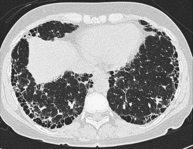
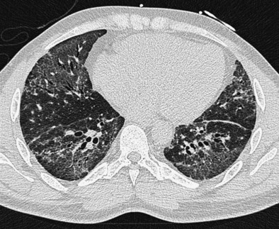
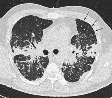
The distinction between predominantly inflammatory and predominantly fibrotic disease can generally be made with reasonable confidence from HRCT. Anatomic distortion and reticular abnormalities are strongly indicative of irreversible fibrotic disease, and this is invariably true of honeycomb change (Figure 46-1). Consolidation is usually reversible, although it may occasionally represent dense fibrosis, especially in sarcoidosis. Ground-glass attenuation is often more difficult to interpret. In early work, this HRCT sign was shown to identify a substantial increase in the likelihood of significant inflammation, especially in the absence of concurrent reticular abnormalities. However, it is now clear that ground-glass attenuation denotes fine fibrosis in many cases, especially in sarcoidosis and nonspecific interstitial pneumonia, in which ground-glass attenuation is the cardinal HRCT feature (Figure 46-2). Traction bronchiectasis is a key HRCT discriminator because it invariably indicates underlying fibrosis. Thus, reversible inflammatory disease is likely only when ground-glass attenuation is not associated with traction bronchiectasis or admixed with reticular abnormalities.

A wide range of HRCT profiles encompassing the distribution and pattern of DLD strongly suggests individual diseases. Box 46-1 summarizes the cardinal findings in common DLDs. As with its other applications, it is essential that HRCT findings be integrated with the pretest diagnostic probability, distilled from the history, clinical signs, previous natural history, or treatment course and the investigative findings, especially chest radiography, pulmonary function tests, and serology for autoimmune disease and environmental antigens. The role of HRCT in diagnosis is critically dependent on the presence or absence of a likely cause. In patients with appropriate environmental antigen or drug exposures (hypersensitivity pneumonitis, pneumoconioses, drug-induced lung disease), malignant disease (lymphangitis carcinomatosis), clinical or serologic evidence of CTD, or a heavy smoking history (Langerhans cell histiocytosis, RBILD), the diagnostic weighting required from HRCT can be reduced. In these contexts, HRCT appearances that are merely compatible (and not classic) often allow a sufficiently confident diagnosis to obviate diagnostic surgical biopsy.

Box 46-1

High-Resolution Computed Tomography (HRCT) Features of Select Diffuse Lung Diseases (DLDs)

Idiopathic pulmonary fibrosis: Lower zone, subpleural predominance, maximal posterobasally, predominantly reticular pattern with associated honeycombing.

Nonspecific interstitial pneumonia: Two typical appearances:

Desquamative interstitial pneumonia: Ground-glass attenuation, sometimes diffuse, sometimes basal and peripheral–centered, frequent associated fibrotic cysts with anatomic distortion and traction bronchiectasis.

Acute interstitial pneumonia: Widespread ground-glass attenuation admixed with features of fibrosis, usually with air space consolidation and occasionally with emphysema.

Respiratory bronchiolitis interstitial lung disease: Patchy ground-glass attenuation, poorly defined centrilobular nodules, occasional mosaic attenuation, prominent bronchial wall thickening.

Sarcoidosis: Highly variable; nodules distributed along bronchovascular bundles, interlobular septae, and subpleurally, including the fissure; ground-glass attenuation that may represent either inflammation or fine fibrosis; reticular abnormalities representing fibrosis; distortion most often in upper zones with posterior displacement of upper lobe bronchus; air trapping; associated hilar and mediastinal lymphadenopathy.

Subacute hypersensitivity pneumonitis: Widespread ground-glass attenuation, often containing poorly defined centrilobular nodules, admixed with areas of “black lung” (mosaic attenuation), representing air trapping and enhanced on expiratory HRCT.

Cryptogenic organizing pneumonia: Bilateral patchy consolidation, subpleural and predominantly basal in most cases; occasional peribronchial distribution; associated, often-sparse nodules up to 1 cm in diameter.

Constrictive bronchiolitis: Patchy areas of hyperlucency enhanced on expiration, which may not change in cross-sectional diameter on full expiration; associated bronchiectasis and bronchial wall thickening.

Langerhans cell histiocytosis: Bizarre cyst shapes and associated nodules throughout the lung fields but sparing costophrenic angles and tips of lingula and middle lobes; associated emphysema often seen.

Pulmonary lymphangioleiomyomatosis: Homogeneously distributed, thin-walled parenchymal cysts, varying from a few millimeters to several centimeters in diameter; associated with retrocrural adenopathy, pleural effusion, thoracic duct dilation, pericardial effusion, and pneumothorax.

By contrast, as discussed, the diagnostic role of HRCT is much greater in idiopathic disease, but unless the pretest diagnostic probability is high, confident diagnosis requires the presence of typical HRCT appearances. The optimal diagnostic use of HRCT in apparently idiopathic DLD can be distilled in a simple pragmatic algorithm, as follows:

1. The first step is to determine whether HRCT abnormalities are predominantly fibrotic, based on the presence of honeycombing, reticular abnormalities, anatomic distortion, or in patients with prominent ground-glass attenuation, the presence of traction bronchiectasis.

2. If disease is fibrotic, as in a large majority of cases, the next important question is whether HRCT findings are typical of idiopathic pulmonary fibrosis. As stated in the 2011 American Thoracic Society/European Respiratory Society (ATS/ERS) recommendations, IPF can be diagnosed confidently on HRCT when there is honeycombing with minimal ground-glass attenuation in a predominantly basal and subpleural distribution. It is logical to focus on IPF because it is the most prevalent idiopathic fibrotic disease among patients in whom the diagnosis is not obvious from clinical and chest radiographic findings (most cases of sarcoidosis are diagnosed without recourse to HRCT) (Figure 46-3). Further, IPF has a much worse prognosis than other fibrosing processes and therefore is the most important diagnosis to confirm or exclude from the outset. It is now known that HRCT appearances considered typical of IPF by experienced thoracic radiologists have a PPV greater than 95%.

3. If appearances are not typical of IPF, the five most important differential diagnoses (based on prevalence in routine practice) are (a) IPF with atypical HRCT appearances, (b) sarcoidosis, (c) nonspecific interstitial pneumonia, (d) hypersensitivity pneumonitis (with the antigen unknown), and (e) the fibrotic sequelae of cryptogenic organizing pneumonia. Among these, IPF with atypical HRCT appearances is the most prevalent disorder in most populations; up to 30% of IPF patients have atypical HRCT features. Atypical IPF is especially likely when HRCT appearances are not typical of any of the other four disorders (b-e).

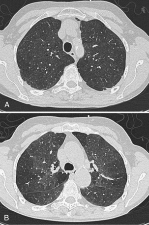
The weighting given to HRCT in the diagnosis of DLD varies from case to case but can usefully be considered in three categories. In some patients, HRCT appearances are virtually pathognomonic; this includes many cases of IPF, Langerhans cell histiocytosis, sarcoidosis, and lymphangitis carcinomatosis. Often, HRCT findings are diagnostic when combined with clinical information. A good example is the combination of widespread ground-glass attenuation (often with poorly defined centrilobular nodules) in combination with mosaic attenuation, which may be strongly indicative of hypersensitivity pneumonitis in nonsmokers with a compatible exposure history but may also represent RBILD in smokers (Figure 46-4). Third, even when not conclusive alone, HRCT may be invaluable when considered with diagnostic surgical biopsy. The histologic entity of nonspecific interstitial pneumonia (NSIP) is found in a variety of clinicoradiologic contexts, including entities overlapping clinically with IPF, fibrosing organizing pneumonia, and hypersensitivity pneumonitis; HRCT evaluation is key to distinguishing among these variants.

Bronchoscopic Procedures

Although endobronchial and transbronchial biopsies are straightforward and relatively noninvasive procedures, the volume of tissue taken is small, with only bronchial and peribronchial tissue sampled. Thus, both procedures have a high yield in diseases with peribronchial distribution, especially sarcoidosis and lymphangitis carcinomatosis. Occasionally, transbronchial biopsy findings can help confirm a diagnosis of hypersensitivity pneumonitis, although bronchoalveolar lavage fluid tends to be more rewarding in this regard. Bronchoscopic biopsy procedures have little or no diagnostic value in the idiopathic interstitial pneumonias.

Hemorrhage and pneumothorax (with transbronchial biopsy) are the important risks associated with bronchoscopic procedures. Major hemorrhage is rare, but pneumothoraces complicate transbronchial biopsies in 1% to 2% of procedures, although intercostal tube drainage is not always required.

In the 1980s, many regarded bronchoalveolar lavage (BAL) as an important part of the diagnostic algorithm in DLD. The distinction between a BAL neutrophilia (suggestive of IPF) and a BAL lymphocytosis (as in sarcoidosis or hypersensitivity pneumonitis) was held to be particularly useful. With experience, however, it became apparent that diagnostic distinctions based on BAL fluid in large groups of patients were insufficiently reliable in individual patients. The advent of HRCT also limited the role of BAL, which had been more influential in the pre-HRCT era, when most noninvasive diagnoses were tentative. There are currently no published evaluations of the diagnostic value added by BAL, once HRCT findings are taken into account.

In recent years, however, use of BAL has increased in some centers. In the 2002 ATS/ERS recommendations, compatible BAL findings (i.e., no lymphocytosis) are a requirement for the noninvasive diagnosis of IPF. Although not formally retained in the 2011 recommendations, this criterion reflects the diagnostic value of BAL findings when HRCT appearances are suggestive of IPF but not definitive. However, a BAL lymphocytosis has an even greater diagnostic impact in some patients with sarcoidosis or hypersensitivity pneumonitis. When significant fibrosis supervenes in these disorders, HRCT appearances often become atypical, and IPF is frequently the preferred diagnosis, before performance of BAL. A BAL lymphocytosis in the setting of fibrotic DLD may be an important justification for diagnostic surgical biopsy. Thus, BAL continues to play a useful diagnostic role in a significant subset of patients, when clinical and HRCT features are inconclusive, although adding little to diagnosis in the majority of DLD patients.

Surgical Biopsy

Surgical biopsy, formerly performed as an open procedure but now widely obtained using video-assisted thoracoscopic surgery (VATS), was once regarded as the diagnostic reference standard and, until recently, was advocated as a routine diagnostic procedure by some authorities. However, routine surgical biopsy is impractical outside referral centers, and in the 1980s, even before HRCT had a significant diagnostic impact, it was performed in less than 15% of patients with IPF in the United Kingdom. Even in referral centers, number of diagnostic surgical biopsies has been radically reduced with the application of HRCT. In one UK referral center, the Royal Brompton Hospital, biopsy was performed in more than 50% of IPF patients in the 1980s but in less than 25% in the mid-1990s. Moreover, it is increasingly clear that open or VATS biopsy is not a true diagnostic gold standard. Variation between 10 thoracic pathologists in assigning a histologic diagnosis in DLD was recently found to be considerable, with agreement only moderate (kappa coefficient of agreement of 0.38). In more than 20% of biopsies, the first-choice diagnosis was assigned with low confidence.

Thus, although biopsy procedures add invaluable information in select patients, and a pattern of usual interstitial pneumonia is usually diagnostically definitive, a histologic diagnosis alone should no longer be viewed as a diagnostic gold standard.

Sometimes, the histologic diagnosis is at odds with clinical and HRCT information and must be integrated with other information. In some patients with fibrotic hypersensitivity pneumonitis, a histologic pattern of usual interstitial pneumonia (which is normally indicative of IPF) is disclosed at biopsy, despite clinical, HRCT, and BAL features of hypersensitivity pneumonitis and an indolent course during follow-up. Similarly, in a cohort of more than 100 patients with IPF or fibrotic NSIP (Flaherty et al., 2003), a combination of histologic and HRCT findings provided more accurate prognostic information than either modality alone. Thus, surgical biopsy is now best viewed, as with HRCT, as a diagnostic “silver standard” that can often be avoided when HRCT and clinical features are typical of an individual DLD.

The morbidity and mortality associated with surgical biopsy in DLD are low in patients with an adequate pulmonary reserve but increase significantly in those with severe disease. In one series (Utz et al.), patients with advanced IPF (mean gas transfer <35% predicted) had a mortality ascribable to biopsy of 15%. Although this figure is generally regarded as an overstatement of the risk of the procedure, based on other series and widespread anecdotal experience, a surgical biopsy should not be performed unless central to management if the gas transfer is less than 30% of predicted. Moreover, in advanced idiopathic fibrotic disease, the prognostic value of a histospecific diagnosis diminishes. In another series (Latsi et al.), mortality was identical in IPF and fibrotic NSIP when the gas transfer was less than 35%, despite major differences in survival in less severe disease.

Thus, in younger patients (<60) presenting with a typical clinical picture of severe IPF and HRCT features suggestive of fibrotic NSIP, immediate referral for consideration of lung transplantation is warranted, without a histologic diagnosis. The exception is the patient presenting with overwhelmingly severe acute DLD, in which the diagnosis is unclear, and realistic differential diagnoses include acute interstitial pneumonia, severe infection (e.g., opportunistic infection), and malignancy. BAL fluid may be required to exclude infection, and occasionally a histologic diagnosis is required to plan management. Both procedures can be performed in ventilated patients, and if disease is slightly less severe, elective mechanical ventilation may be warranted to investigate appropriately.

Integrated Diagnosis in Diffuse Lung Disease

The central diagnostic challenge for the clinician is to integrate the clinical and diagnostic information into a final diagnosis, without overemphasizing any single clinical or investigative feature. Indeed, only the clinician is able to play this latter role, because both histologic and HRCT diagnoses made without reference to other information are seriously flawed in a significant proportion of patients. The most difficult dilemma, when noninvasive evaluation discloses two or more realistic diagnoses, is whether to accept diagnostic uncertainty, without investigating further, or to resort to invasive (surgical biopsy) or semi-invasive (BAL fluid, transbronchial biopsy) procedures.

This decision should be made pragmatically and not by protocol. The value of a specific diagnosis in diffuse idiopathic lung disease is that the clinician is informed of the probable natural history and the likelihood that treatment will play a useful role. From these considerations, the optimal approach to monitoring disease during follow-up usually becomes apparent. Thus, the essential purpose of pursuing a diagnosis is to identify probable disease behavior with and without treatment. Broadly, with occasional exceptions, longitudinal disease behavior in DLD can be subdivided into five patterns (Table 46-3). When a patient can be subclassified confidently into one of these groups, invasive investigation will often add little to short-term and long-term management. Three strands of information are of particular value in making these distinctions: the underlying cause (if any), a morphologic assessment using HRCT (and histologic evaluation in select patients), and observed longitudinal disease behavior.

Table 46-3 Most Common Patterns of Longitudinal Disease Behavior in Diffuse Lung Disease (DLD) with Select Underlying Diagnoses*

Pattern Select Diagnoses
Self-limited inflammation

Stable fibrotic disease Major inflammation, risk of fibrotic progression Inexorably progressive fibrosis Explosive acute DLD

* May appear in several categories; excluding idiopathic interstitial pneumonias (see Table 46-4).

The identification of an underlying cause is vital because considered with HRCT appearances, it may allow disease to be classified confidently as self-limited inflammation (e.g., acute drug-induced lung disease, hypersensitivity pneumonitis, RBILD in smokers), with a good outcome, provided the offending agent is removed. In long-standing disease, knowledge of a cause often allows the clinician to classify fibrotic abnormalities on HRCT as stable fibrotic disease (e.g., fibrotic sequelae of nitrofurantoin lung, silicosis, other pneumoconioses); the confidence of this conclusion is increased by the documentation of stable longitudinal disease behavior, based on previous chest radiographs, symptoms, and occasionally pulmonary function tests. In both self-limited inflammation and stable fibrotic disease, invasive diagnostic investigations are seldom warranted, and potentially toxic treatments can usually be minimized.

A histologic diagnosis is required more frequently in apparently idiopathic disease, to draw two essential distinctions: between inherently stable and potentially progressive fibrotic disease, and between major inflammation (with high risk of evolution to fibrosis with undertreatment) and inexorably progressive fibrotic disease. In both scenarios, therapeutic intervention may be the key to a substantially better outcome. Knowledge of likely intrinsic disease behavior is invaluable because it allows decisive management and increases patient confidence considerably. A confident diagnosis of hypersensitivity pneumonitis, NSIP, or cryptogenic organizing pneumonia associated with significant fibrosis justifies aggressive intervention with a higher risk of drug toxicity, because the treated outcome is often good. By contrast, in IPF, in which the benefits of treatment may be marginal, a more cautious therapeutic approach is often warranted, and in younger patients, the timing in considering transplantation can be planned. As a general principle, BAL and surgical biopsy should always be pursued in patients fit for these procedures, as determined by age, disease severity, and comorbidity, if a confident management strategy based on disease behavior cannot be devised from noninvasive evaluation.

These principles apply especially to the most common presentation of nongranulomatous idiopathic DLD: the cryptogenic fibrosing alveolitis (CFA) clinical syndrome. In previous decades, underlying histologic appearances have tended to be lumped together, but the recent ATS/ERS reclassification of the idiopathic interstitial pneumonias has provided a framework for the separation of a number of disease entities with strikingly diverse natural histories and treated outcomes (Table 46-4). In evaluation of biopsy diagnoses in the 1980s of “cryptogenic fibrosing alveolitis” in patients presenting with the CFA clinical syndrome, an alternative histologic diagnosis associated with a much better observed outcome was evident on review in more than 50% of cases. The ATS/ERS classification system is logical and pragmatic because each entity tends to fall into a particular category of longitudinal disease behavior, although a degree of overlap is inevitable. Thus, when a confident noninvasive diagnosis is unattainable in patients with idiopathic interstitial pneumonia, surgical biopsy should always be considered.

Table 46-4 Idiopathic Interstitial Pneumonias*

Clinicopathologic Diagnosis Likely Longitudinal Behavior
Idiopathic pulmonary fibrosis/cryptogenic fibrosing alveolitis Inexorably progressive fibrosis
Nonspecific interstitial pneumonia (NSIP) Cellular NSIP: self-limited or major inflammation
Fibrotic NSIP: stable or progressive fibrosis
Cryptogenic organizing pneumonia Self-limited or major inflammation
Desquamative interstitial pneumonia Self-limited or major inflammation
Respiratory bronchiolitis–associated Self-limited inflammation interstitial lung disease
Lymphocytic interstitial pneumonia Self-limited or major inflammation

* American Thoracic Society/European Respiratory Society (ATS/ERS) consensus classification, with most common patterns of longitudinal behavior associated with individual diagnoses.

In the influential 2004 study of Flaherty et al., in which the final diagnosis was multidisciplinary, two conclusions highly relevant to routine diagnosis were apparent. When a confident prebiopsy diagnosis of IPF was made by clinicians or radiologists, the diagnosis virtually never changed with the addition of histologic data. By contrast, in the remaining cases, diagnoses made by clinicians and radiologists changed in approximately 50% of cases when biopsy data were considered. However, it should also be stressed that the final diagnosis differed from the histologic diagnosis in 25% of cases. It is now widely accepted that a multidisciplinary diagnosis, negotiated among clinicians, radiologists, and in biopsied cases, pathologists, is the diagnostic reference standard in DLD.

Treatment

Key diagnostic distinctions include those that influence the management algorithm. Treatment regimens can be subdivided into those used in predominantly inflammatory disease, in which a treatment response can be expected, and those in which a response is not expected but the primary goal is to prevent or delay the progression of interstitial fibrosis. The broad pragmatic approaches to therapy can best be considered in relation to the broad patterns of disease behavior, as follows:

1. A policy of careful observation without immediate intervention is appropriate if the pattern of behavior is one of self-limited inflammation or stable fibrotic disease.

2. When inflammatory disease is viewed as intrinsically dangerous because of disease severity or is admixed with progressive fibrotic disease, high-dose initial therapy is necessary, with careful definition of the optimal treatment status, as determined by clinical features, imaging, and pulmonary function tests. In the longer term, after initial treatment, best management consists of establishing the minimum level of treatment that serves to preserve the initial gains. However, the specifics of any follow-up treatment protocol depend on the amplitude of the response and the severity of residual irreversible disease.

3. In purely fibrotic diseases, in which stabilization is a realistic long-term goal, initial high-dose corticosteroid or immunosuppressive therapy would not be expected to achieve regression of disease. The key to management lies in finding “civilized” long-term regimens, with toxicity levels acceptable to patients, which prevent further disease progression.

4. In inexorably progressive fibrotic disease, effectively equating with IPF or an IPF-like treatment course in other fibrosing disorders, a “civilized” nontoxic approach is paramount. Delaying disease progression is a realistic goal in some patients, but not at the cost of major toxicity. The importance of distinguishing between IPF and other fibrosing disorders is that in non-IPF diseases, it may be justifiable to take risks with treatment toxicity to attempt to stabilize disease.

5. A final diagnosis is not possible in many patients. The most likely scenario, based on disease prevalence, is probable IPF, but possible fibrotic NSIP or hypersensitivity pneumonitis. Guideline statements written for “definite IPF” do not address this frequent conundrum, and in many elderly patients with major comorbidity or severe lung disease, a surgical biopsy is not practical. In the absence of guideline statements, arguably the most frequent error is to miss an opportunity, and management should be determined by the most optimistic outcome scenario. If NSIP or hypersensitivity pneumonitis is a realistic possibility, a more aggressive approach, as indicated by those diagnoses, is appropriate, even when IPF is clearly the most probable diagnosis.

The optimal diagnostic approach to DLD, as discussed in this chapter, informs all these treatment considerations. In the end, best management consists of reconciling treatments to the predicted and observed patterns of disease behavior. Careful assessment of the many aspects of the DLDs at presentation and during follow-up is indispensable.

Controversies and Pitfalls

Classification of Disease

Given the pivotal importance to management of identifying the likely or observed pattern of disease behavior, with a view to effective management, clinicians need a formalized disease behavior classification of DLDs, as discussed earlier. This approach (1) provides a better understanding of treatment principles by patients and non-DLD clinicians because this classification sidesteps the opaque terminology of DLD, (2) deals with the problem of diagnostic overlap (e.g., probable IPF, possible NSIP, possible hypersensitivity pneumonitis), and (3) provides a simple framework for rational treatment for the large subset of patients who cannot be classified using the current classification approach. An ATS/ERS committee is constructing a preliminary disease behavior classification along these lines.

To achieve a useful system, clinicians need to confront three uncertainties highly relevant to routine practice. First, this type of approach is not a replacement for the current diagnostic classification. Best diagnosis and management practice requires that the two classifications be viewed as complementary. A confident histospecific diagnosis establishes which patterns of disease behavior are possible. A diagnosis of IPF, for example, is essentially a statement that management should be as for inexorably progressive fibrosis. A diagnosis of hypersensitivity pneumonitis clarifies that all patterns of disease behavior are possible, and therefore best management should be strongly influenced by many other considerations, as discussed earlier.

A second problem is that in DLD, the current ethos is “definite classification at a single point in time.” An approach in which disease behavior is the basis for management is limited because such a system is tentative at presentation and becomes increasingly certain as short-term disease behavior, including change with initial therapy, is taken into account. This is actually an advantage because the use of a disease behavior classification perfectly mirrors the way in which experienced clinicians evaluate DLD, with reappraisal of initial impressions and therapies at follow-up. However, formalization of this approach undoubtedly requires a change in mindset.

Third, it is important to consider that although the inflammation/fibrosis dichotomy is the most therapeutically relevant treatment distinction, new antifibrotic agents may ultimately result in partial reversibility of some fibrotic disorders. It may now be timely to design a disease behavior classification around “reversible” versus “irreversible” disease, rather than inflammation and fibrosis. Such an approach would make a classification of disease behavior relevant to rare disorders, such as pulmonary alveolar proteinosis and Langerhans cell histiocytosis, and to clinicians who manage chronic lung and systemic diseases in general, who will appreciate the straightforward rationale of therapies applied to a framework of reversible/irreversible disease.

Decision to Perform Biopsy

No authoritative guidance on the indications for performing a diagnostic lung biopsy changes that the decision is often nuanced and difficult to make clinically, even before patient concerns are considered. Clinicians know what constitutes a high-risk biopsy and generally know what constitutes a low-risk biopsy. However, many biopsies fall somewhere in-between. Moreover, patients are involved in decisions, especially those associated with increased risk, although expecting a patient to decide whether to undergo a biopsy is unreasonable. One helpful approach is to outline for patients the following five conversations about biopsy and to inform them where they lie in this spectrum:

1. A biopsy is an absolute requirement if a logical management plan can be constructed only with this information, if disease is overtly dangerous, and if there is a real risk of worsening the situation with the wrong treatment approach. Clearly, it is the duty of the physician in this scenario to persuade the patient to undergo the lung biopsy.

2. A biopsy would make management somewhat more precise and might provide benefits in terms of diagnosis and management in the longer term. However, it would be possible to construct a rational, reasonably definitive treatment approach without this information, but without less confidence. The patient can then be informed that a biopsy is strongly recommended, but that the physician could, with some misgivings, construct a logical treatment plan without this information if the patient does not accept the recommendation.

3. The arguments for and against a biopsy are finely balanced (“50/50 call”). In this case, if the patient has strong views as to whether or not to undergo the procedure, the decision is no longer finely balanced.

4. On balance, a biopsy is not needed, and the physician can construct management without this information. However, a biopsy might provide earlier information on the likely outcome, and the patient needs to be informed that a biopsy might therefore reduce uncertainty, while not changing management. Severe anxiety caused by uncertainty is a valid indication for an invasive diagnostic procedure, and it is appropriate for the patient to choose to undergo the procedure for that reason alone.

5. A biopsy should not be performed because the diagnosis is already secure, based on noninvasive data, or because the risks of the procedure are unacceptably high.

The five conversations just summarized do allow a clear statement of the balance of benefit and risk and provide most patients with the information they need to participate in decision making. By contrast, an isolated discussion of the risks of a diagnostic surgical biopsy, although satisfying the demands of clinical governance, fails to make the discussion relevant to the key medical considerations or to engage the patient fully.

Suggested Readings

American Thoracic Society. Idiopathic pulmonary fibrosis: diagnosis and treatment. International consensus statement. Am J Respir Crit Care Med. 2000;161:646–664.

American Thoracic Society, European Respiratory Society. ATS/ERS international multidisciplinary consensus classification of the idiopathic interstitial pneumonias. Am J Respir Crit Care Med. 2002;165:277–304.

American Thoracic Society/European Respiratory Society/Japanese Respiratory Society/Latin American Thoracic Society Committee on Idiopathic Pulmonary Fibrosis. An official ATS/ERS/JRS/ALAT statement: idiopathic pulmonary fibrosis: evidence-based guidelines for diagnosis and management. Am J Respir Crit Care Med. 2011;183:788–824.

Bertorelli G, Bocchino V, Olivieri D. Hypersensitivity pneumonitis. Eur Respir Monogr. 2000;14:120–136.

Coultas DB, Zumwalt RE, Black WC, Sobonya RE. The epidemiology of interstitial lung diseases. Am J Respir Crit Care Med. 1994;150:967–972.

Epler GR, McLoud TC, Gaensler EA, et al. Normal chest radiographs in chronic diffuse infiltrative lung disease. N Engl J Med. 1978;298:934–939.

Flaherty KR, Thwaite EL, Kazerooni EA, et al. Radiological versus histological diagnosis in UIP and NSIP: survival implications. Thorax. 2003;58:143–148.

Flaherty KR, King TEJr, Raghu G, et al. Idiopathic interstitial pneumonia: what is the effect of a multidisciplinary approach to diagnosis? Am J Respir Crit Care Med. 2004;170:904–910.

Hunninghake GW, Zimmerman MB, Schwartz DA, et al. Utility of a lung biopsy for the diagnosis of idiopathic pulmonary fibrosis. Am J Respir Crit Care Med. 2001;164:193–196.

Latsi PI, Du Bois RM, Nicholson AG, et al. Fibrotic idiopathic interstitial pneumonia: The prognostic value of longitudinal functional trends. Am J Respir Crit Care Med. 2003;168:531–537.

Nicholson AG, Colby TV, du Bois RM, et al. The prognostic significance of the histologic pattern of interstitial pneumonia in patients presenting with the clinical entity of cryptogenic fibrosing alveolitis. Am J Respir Crit Care Med. 2000;162:2213–2217.

Utz JP, Ryu JH, Douglas WW, et al. High short-term mortality following lung biopsy for usual interstitial pneumonia. Eur Respir J. 2001;17:175–179.

Wells AU. High resolution computed tomography in the diagnosis of diffuse lung disease: A clinical perspective. Semin Respir Crit Care Med. 2003;24:347–356.