Apparent Life-Threatening Event and Sudden Infant Death Syndrome

Published on 06/06/2015 by admin

Filed under Pediatrics

Last modified 22/04/2025

Print this page

rate 1 star rate 2 star rate 3 star rate 4 star rate 5 star
Your rating: none, Average: 0 (0 votes)

This article have been viewed 1679 times

11 Apparent Life-Threatening Event and Sudden Infant Death Syndrome

Apparent Life-Threatening Event

Apparent life-threatening events are frightening events often of sudden onset, affecting predominantly young infants. A National Institutes of Health (NIH) consensus conference in September 1986 defined an apparent life-threatening event (ALTE) as “an episode that is frightening to the observer and that is characterized by some combination of apnea (central or occasionally obstructive), color change (usually cyanotic or pallid but occasionally erythematous or plethoric), marked change in muscle tone (usually marked limpness), choking, or gagging. In some cases, the observer fears that the infant has died.”

The community incidence of ALTE has been reported as 2.4 to 9.4 per 1000 live births. It accounts for 0.6% to 1% of emergency department (ED) visits by infants. The median age of infants with ALTE is about 50 days. The incidence is similar in boys and girls.

Clinical Presentation

ALTE is a presenting complaint and not a diagnosis. It represents a heterogeneous group of potential underlying disorders. As the NIH definition indicates, it encompasses a broad range of conditions, varying from choking or gagging to obstructive apnea to central apnea.

Because ALTE is a diagnosis based on symptomatology rather than pathophysiology, the differential diagnosis and recommended medical evaluation can be broad. A careful history is often the most helpful part of the evaluation. Determination of whether the infant has been chronically ill or previously well is of utmost importance because a history of previous similar episodes, failure to thrive, poor feeding, and prematurity may provide important clues as to an identifiable cause for the observed symptoms.

A clear description of the event from the caregiver who witnessed the event often contains valuable insight into an underlying cause. Normal infant behaviors such as irregular breathing during REM (rapid eye movement) sleep, periodic breathing, respiratory pauses (5–15 sec), and transient coughing or gagging during feeding may be misinterpreted as abnormal behavior. It is important to carefully use the history and physical examination to distinguish these normal behaviors from underlying pathology.

Infection, gastrointestinal pathology, toxic ingestion, metabolic decompensation, and trauma (both accidental and nonaccidental) are among some of the serious conditions that may initially be identified as an ALTE. Chronic conditions may also present initially as an ALTE (Box 11-1).

Evaluation and Management

Diagnostic evaluation and the need for laboratory or radiographic studies should be directed by a thorough history and physical examination. The importance of a detailed history and examination was highlighted in a 2005 study that found the diagnosis of ALTE was suggested or made by historical or physical examination findings in approximately 70% of cases.

Both the caretaker who witnessed the episode and any emergency personnel or first responders involved in the case should be interviewed. Key historical elements include:

A careful physical examination should pay particular attention to any abnormalities identified when obtaining the history. Growth parameters, including height, weight, and head circumference should be obtained and compared with age- and gender-appropriate standards. A comprehensive set of vital signs, including body temperature, heart rate, respiratory rate, and pulse oximetry should be obtained. During both the history and physical examination, it is also important to observe the interaction between the caregiver and the infant, as nonaccidental trauma is part of the differential diagnosis.

There is no consensus statement on what laboratory or radiographic studies to obtain for infants presenting with ALTE. Thus, further diagnostic testing should be based on the information obtained during the initial history and physical examination. A retrospective study of ED evaluation of infants presenting with ALTE showed that of the 81% of the patients who underwent some diagnostic test in the ED, fewer than 3% had a positive result. Infants presenting with ALTE do not routinely require evaluation for serious bacterial infections. In well-appearing infants, blood culture and cerebrospinal fluid studies may not be needed. This evaluation may, however, be considered if there are clear signs of infection and should include viral studies.

Multichannel polysomnography may be helpful in infants with recurring ALTEs or infants who experience a particularly severe episode without an identifiable cause or explanation. This test typically spans the course of 12 to 24 hours and includes:

Polysomnography may assist in identifying the underlying cause of an ALTE; however, it cannot predict the risk of future ALTE episodes or of sudden infant death syndrome (SIDS).

Infants with an ALTE have historically been considered a high-risk group for sudden subsequent death with most reports recommending a mandatory period of inpatient observation. More recent studies have, however, found the natural history of an ALTE to be more benign and have questioned its association with SIDS. In a recent prospective study that enrolled 300 infants with ALTE, no infant died during hospital stay or within 72 hours of discharge. None was diagnosed with serious bacterial infection (bacterial meningitis, bacteremia, or urinary tract infection). Only 12% of infants in this cohort had a significant intervention warranting hospital admission, thus questioning the need for mandatory admission for all infants presenting to EDs with ALTE. Criteria predicting an admission that was truly warranted were prematurity, absence of history of choking, color change to blue, and abnormal examination findings in the ED. Inpatient observation with cardiorespiratory monitoring should also be considered in cases in which concern for continued physiologic compromise exists or if the event is so distressing to the caregiver that a brief period of observation in the ED is not sufficient to alleviate his or her fears. Specific medical or surgical treatment should be reserved for infants with an identifiable cause. In all cases of ALTE episodes requiring hospital admission, the caregivers should receive appropriate training in infant CPR.

Sudden Infant Death Syndrome

SIDS is defined as the sudden death of an infant younger than 1 year of age during sleep whose cause remains unexplained despite a complete investigation, including history, examination, autopsy, and death scene investigation. Babies with SIDS appear to be healthy before death. Because most SIDS deaths occur while the infants are sleeping, the disorder is also referred to as “crib death” or “cot death.”

SIDS is the leading cause of infant mortality between 1 month and 1 year of age in the United States. In a study by the National Institute of Child Health and Development, the median age for SIDS deaths was 11 weeks; the peak incidence was between 2 and 4 months, and 90% occurred before 6 months of age. The SIDS rate in industrialized countries varies from 0.1 to 0.8 per 1000 infants. Preterm infants are at a higher risk for SIDS than term infants, with the postmenstrual age of peak vulnerability for SIDS occurring 4 to 6 weeks earlier among preterm than term infants. The risk of SIDS is also higher among African American and American Indian babies, in infants born to women who smoked during pregnancy, infants born to very young women, and in male infants.

Etiology and Pathogenesis

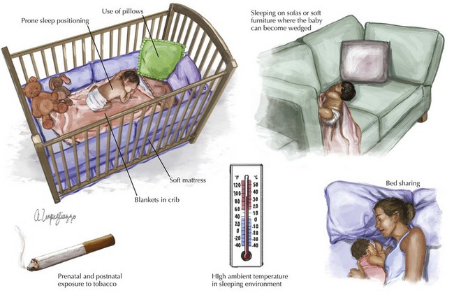
The most recent research suggests that SIDS is a polygenic, multifactorial condition inclusive of genetic, environmental, behavioral, and sociocultural factors. Well-established extrinsic risk factors for SIDS include prone sleep positioning; use of pillows, soft mattresses, or blankets in cribs; sleeping on sofas or other soft furniture in which the infant could become wedged; bed sharing; high ambient temperature in the sleeping environment; and prenatal and postnatal exposure to tobacco (Figure 11-1).

For many years, an ATLE was believed to be the predecessor of SIDS. This led to the widespread use of home apnea monitors in an attempt to prevent SIDS. However, studies such as the Collaborative Home Infant Monitoring Evaluation (CHIME) have demonstrated that ALTE neither precedes nor predicts SIDS. This has led to a change in focus from home apnea monitoring to the proven efficacy of the prevention methods outlined below.

Several factors argue against a causal relationship between ALTE and SIDS. Most notably, the timing of ALTE events versus SIDS deaths is distinctly different. Eighty percent of SIDS deaths occur between midnight and 6 AM, 82 percent of ALTE episodes occur between 8 AM and 8 PM. Upon review of effective preventive measures, interventions proven to reduce the incidence of SIDS (most notably supine sleep position) have not resulted in a decreased incidence of ALTE. In fact, the vast majority of SIDS victims do not experience ALTE before death. Further statistical analysis reveals that prior ALTE episodes were reported in only 5% of SIDS victims.

Because neither ALTE nor SIDS appears to exist beyond infancy, emerging evidence suggests that underlying brain immaturity may play a role in the pathogenesis. As noted above, the highest incidence of SIDS occurs between the second and fourth months of life, a period of intensive developmental changes in ventilatory, cardiac, and sleep–wake patterns in normal infants. The coincidence of timing suggests that infants are vulnerable to sudden death during a critical period of autonomic maturation. Additionally, classic studies on the infant nervous system show profound cardiovascular compromise in infants upon stimulation of the immature autonomic nervous system in the presence of apnea or hypoxia during sleep. This compromise was not noted in adult models. Currently, abnormalities in neurologic serotonin signaling and brainstem functioning as well as genetic polymorphisms interacting with specific environmental risk factors remain at the heart of ongoing research.

The final common pathway that seems to explain most cases of SIDS involves (1) a life-threatening event that causes asphyxia and brain hypoperfusion (e.g., rebreathing exhaled gases in prone position, gastroesophageal regurgitation causing obstructive apnea or activation of laryngeal receptors causing reflex apnea); (2) failure of arousal in response to asphyxia so that the infant does not turn his or her head and recover from the apnea; (3) hypoxic coma as a consequence of the continued asphyxia; (4) extreme bradycardia and gasping, and (5) failure of autoresuscitation because of ineffectual gasping, resulting in uninterrupted apnea and death.

Evaluation and Management

An infant coming to the ED in cardiopulmonary arrest should be managed as per the principles of Pediatric Advanced Life Support (PALS) with a brief period of well-executed CPR. CPR is a series of interventions aimed at restoring and supporting vital functions after apparent death (see Chapter 1). This involves cardiorespiratory monitoring, careful management of airway and breathing (including definitive airway management with artificial ventilation), vigorous monitored chest compressions, intraosseous and/or intraventricular access, and two to three doses of epinephrine. During this period, the patient’s history should be reviewed with the parent, if available, and with the emergency medical services personnel, and the infant should be examined thoroughly with a primary and secondary survey. The examination should include evaluation for signs of prolonged death such as rigor mortis, corneal clouding, and dependent lividity. The infant should be transferred to the pediatric intensive care unit if resuscitative efforts achieve cardiorespiratory stability. Infants who arrive in the ED in asystolic arrest have a poor prognosis. Prolonged resuscitation efforts past 20 minutes, without return of spontaneous circulation, are usually futile in the absence of treatable problems such as hypothermia, drug overdose, or ventricular tachycardia or fibrillation. The team leader should make the diagnosis of death and decide about discontinuation of resuscitative efforts based on the foregoing.

Family presence in the resuscitation room is gradually becoming the norm in pediatric EDs. This is because many parents want to be with their children during what may be the last moments of life. They also want to be sure that they and the ED staff have done all that is possible to resuscitate the child. If parents want to be in the resuscitation area, a nurse or social worker who can serve as a support person and interpret the ongoing resuscitative efforts should accompany them.

A well-prepared ED should have a plan in place for issues such as bereavement measures and postmortem care and notification of medicolegal authorities, the infant’s pediatrician, and any referring physicians and consultants.

First-response teams should be trained to make observations at the scene, including the position of the infant, any marks on the body, body temperature and rigor, type of bed and position of clothing and bedding, room temperature, type of ventilation and heating, and reactions of caretakers.

The loss of an infant is devastating for all concerned. Families who suffer such a loss from SIDS may encounter a police investigation, a waiting period for autopsy results, and a lack of emotional closure. It is important for the professional response teams to remain supportive, empathic, and nonaccusatory while obtaining essential information surrounding the death of the infant. Emotional support should be offered to the caregivers in the ED by both the medical staff and by a social worker or a grief counselor. The parents may be informed that a SIDS death happens quickly and silently without causing any pain and suffering to the infant. Additionally, information obtained in the acute care setting should be relayed to the family’s pediatrician or family practice physician so that appropriate follow-up can be arranged, including age-appropriate support for surviving siblings.

Prevention and Future Directions

Because of the lack of evidence available to support the use of home monitoring, the current and future management of SIDS focuses on risk reduction strategies. Infants should be placed in the supine position every time they are laid down for sleep. The prone sleeping position is associated with an increased risk of SIDS in a number of observational studies. Similarly, the AAP recommends against placing infants on their side for sleep because of the instability of this position. As a result of the national Back to Sleep campaign launched in 1994, the SIDS rate in United States has fallen by more than 50%.

In addition to sleep position, the sleep environment (inclusive of sleep surface, sleepwear, bedding and the incidence of co-sleeping) appears to affect the risk of SIDS. It is recommended that infants sleep on a firm surface, such as on a safety-approved crib mattress covered with a fitted sheet without blankets, pillows, wedges, rolls, or toys in their bassinet or crib, since these objects increase the risk of suffocation. Infants should sleep in a shared room but in a separate bed. Bed sharing has benefits such as bonding and promoting breast feeding, but has been linked to higher rates of SIDS, especially for infants younger than 4 months of age, if either parent smokes, drinks alcohol before bedtime, or is abusing prescription or illicit drugs. Additionally, there is a consistent association between increased risk of SIDS and sharing a sofa or couch with parents.

Studies have suggested that pacifier use may help reduce the risk of SIDS. The mechanism is unknown, but it is hypothesized that it may lower the arousal threshold in sleeping infants. Overheating the room should be avoided because it increases the SIDS risk. A recent study suggested that having a fan in the room may reduce the risk of SIDS by 70%. Mothers should avoid smoking during pregnancy and around the infants. Home cardiorespiratory monitoring of apnea and bradycardia has not been shown to prevent SIDS and is therefore not recommended.

About 20% of SIDS deaths occur while the infant is under the care of a nonparent caregiver. Accordingly, it is important that all childcare providers be apprised of the above precautions and safe practices to further lower the SIDS rate.

Suggested Readings