Chapter 30
Aortic Valve Disease
1. What is the most common cause of aortic stenosis (AS) in developed countries today, and what is the current thinking about its pathogenesis?
Although rheumatic fever was once the most common cause of AS, today calcific disease of either bicuspid or tricuspid AS is the leading cause. Once considered a degenerative disease, it is now clear that calcific AS is an inflammatory process with many similarities to atherosclerosis. A normal aortic valve, and AS as a result of congenital bicuspid aortic valve, rheumatic AS, and calcific AS, are shown in Figure 30-1.
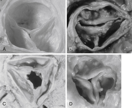
Figure 30-1 Normal and stenotic aortic valves. A, Normal aortic valve. B, Congenital bicuspid aortic stenosis. A false raphe is present at 6 o’clock. C, Rheumatic aortic stenosis. The commissures are fused with a fixed central orifice. D, Calcific degenerative aortic stenosis. (From Libby P, Bonow RO, Mann DL, Zipes DP: Braunwald’s heart disease: a textbook of cardiovascular medicine, ed 8, Philadelphia, 2008, Saunders.)
2. What is the pathophysiology of AS, and what effect does it have on the left ventricle (LV)?
AS exerts a pressure overload on the LV. Normally, pressure in the LV and aorta are similar during systole, as the normal aortic valve permits free flow of blood from LV to aorta. However, in AS, the stenotic valve forces the LV to generate higher pressure to drive blood through the stenosis, causing a pressure difference (gradient) from LV to aorta. The LV compensates for this pressure overload by increasing its mass (left ventricular hypertrophy [LVH]). The ways in which the transvalvular gradient are measured and quantified are shown in Figure 30-2.
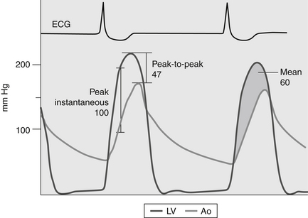
Figure 30-2 Various methods of describing an aortic transvalvular gradient. The figure shows representative pressure tracings measured during cardiac catheterization in a patient with aortic stenosis. One pressure transducer is placed in the left ventricle and a second pressure transducer is positioned in the ascending aorta. The peak-to-peak gradient (47 mm Hg) is the difference between the maximal pressure in the aorta (Ao) and the maximal left ventricle (LV) pressure. The peak instantaneous gradient (100 mm Hg) is the maximal pressure difference between the Ao and LV when the pressures are measured in the same moment (usually during early systole). The mean gradient (shaded area) is the integral of the pressure difference between the LV and Ao during systole (60 mm Hg). (From Bashore TM: Invasive cardiology: principles and techniques, Philadelphia, 1990, BC Decker, p. 258.) ECG, Electrocardiogram.
3. How is left ventricular hypertrophy compensatory?
The Law of Laplace states that systolic wall stress (σ) is equal to:

4. Are there downsides to LVH?
5. What are the classic symptoms of AS, and why are they important?
The classic symptoms of AS are angina and syncope and those of heart failure (dyspnea, orthopnea, paroxysmal nocturnal dyspnea, edema, etc.). Their importance is graphically displayed in Figure 30-3. In the absence of symptoms, survival is nearly the same as that of an unaffected population. However, at the onset of symptoms there is a dramatic demarcation such that mortality increases to 2% per month, so that three-quarters of all AS patients are dead within 3 years of symptom onset unless proper therapy is instituted. It should be noted that when the data for this figure were compiled, the etiology of AS was usually rheumatic or congenital heart disease and the average age of the patients was 48 years. Since then the etiology of AS has changed (as noted above) and with it the age at symptom onset has increased by about 15 years.
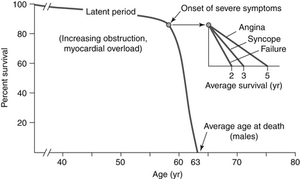
Figure 30-3 The natural history of medically treated aortic stenosis. Once symptoms develop in a patient with aortic stenosis, the average survival with medical treatment (e.g., no curative surgery) is only 2 to 5 years. (From Townsend CM Jr, Beauchamp RD, Evers BM, Mattox KL: Sabiston textbook of surgery: the biological basis of modern surgical practice, ed 18, Philadelphia, 2008, Saunders.)
6. What are findings of AS on physical examination?
7. How is echocardiography used to assess the patient with AS?

as the valve area decreases, velocity of flow must increase for flow to remain constant (Fig. 30-4). This increase in blood velocity at the valve orifice is detected by Doppler ultrasound. The severity of AS is assessed using the factors given in Table 30-1. In general, if a patient has the symptoms of AS and assessment indicates severe disease, then the symptoms are attributed to AS. However, it must be emphasized that the benchmarks listed here are only guidelines to severity, and some patients exhibit exceptions to them.
TABLE 30-1
ECHOCARDIOGRAPHIC CRITERIA FOR THE DEGREE OF AORTIC STENOSIS
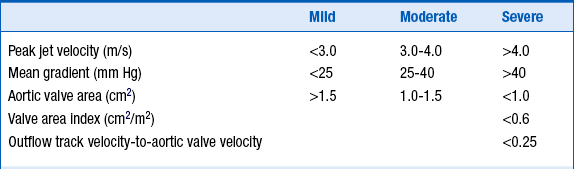
Modified from Bonow RO, Carabello BA, Chatterjee K, et al: ACC/AHA 2006 guidelines for the management of patients with valvular heart disease. J Amer Coll Cardiol 48:e1-e148, 2006.
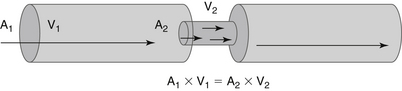
Figure 30-4 Determination of aortic valve area using the continuity equation. For blood flow (A1 × V1) to remain constant when it reaches a stenosis (A2), velocity must increase to V2. Determination of the increased velocity V2 by Doppler ultrasound permits calculation of both the aortic valve gradient and solution of the equation for A2. (From Townsend CM Jr, Beauchamp RD, Evers BM, Mattox KL: Sabiston textbook of surgery: the biological basis of modern surgical practice, ed 18, Philadelphia, 2008, Saunders.) A, Area; V, velocity.
8. What other tests are useful in assessing AS?
As noted earlier, the presence or absence of symptoms is a key determinant of outcome, yet in some patients an accurate history may be difficult to obtain. In such cases, more objective evidence of cardiac compromise, such as exercise intolerance, may be helpful. Although exercise stress testing should never be performed in symptomatic AS patients, stress testing may be very helpful in establishing more objective evidence of symptomatic status when the history is unclear. As many as one-third of AS patients may become symptomatic for the first time during stress testing. This phenomenon probably indicates a previous denial of symptoms or a lifestyle altered to avoid symptoms. Such stress testing, if undertaken, should only be done with careful physician supervision.
In some cases, the severity of AS is still uncertain following echocardiography. Cardiac catheterization to obtain invasive hemodynamics, including the transvalvular pressure gradient and cardiac output, is then used to derive the valve area (see Chapter 14).
9. What is the therapy for AS?
No effective medical therapy is effective in the chronic treatment of this disease. The mainstay of therapy for this mechanical problem is mechanical relief of the obstruction, in the form of AVR. In most patients, AVR is performed surgically. However, for selected patients at prohibitive or high risk for surgical AVR, transcatheter placement of the aortic valve (“TAVI” or “TAVR”) is now approved from the femoral and transapical approaches in the US (see Chapter 32, Prosthetic Heart Valves). It is almost certain that this field will evolve to have broader indications as devices become safer and easier to employ.
10. What are the class I indications for aortic valve replacement?
 Symptomatic patients with severe AS
 Severe AS with LV systolic dysfunction (LV ejection fraction [LVEF] less than 50%)

11. What is the outcome after AVR?
12. What are the causes of aortic regurgitation?
13. What is the pathophysiology of aortic regurgitation?
14. What are the symptoms of aortic regurgitation?
15. What are the findings of AR on physical examination?
The widened pulse pressure and high total stroke volume cause several physical signs of AR.
 The head may bob with each heartbeat (de Musset sign).
 Auscultation of the femoral artery may produce a sound similar to a pistol shot (pistol shot pulse).


16. How is the diagnosis of AR confirmed?
Although a chest radiograph is helpful in evaluating heart size and pulmonary congestion, echocardiography remains the mainstay of diagnosis. Cardiac size and function are evaluated using this technique. Often the anatomic abnormality responsible for the patient’s AR can be established. As shown in Figure 30-5, Doppler interrogation of the valve reveals the jet of blood leaking across the valve in diastole. Table 30-2 displays current criteria for establishing the severity of AR. Cardiac magnetic resonance imaging may also be useful in quantitating the magnitude of volume overload and in assessing left ventricular volume and mass.
TABLE 30-2
CARDIAC CATHETERIZATION AND ECHOCARDIOGRAPHIC CRITERIA FOR THE DEGREE OF AORTIC REGURGITATION
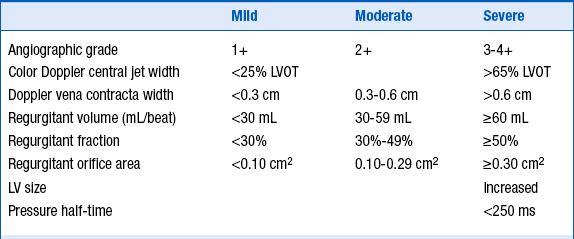
LV, Left ventricle; LVOT, left ventricular outflow tract.
Modified from Bonow RO, Carabello BA, Chatterjee K, et al: ACC/AHA 2006 guidelines for the management of patients with valvular heart disease. J Amer Coll Cardiol 48:e1-e148, 2006.
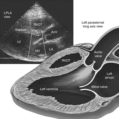
Figure 30-5 Aortic regurgitation. Left parasternal long-axis view of the heart, demonstrating aortic regurgitation seen on Doppler echocardiogram, along with the corresponding anatomic illustration. In actuality, the regurgitant jet is displayed in color, reflecting the direction of blood flow. (From Yale School of Medicine: Yale atlas of echocardiography. Available at: http://www.yale.edu/imaging/echo_atlas/entities/aortic_regurgitation.html. Accessed August 30, 2008.) AoV, Aortic valve; LA, left atrium; LPLA, left parasternal long axis: LV, left ventricle; MV, mitral valve; RVOT, right ventricular outflow tract.
 Symptomatic patients with severe AR, irrespective of LV systolic function
 Asymptomatic patients with chronic severe AR and LV systolic dysfunction (LVEF 50% or less)

18. Is the presentation of acute AR different from that of chronic AR?
Bibliography, Suggested Readings, and Websites
1. Bekeredjian, R., Grayburn, P.A. Valvular heart disease: aortic regurgitation. Circulation. 2005;112:125–134.
2. Bonow, R.O., Carabello, B.A., Chatterjee, K., et al. ACC/AHA 2006 guidelines for the management of patients with valvular heart disease. J Am Coll Cardiol. 2006;48:e1–e148.
3. Carabello, B.A. Clinical practice. Aortic stenosis. N Engl J Med. 2002;346:677–682.
4. Carabello, B.A. Evaluation and management of patients with aortic stenosis. Circulation. 2002;105:1746–1750.
5. Enriquez-Sarano, M., Tajik, A.J. Clinical practice. Aortic regurgitation. N Engl J Med. 2004;351:1539–1546.
6. Gaasch, W.H., Course and management of chronic aortic regurgitation in adults. Basow D.S., ed. UpToDate, Waltham, MA 2013 UpToDate. Available at: http://www.uptodate.com/contents/course-and-management-of-chronic-aortic-regurgitation-in-adults Accessed March 26, 2013
7. Wang, S.S. Aortic regurgitation. Available at: http://www.emedicine.com. Accessed March 26, 2013
8. Leon, M.B., Smith, C.R., Mack, M., et al. Transcatheter aortic-valve implantation for aortic stenosis in patients who cannot undergo surgery. N Engl J Med. 2010;363:1597–1607.
9. Otto, C.M., Pathophysiology and clinical features of aortic stenosis in adults. Basow D.S., ed. UpToDate, Waltham, MA 2013 UpToDate. Available at: http://www.uptodate.com/contents/clinical-features-and-evaluation-of-aortic-stenosis-in-adults Accessed March 26, 2013
10. Vahanian, A., Baumgartner, H., Bax, J., et al. Guidelines on the management of valvular heart disease: the Task Force on the Management of Valvular Heart Disease of the European Society of Cardiology. Eur Heart J. 2007;28:230–268.

