Anterior Sciatic Nerve Block

Published on 06/02/2015 by admin

Filed under Anesthesiology

Last modified 22/04/2025

Print this page

rate 1 star rate 2 star rate 3 star rate 4 star rate 5 star
Your rating: none, Average: 0 (0 votes)

This article have been viewed 1249 times

44 Anterior Sciatic Nerve Block
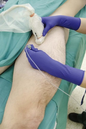
The anterior approach to proximal sciatic nerve block can be used in patients who are difficult to position lateral or prone. This supine approach is deeper than other approaches because the sciatic nerve lies far from the anterior surface of the thigh and is therefore primarily used in thin patients.

Suggested Technique

First obtain a long-axis view of the femur with the transducer placed on the anterior aspect of the thigh. The femur is easily identified by its bright cortical surface and acoustic shadowing. Next, slide the transducer medially to obtain a long-axis view of the sciatic nerve at approximately twice the depth of the femur. The sciatic nerve is wide and straight and therefore appears as an echogenic linear structure lying deep to the adductor magnus muscle. If the femoral artery is visible, then the transducer has slid too far medially.

The block needle approaches in-plane with the sciatic nerve in this long-axis view. The approach can be from proximal to distal or distal to proximal depending on the side of the block and the handedness of the operator. The sciatic nerve will bow like a string as the block needle approaches. When the local anesthetic is within the correct tissue plane the injection will track along the proximal-distal course of the nerve and ideally on both the anterior and posterior sides of the nerve. Some practitioners elect to combine ultrasound imaging with nerve stimulation to confirm nerve identity for this block. Even though the sciatic nerve has a large diameter and a relatively straight course, it can still be difficult to simultaneously maintain a long-axis view of the nerve with the needle in-plane.

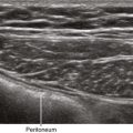
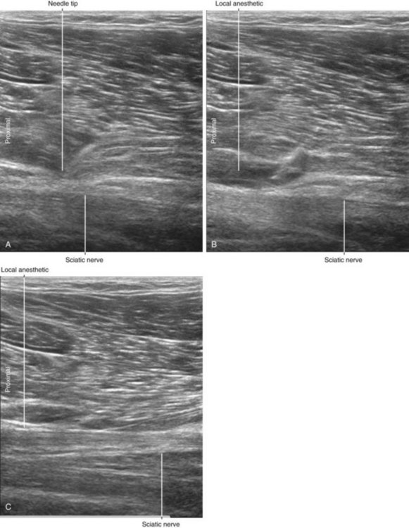
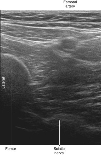
In many patients the sciatic nerve is easy to identify because it appears as a hyperechoic, linear (cable-like) structure deep to the clearly delineated border of the adductor magnus muscle that is formed by the intermuscular septum.1 Because this block is performed distal to the lesser trochanter of the femur, external rotation of the leg promotes access to the sciatic nerve.2

The long-axis in-plane approach constrains the needle to a trajectory that can potentially puncture the nerve. Nerve stimulation may be used to help rule out intraneural needle tip placement by verifying evoked motor responses are eliminated at low stimulation currents (e.g., ≤0.2 mAmp via cathodal stimulation).