Antenatal screening

Published on 01/03/2015 by admin

Filed under Basic Science

Last modified 22/04/2025

Print this page

rate 1 star rate 2 star rate 3 star rate 4 star rate 5 star
Your rating: none, Average: 0 (0 votes)

This article have been viewed 1404 times

77

Antenatal screening

There are approximately 700,000 pregnancies per annum in the UK and 200 to 250 million world wide. Most result in the birth of a healthy baby, though in a few cases there may be problems affecting delivery or a baby’s development. Antenatal screening is a way of assessing whether the fetus could potentially develop, or indeed has developed, an abnormality during pregnancy. If the risk is high the mother may be offered prenatal diagnosis to find out the likelihood of developing the abnormality. Prior knowledge of problems can help parents plan how best to deal with them: by preparing for special care, or choosing to terminate the pregnancy.

First trimester screening

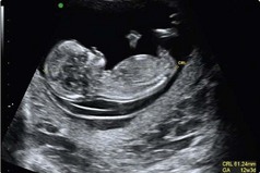
Although second trimester screening has been common practice, combined first trimester screening is currently considered to be best practice as it provides a higher detection rate and lower false positive rate. It uses a combination of ultrasound measurement of fetal nuchal translucency (NT), and measurement of the maternal serum markers free beta HCG (FβHCG) and pregnancy-associated plasma protein A (PAPP-A), to derive a combined risk for Down’s syndrome. Each of these markers, including NT, varies with gestation and an accurate measurement of fetal maturity is required for accurate interpretation of results. For first trimester screening, ultrasound measurement of fetal crown rump length (CRL; Fig 77.1), carried out at the same time as the NT measurement, is used as the basis of the calculation of gestation for conversion of marker concentrations into a multiple of the median (MoM). An MoM is a measure of how far an individual test result deviates from the median. MoM is commonly used to report the results of medical screening tests, particularly where the results of the individual tests are highly variable.

Nuchal translucency

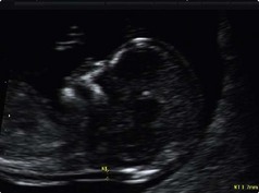
Nuchal translucency (Fig 77.2) is the fluid-filled area that is present at the back of the fetal neck and measures around 1.0 mm in unaffected pregnancies at 11–13 weeks’ gestation. It tends to be increased in Down’s syndrome and can be measured accurately by ultrasound (to the nearest 0.1 mm). The NT measurement is converted to a multiple of the median NT size at the appropriate CRL and a risk estimated.

For combined screening, NT measurements should be taken only when the fetal CRL falls within the range of 45–84 mm (equivalent to 11+2 – 14+1 weeks of gestation). Before this, the fetus is too small to allow accurate NT measurement, and after 14 weeks the strength of the association between NT and fetal chromosome abnormalities is not strong enough to be used in this respect.

Risk calculation

The results (MoM) of NT and biochemical markers are used to calculate the risk of Down’s syndrome. A cut-off of 1 in 150 is used to define whether a pregnancy is ‘low risk’ or ‘high risk’ on combined screening. All screening risk results equal to or greater than 1 in 150 are considered high risk, and these women are offered further counselling and diagnostic testing. About 2% of results fall into this category. All screening risk results less than 1 in 150 are considered low risk and no further action is usually indicated in these cases. It is also worth noting that as maternal age is a component of the screening risk calculation, there are a higher proportion of ‘positive’ tests in older women and the test detects an increasing proportion of Down’s syndrome pregnancies as maternal age advances. Multi-stage testing is strongly discouraged. Women who have had first trimester combined screening should not subsequently go on to have a second trimester biochemical screening test for Down’s syndrome.

Second trimester screening

Although all women, who wish to be screened, should be encouraged to do so in the first trimester, second trimester screen provides an opportunity for those who present too late for first trimester screening. Maternal blood sample should be taken between 14+2 weeks and 20+0 weeks for measurement of alpha fetoprotein (AFP), human chorionic gonadotrophin (HCG), unconjugated oestriol (UE3) and Inhibin A. Pregnancies affected by Down’s syndrome have elevated levels of hCG and Inhibin A to around twice the levels found in unaffected pregnancies (2.0 MoM), while AFP and UE3 are reduced to around three-quarters of normal levels (0.75 MoM). These results along with maternal age are used to calculate the risk of Down’s syndrome. As with first trimester, a cut-off of 1 in 150 is used to define risk. All screening risk results equal to or greater than 1 in 150 (around 3–4% of pregnancies fall in this category) are high risk and these women are offered further counselling and diagnostic testing.

Other factors affecting interpretation of biochemical markers

Several factors have been identified which affect serum marker concentrations and therefore the risk estimate derived from them. Corrections, to take account of these variables, can be made to provide a more accurate estimate of risk for individual women.

image Gestation. As all serum marker concentrations vary with gestation, they (and NT) are interpreted by expressing results as a multiple of the appropriate gestational median level in unaffected pregnancies, but the precision of this estimate depends on the accuracy of the gestational estimate. Screening results cannot and should not be interpreted without an accurate estimate of gestation. An ultrasound estimate of gestation is used in preference to that calculated from last menstrual period.

image Maternal weight. Women who weigh more than 65 kg tend to have increased blood volume, resulting in a dilutional lowering of serum concentration of various markers. The opposite effect is found in women of lower than average maternal weight. The effect of maternal weight is particularly marked at the extremes of the weight range and a correction factor is usually applied. Maternal weight has no effect on NT measurements.

image Maternal smoking. Smoking in pregnancy affects placental function resulting in reduced secretion of PAPP-A, hCG and UE3, and increased secretion of AFP and Inhibin A. Correction for smoking status allows for a more accurate risk calculation.

image Assisted reproduction. An important practical consideration in in vitro fertilization pregnancies is that the age of an egg donor (if applicable) must be used to derive the maternal age risk, while for frozen embryos, the age at conception should be used.

image Previous affected pregnancy with Down’s syndrome. A previous pregnancy with Down’s syndrome increases the risk in subsequent ones. This is in addition to increased maternal age and significantly increases the probability that a screening result will fall into the high-risk category.

image Ancestry/ethnicity. If this information is available then appropriate median concentration should be used to calculate the MoMs and produce a more accurate risk estimate.