Hematometra and Hematocolpos

Published on 10/03/2015 by admin

Filed under Obstetrics & Gynecology

Last modified 22/04/2025

Print this page

rate 1 star rate 2 star rate 3 star rate 4 star rate 5 star
Your rating: none, Average: 5 (1 votes)

This article have been viewed 10923 times

Hematometra and Hematocolpos

Synonyms/Description

Hydrometra and hydrocolpos
Pyometra and pyocolpos

Etiology

The most common etiology is cervical or vaginal obstruction, resulting in a collection of blood, pus, or fluid that distends the uterus or vagina.

Hematometra/Hydrometra

Cervical stenosis can develop for multiple reasons (treatment for cervical dysplasia, prior ablation, postmenopausal effect), blocking the cervical canal and resulting in accumulation of fluid/blood in the uterine cavity. Advanced endometrial cancer can either block the canal or produce copious amounts of fluid/blood filling the uterine cavity. Many years ago, fluid in the endometrial cavity was considered a sonographic sign of cancer. More recently, Dr. Steven Goldstein showed that it is not the intracavitary fluid that conveys the risk of cancer, rather the appearance of the wall around the fluid. If the endometrium surrounding the fluid is smooth and thin, endometrial cancer is unlikely, whereas an irregular wall or mass protruding into the fluid indicates the presence of a tumor.
Benign causes of hematometra include an intracavitary fibroid or polyp in the cervix or lower uterine segment, and adhesions such as those encountered after an incomplete endometrial ablation. If the ablation seals the lower uterine segment but leaves intact endometrium at the fundus, cyclical hematometra with bilateral hematosalpinges may result. Patients with Müllerian duct anomalies who have a uterine duplication may have an obliterated horn, which will fill with blood and cause cyclical pain, typically presenting at puberty. This may pose a diagnostic dilemma if the uterine anomaly is not known, because these patients will present with a painful cystic mass in the pelvis often mistaken for a degenerating fibroid or adnexal mass. If the Müllerian duct anomaly is severe, the hematometra/hydrometra may even be visible prenatally as an intra-abdominal cystic mass in the fetus.

Hematocolpos/Hydrocolpos

Obstruction within the distal vagina is usually responsible for the development of a hematocolpos. If there is a backup of blood or fluid in the vagina, this may extend into the uterine cavity and result in an associated hematometra. Obstructions of the distal vagina are commonly owing to congenital Müllerian duct anomalies such as a transverse vaginal septum or an imperforate hymen. Patients with congenital anomalies of the vagina often have associated anomalies of the uterus; however, patients with vaginal obstruction may not become symptomatic until menarche.

Ultrasound Findings

The sonographic appearance of a hematometra/hydrometra is the presence of fluid distending the uterine cavity. Similarly hematocolpos/hydrocolpos is the presence of fluid in an obstructed vagina. The fluid often contains low-level echoes much like the texture of an endometrioma, indicating the presence of unclotted blood. The presence of more than a sliver of fluid in the uterus and/or vagina needs to be further investigated sonographically. Consider the patient’s history: Has the patient had an ablation, and does she have cyclical symptoms? Is there a known Müllerian duct anomaly? Did the patient become symptomatic at menarche or is she asymptomatic? The ultrasound exam should include a 3-D evaluation of the uterine shape looking for Müllerian uterine anomalies (see Müllerian Duct Abnormalities). The tubes should be evaluated for the presence of fluid, to determine if the obstruction has extended to the fallopian tubes. The inner lining of the uterus (endometrial surface) should be evaluated carefully for any masses indicating polyps or malignancy. The cervix should be assessed for the presence of obstructing lesions such as fibroids or large polyps. It may be difficult to see a small cervical carcinoma sonographically; however, the type of cervical lesion that obstructs the uterus is likely large enough to be visualized using a high-frequency transvaginal probe.
The vagina is often easier to evaluate sonographically by placing the vaginal probe on the introitus and looking down the length of the vagina and urethra (see Vaginal Masses and also Bladder Masses). Any fluid collection along the vagina may represent an obstructed hemivagina, a finding often associated with congenital uterine anomalies.

Differential Diagnosis

Fluid inside a normal uterine cavity does not have a differential diagnosis and is not always pathologic. It is important to note that a small amount of clear fluid in a postmenopausal uterus is not abnormal. The different causes of hematometra/hydrometra (described in Etiology, earlier) are the more diagnostically challenging step. The presence of fluid in an abnormal uterine cavity such as a rudimentary uterine horn can be confused with a cystic adnexal mass, including those of ovarian origin. If the ipsilateral ovary is normal sonographically, then fluid in a rudimentary horn may be confused with a degenerating fibroid or a hydrosalpinx. Recognizing the uterus as unicornuate using 3-D ultrasound would be an important clue to the correct diagnosis of a Müllerian duct abnormality. The presence of such a uterine anomaly is also vital to making the correct diagnosis of an obstructed hemivagina. Otherwise the vaginal fluid could be confused with a Gartner’s duct cyst or urethral diverticulum (see Vaginal Masses and also Bladder Masses). The patient’s age, pain, and menstruation history are likely to be important factors in arriving at the correct diagnosis.

Clinical Aspects and Recommendations

The cause of the hematometra/hydrometra or hematocolpos/hydrocolpos will guide the management. Patients who are asymptomatic and simply have a small amount of clear intrauterine fluid with a normal endometrium do not require treatment because this is not considered a clinically significant finding. Pediatric patients are likely to have Müllerian duct abnormalities that need surgical intervention. Postmenopausal patients who are bleeding and have a fluid collection in the uterus require evaluation of the cervix and endometrium to rule out malignancy. Those with a previous ablation that somehow spared the fundus of the uterus are a challenge and should be evaluated on an individual basis.

Figures

image
image
image

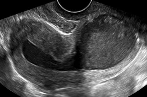
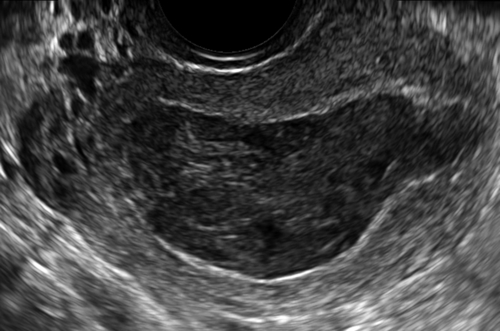
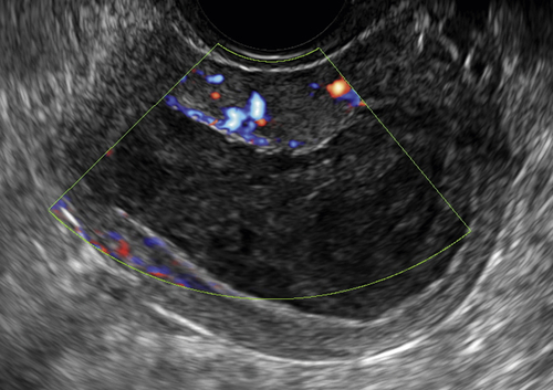
Figure H1-1 A and B, Longitudinal and transverse views of the uterus with fluid present in the cavity, including the cervix. Note that the fluid has low-level echoes, which is characteristic of unclotted blood. This was the result of an obstruction at the level of the external os. C, Blood flow in the myometrium but no flow within the uterine cavity.

image

Figure H1-2 Large hematometra. Note the large fluid collection filling the uterus with a closed cervix.

image

Figure H1-3 Small amount of fluid in the uterine cavity of an asymptomatic patient. With a normal-appearing endometrium, this is not considered to be clinically significant.


image

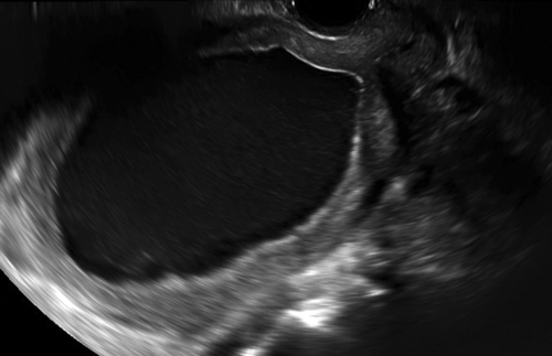
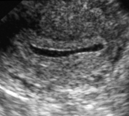
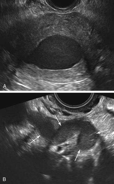
Figure H1-4 This is a patient post-endometrial ablation with pelvic pain. A, A transverse view through the uterine fundus showing a hematometra (centrally). B, A dilated fallopian tube full of fluid (arrows) with debris consistent with a hematosalpinx. The contralateral tube had the same appearance, suggesting that there is residual cycling endometrium at the uterine fundus, above the level of the ablation.


image

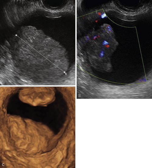
Figure H1-5 Postmenopausal patient with endometrial cancer and a 5-week history of postmenopausal bleeding. A and B, An endometrial mass (calipers) with abundant vascularity, located in the uterus and outlined by intracavitary fluid. C, A 3-D rendered image of the uterine cavity distended with fluid, showing two separate irregular masses in the endometrium and protruding into the cavity.


image

Figure H1-6 Translabial view of the vagina of a young patient with a known complex Müllerian duct abnormality. Note the obstructed hemivagina containing fluid (arrows).


Suggested Reading

Goldstein S.R. Postmenopausal endometrial fluid collections revisited: look at the doughnut rather than the hole. Obstet Gynecol. 1994;83:738–740.

Drakonaki E.E., Tritou I., Pitsoulis G., Psaras K., Sfakianaki E. Hematocolpometra due to an imperforate hymen presenting with back pain: sonographic diagnosis. J Ultrasound Med. 2010;29:321–322.

Shaked O., Tepper R., Klein Z., Beyth Y. Hydrometrocolpos—diagnostic and therapeutic dilemmas. J Pediatr Adolesc Gynecol. 2008;21:317–321.

Verma S.K., Baltarowich O.H., Lev-Toaff A.S., Mitchell D.G., Verma M., Batzer F. Hematocolpos secondary to acquired vaginal scarring after radiation therapy for colorectal carcinoma. J Ultrasound Med. 2009;28:949–953.